Carl A. Germann
Orthopedic Emergencies, ed. Michael C. Bond, Andrew D. Perron, and Michael K. Abraham. Published by Cambridge University Press. © Cambridge University Press 2013.
Distal radius and ulnar injuries
PEARL: Fractures of the distal radius and ulna are the most common type of fractures in patients younger than 75 years.
PEARL: Distal radius and ulnar injuries are often associated with median and ulnar neuropathies.
Distal radius fracture
Key facts
· A Colles fracture (Figures 1.1 and 1.2): A transverse fracture of the distal radial metaphysis with dorsal displacement and angulation, often caused by a fall on an outstretched hand
· A reverse Colles or Smith fracture (Figure 1.3): A transverse fracture of the metaphysis of the distal radius, with associated volar displacement and volar angulation. The mechanism of injury is often a fall on to the dorsum of the hand with the wrist in flexion
· Barton fracture (Figure 1.4): A distal radius fracture with dislocation of the radiocarpal joint
o A volar Barton fracture occurs when the wrist is volarly flexed, and affects the volar rim of the radius.
o A dorsal Barton fracture occurs with dorsal flexion and affects the dorsal rim of the radius
· Hutchinson fracture (Figure 1.5): An intra-articular transverse fracture of the radial metaphysis with extension through the radial styloid, often caused by a direct blow or a fall on the radial side of the wrist
o Also termed a Chauffer’s fracture
· Clinical presentation: distal radius fracture patterns usually present with pain, swelling, and deformity of the wrist
· On physical examination, Colles fractures have a dinner-fork deformity caused by the dorsal displacement and angulation of the radius
· Smith fractures often have fullness on the volar aspect of the wrist
· Median nerve injury can occur with Colles and Smith fractures and a careful neurovascular examination both on initial presentation and following treatment is required

Figure 1.1 Colles fracture. Note the dorsal angulation of the distal radius as shown in Figure 1.2. (Image courtesy of Carl Germann, MD.)

Figure 1.2

Figure 1.3 Smith fracture. The hand and wrist is volarly displaced with respect to the forearm. (Image courtesy of Carl Germann, MD.)

Figure 1.4 Volar Barton fracture. A fracture of the volar margin of the carpal surface of the radius. (Image courtesy of Carl Germann, MD.)

Figure 1.5 Hutchinson fracture: An intra-articular fracture through the radial styloid process. (Image courtesy of Carl Germann, MD.)
Diagnostic testing
· For Colles and Smith fracture patterns, radiographs of the wrist will demonstrate the fracture through the radial metaphysis. The lateral radiograph is the best view to determine the degree of dorsal or volar displacement and angulation
· The lateral radiograph is the best view for revealing an intra-articular fracture of the radius and any associated carpal displacement in Barton fractures. A posteroanterior (PA) radiograph often shows a comminuted fracture of the distal radius
· PA radiographs of the wrist are best to see a Hutchinson fracture
Treatment
· Colles fractures should undergo closed reduction. This can be facilitated by the use of a hematoma block and finger traps. After successful reduction, patients should be immobilized in a long-arm splint in neutral position or pronation with orthopedic follow-up in 7 to 10 days. Emergent orthopedic consultation is necessary if initial attempts at closed reduction are unsuccessful, if there is neurovascular compromise, or if there is an open fracture
· Smith fracture should undergo closed reduction. Following reduction, patients should be placed in a long-arm splint in supination. Emergent orthopedic/hand-specialist consultation is recommended for these fractures because they are more likely to be unstable and urgent surgical management is more often necessary
· Barton fractures require emergency orthopedic/hand-specialist consultation for early operative management
· Non-displaced Hutchinson fractures can be managed with a short-arm splint and routine orthopedic/hand-specialist follow-up. Displaced fractures require reduction and immobilization. Accurate anatomic alignment following reduction is essential because multiple ligaments of the wrist attach to the radial styloid process and inappropriate alignment can cause future complications
Prognosis
· Complications include:
o Malunion
o Radioulnar and radiocarpal instability
o Arthritis
o Chronic pain
o Non-union
· However, good to excellent results are often achieved in most patients
Distal radioulnar joint disruption (DRUJ)
Key facts
· Disruption of the distal radioulnar joint (DRUJ) may be seen as an isolated injury, or more commonly, in association with distal radius fractures
o Initially unrecognized in up to 50% of cases
· Dorsal dislocations are the most common and are typically the result of a fall on to an outstretched arm with a rotational pronation force to the impact
· Volar dislocations are typically the result of a fall on to an outstretched arm with a rotational supination force to the impact
Clinical presentation
· Often overshadowed by more apparent injuries
· On physical examination a dorsal dislocation reveals excessive prominence of the ulnar head and lack of forearm rotation secondary to pain when the wrist is supinated
· Volar dislocations will have a loss of the typical dorsal prominence of the ulnar head and lack of forearm rotation secondary to pain when the wrist is pronated
Diagnostic testing
· PEARL: In DRUJ injuries, the lateral radiograph usually demonstrates volar or dorsal displacement of the ulna that normally overlap the radius. Standard radiographs of a DRUJ dislocation demonstrate overlap of the distal ulna with the distal radius on the PA view. On the lateral view the ulnar head will be displaced:
o Dorsally for dorsal dislocations
o Volarly with volar dislocations
· Radiographic signs of DRUJ instability are:
o Ulnar styloid fracture involving the base with more than 2 mm displacement
o Irreducible dislocation of the DRUJ
o Fractures involving the sigmoid notch of the radius
o Wide displacement of the DRUJ
o Radial shortening
Treatment
· If DRUJ instability is suspected, based on clinical examination or radiographic studies, an emergent orthopedic or hand-specialist consultation should be obtained for reduction and immobilization
Prognosis
· DRUJ injuries have a high recurrence rate and may require reconstructive surgery
Carpal bone fractures and dislocations
Scaphoid fracture
PEARL: The scaphoid is the most commonly fractured carpal bone yet one of the most commonly missed wrist injuries. A thorough history and physical examination, coupled with a high index of suspicion, are necessary to make the diagnosis.
Key facts
· Scaphoid fractures account for 60–70% of all diagnosed carpal injuries
· Radiographic findings (Figure 1.6) can be subtle or absent, rendering the diagnosis difficult to make
· Accurate early diagnosis of scaphoid fractures is critical, as a missed or delayed diagnosis can result in long-term pain, loss of mobility, and decreased function
· The scaphoid has a high rate of non-union
· Avascular necrosis of the scaphoid is because its blood supply arises distally from small branches of the radial artery and the palmar and superficial arteries. The proximal portion of the scaphoid is completely dependent on this distal blood supply, thus it is at risk of avascular necrosis following fracture
· In general, the more proximal, oblique, or displaced the fracture, the greater the risk of interrupting the blood supply

Figure 1.6 Scaphoid fracture: An acute non-displaced fracture is shown in anteroposterior view. (Image courtesy of Carl Germann, MD.)
Clinical presentation
· Snuff box tenderness is classically cited as the most common finding, although the sensitivity of this test is disputed
· Many authors feel a better test for scaphoid injury is axial compression of the thumb along its longitudinal axis
· The examining physician (EP) should remain vigilant for associated injuries that can be found on physical examination
o Common associated injuries include fractures of the distal radius, lunate, or radial head at the elbow
o Median nerve injury has also been described in association with scaphoid fractures
Diagnostic testing
PEARL: Even with appropriate films, fractures of the scaphoid can be subtle and difficult to visualize. Conservative estimates suggest that 10–20% of these fractures will not be visible on any view in the acute setting.
· A typical wrist series includes a PA and lateral radiograph of the wrist
· In cases where there is high clinical suspicion, a scaphoid view of the wrist can also be obtained
o This reduces the foreshortening of the scaphoid that occurs on a normal PA view, and displays the entire length of the scaphoid
o However, even with excellent radiographic technique, a fracture may not be visualized
· Magnetic resonance imaging (MRI) and computed tomography (CT) have much better sensitivity and specificity in detecting scaphoid fractures. However, these are not routinely done in the ED as it does not affect the initial treatment, which consists of immobilization and orthopedic follow-up for clinically suspected scaphoid injury
Treatment
· Reduce swelling in the extremity (i.e., elevate, apply ice)
· Provide adequate pain control
· Remove any restrictive clothing, splints, casts, jewelry, etc
· Confirmed or suspected scaphoid fractures with normal radiographs require a thumb spica splint
PEARL: Confirmed or suspected scaphoid fractures require that the patient be placed in a thumb spica splint.
Prognosis
· The most common complication of scaphoid fractures is non-union, which has an overall occurrence rate of 8%–10%
· The rate of non-union varies with the actual fracture site
o Non-union complicates up to 20%–30% of proximal- third fractures, and 10% –20% of middle-third fractures
o Non-union of distal-third fractures is relatively rare
· Besides non-union, patients are also at risk to develop avascular necrosis (AVN) of the scaphoid, which occurs in approximately 10% of proximal pole fractures, and 5% of middle-third fractures
Lunate fracture
Key facts
· Lunate fractures account for 3.9% of all carpal bone fractures
· Isolated lunate fractures are uncommon except in the case of Kienböck’s disease, also known as idiopathic avascular necrosis of the lunate
· Associated injuries of the radius, carpal bones, or metacarpals occur 50% of the time
Clinical presentation
· The typical mechanism of injury for a lunate fracture is a fall on to an outstretched hand
· Patients with lunate fractures will present with pain over the dorsum of the wrist that is exacerbated by palpation of the dorsal aspect of the lunate
· Axial loading of the third metacarpal can also accentuate the pain
Diagnostic testing
· Standard wrist radiographs often fail to demonstrate lunate fractures because visualization of the lunate is often obscured by superimposed bones
· CT has been found to be more sensitive than plain radiography at identifying fractures of the lunate
Treatment
· Early identification and management of these fractures is essential to prevent AVN, carpal instability, and non-union
· Patients with suspected or diagnosed lunate fractures should be immobilized in a thumb spica splint with the hand and thumb in neutral position
· Lunate fractures require a hand-specialist follow-up in 1 to 2 weeks
Prognosis
· Lunate fractures are at risk of avascular necrosis leading to:
o Osteoarthritis
o Chronic pain
o Decreased grip strength
Triquetral fracture
Key facts
· Third most common carpal bone fracture following scaphoid and lunate fractures
· A fall can lead to impingement of the hamate or ulnar styloid process on to the triquetrum
Clinical presentation
· Patients often present following a direct blow to the wrist or a fall on to an outstretched hand
· Localized tenderness should be present over the dorsum of the wrist distal to the ulnar styloid
Diagnostic testing
· Lateral wrist radiographs may show a dorsal chip fracture of the triquetrum
· A pronated lateral view often projects the dorsal triquetrum away from other carpal bones
· Triquetral body fractures are best visualized on anteroposterior (Figure 1.7) and oblique radiographs

Figure 1.7 Triquetral fracture: Anteroposterior radiograph of a subtle triquetral fracture. (Image courtesy of Timothy Sweeney, MD.)
Treatment
· Immobilization of the wrist with a short-arm splint and prompt orthopedic follow-up is recommended
· Displaced fractures often require internal fixation
Prognosis
· The deep branch of the ulnar nerve lies in close proximity to the triquetrum and may cause motor impairment
· Non-union, malunion may occur
Pisiform fracture
Key facts
· The pisiform is rarely fractured and accounts for only 1.3% of all carpal bone fractures
· Pisiform fractures are most often caused by a direct blow or fall on to an outstretched hand
· Less commonly the pisiform can be avulsed by the flexor carpi ulnaris during forced wrist hyperflexion or from the strain of lifting a heavy object
Clinical presentation
· Patients with fractures of the pisiform complain of ulnar-sided wrist pain that is accentuated by resisted wrist flexion
· Physical examination demonstrates pain over the pisiform
· Occasionally, ulnar nerve palsy may result from compression by a fragment of the pisiform which serves as the ulnar wall of the Guyon’s canal, which the ulnar nerve transverses
Diagnostic testing
· Diagnosis of a pisiform fracture is difficult on standard radiographs because adjacent and overlying bones prevent an unobstructed view of the pisiform
· If a pisiform fracture is suspected special views, such as a carpal tunnel view or a reverse oblique view with the wrist in 30° of supination, can be helpful in imaging the pisiform
· CT can be used if clinical suspicion of pisiform fracture persists despite normal or non-diagnostic plain radiographs
Treatment
· Immobilize in an ulnar gutter splint for 3 to 4 weeks
· If ulnar nerve palsy is present, hand-specialist consultation should be obtained for possible surgical decompression
Prognosis
· Most ulnar nerve palsies that are present at initial presentation will resolve in 8 to 12 weeks and require only close observation
· Pisiform fractures have an excellent prognosis
Carpal bone dislocations
Key facts
· Perilunate and lunate dislocations result from hyperextension
· Perilunate dislocations are more common, and lunate dislocations are more severe
· Perilunate and lunate dislocations generally are the result of high-energy trauma to the wrist, with the most common mechanism being a fall on to the outstretched hand, followed by motor vehicle and motorcycle crashes
· Carpal bone dislocations are a progressive pattern of carpal ligamentous injuries caused by wrist hyperextension and ulnar deviation
· Mayfield’s study of the pathomechanics of these injuries led to the classification of carpal bone dislocations into four distinct stages with each stage representing a sequential intercarpal injury beginning with scapholunate joint disruption and proceeding around the lunate, creating progressive ligamentous injury and progressive carpal bone instability
PEARL: The intracarpal distance between the scaphoid and lunate should not be more than 2 mm.
· Stage I injury (scapholunate dissociation):
o Results in a characteristic widening of the scapholunate joint on the PA view – Terry Thomas sign (Figure 1.8)
o A gap of 2 mm or less between the scaphoid and lunate is considered normal on the PA view
o Scapholunate dissociation can be associated with a rotatory subluxation of the scaphoid, where the scaphoid is seen on end with the cortex of the distal pole appearing as a ring shadow superimposed over the scaphoid; this is known as the “signet ring sign”
o Standard radiographs are usually normal, so when a scapholunate ligament injury is suspected clinically, additional stress views can be obtained
o Views taken in ulnar deviation with a clenched fist (the clenched fist AP view) will accentuate widening of the scapholunate joint
· Stage II injury (perilunate dislocation):
o Seen best on the lateral view of the wrist (Figure 1.9)
o Although the lunate remains in normal position in relation to the distal radius, the capitate is dislocated, usually in a dorsal direction
o The PA view often will show overlap of the distal and proximal carpal rows and may also demonstrate an associated scaphoid fracture or subluxation (Figure 1.10)
· Stage III injury:
o Appears similar to a stage II injury but with the addition of a dislocation of the triquetrum, best seen on the PA view, with overlap of the triquetrum on the lunate
o The stage III injury is frequently associated with a volar fracture of the triquetral bone
· Stage IV injury (lunate dislocation):
o Results in a characteristic triangular appearance of the lunate on the PA view, also known as the “piece of pie” sign (Figure 1.11)
o This is caused by the rotation of the lunate in a volar direction
o The triangular appearance of the lunate when dislocated is in stark contrast to its normal quadrangular appearance (Figure 1.12)
o This rotation is also visible on the lateral view of the wrist, where the lunate looks like a tea cup tipped in a volar direction that has spilled its contents (“spilled teacup sign”) into the palm (Figure 1.13)
o On the lateral view, the capitate will lie posterior to the lunate and can even migrate proximally and make contact with the distal radius.
PEARL: In a lunate dislocation, the lunate will appear as a “spilled teacup” on the lateral wrist radiograph.

Figure 1.8 Scapholunate dislocation: Terry Thomas sign. (Image courtesy of Timothy Sweeney, MD.)

Figure 1.9 Perilunate dislocation: Dorsal displacement of the capitate is identified on the lateral view. (Image courtesy of Timothy Sweeney, MD.)

Figure 1.10 Perilunate dislocation: Posteroanterior radiograph showing overlap of the distal and proximal carpal rows. This is often referred to as a “jumbled carpus.” (Image courtesy of Timothy Sweeney, MD.)
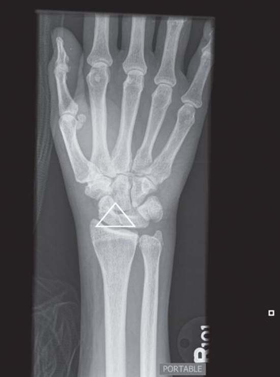
Figure 1.11 Lunate dislocation: Rotation of the lunate resulting in a triangular appearance on posteroanterior view. (Image courtesy of Carl Germann, MD.)

Figure 1.12 Lunate dislocation: Normal quadrangular appearance of the lunate. (Image courtesy of Carl Germann, MD.)

Figure 1.13 Lunate dislocation: Volar displacement of the lunate (“spilled teacup sign”). (Image courtesy of Carl Germann, MD.)
Clinical presentation
· Carpal bone dislocation injuries typically are the result of a high-energy mechanism such as fall from a height on to the outstretched hand, or a motor vehicle crash
· The mechanism of injury is ulnar deviation of the wrist coupled with dorsiflexion
· The patient will complain of pain and swelling over either the dorsum or volar aspect of the wrist and have limited range of motion
· On physical examination there will likely be palpable tenderness over the dorsum of the wrist, particularly in the region of the scapholunate ligament, located just distal to Lister’s tubercle
· With palpation alone it is often difficult to distinguish one source of wrist pain from other causes, including scapholunate strain, scaphoid fracture, triangular fibrocartilage complex tears, and other disorders
Diagnostic testing
· Plain radiographs of the wrist consisting of PA and lateral views are essential to diagnose wrist dislocations (as well as other carpal bone instabilities)
· The PA view should be obtained with the wrist in a neutral position
· A relatively constant 2 mm intercarpal joint space should be seen on a normal PA view. An increase in this distance suggests ligamentous interruption, or a stage I injury (scapholunate dissociation)
· On the AP view, three arcs should be identified (Figure 1.14)
o The first arc consists of the radiocarpal row, which should be both smooth and continuous. Disruption of this arc is suggestive of a lunate dislocation
o The second arc consists of the mid-carpal row, which should similarly be smooth and continuous. Disruption of this arc is suggestive of a perilunate dislocation
o The third arc outlines the proximal surface of the distal carpal row. Disruption of any of these arcs is a sign of carpal dislocation or fracture
· On the lateral view the radius, lunate, and capitate should all line up in a row (Figure 1.15)
o The lunate should lie within the radius cup and the capitate should rest within the lunate cup
o Loss of this normal column configuration implies lunate or perilunate dislocation
o Stress x-rays obtained with radial and ulnar deviation of the hand may demonstrate scapholunate dissociation

Figure 1.14 Normal AP arcs: Disruption suggests carpal dislocation or fracture. (Image courtesy of Carl Germann, MD.)

Figure 1.15 Normal lateral arcs: Disruption suggests lunate or perilunate dislocation. (Image courtesy of Carl Germann, MD.)
Treatment
· Reduce swelling in the extremity (i.e., elevate, apply ice)
· Provide adequate pain control
· Remove any restrictive clothing, splints, casts, jewelry, etc
· Carpal bone dislocations usually mandate the consultation of a hand surgeon in the ED for reduction and stabilization
· Open fractures and open dislocations require temporary splinting, IV antibiotics and prompt operative intervention (keep NPO)
· Closed reduction and long-arm splint immobilization may be attempted but is frequently unsuccessful. If attempted, it is more likely to be successful with a perilunate rather than lunate dislocation because of the extent of ligamentous disruption in the latter
· If the dislocation is irreducible or the result is unstable, then open reduction with internal fixation is required
· Many authors believe immediate open reduction with internal fixation is the treatment of choice, citing the extensive ligamentous injury inherent in such injuries, and frequent unstable results that come with closed reduction
· A lunate or perilunate injury with median nerve symptoms requires immediate operative reduction, carpal tunnel release, and ligamentous reconstruction
Prognosis
· Complications of carpal dislocation include median nerve injury resulting in an acute or subacute carpal tunnel syndrome
· Other complications include chronic carpal bone instability with resultant degenerative arthritis, chronic pain, and limitation in range of motion
· Scapholunate advanced collapsed deformity (“SLAC wrist”) is the end-stage result for many patients
Metacarpal bone fractures and dislocations
Metacarpal head fracture
Key facts
· Rare
· Usually from a direct blow or crush
· Considered as intra-articular fractures
Clinical presentation
· A direct blow often causes a comminuted fracture
· Examination reveals pain and swelling over the involved MCP joint
· Pain is produced by applying axial pressure to the associated digit
· Lacerations suggest an open fracture or “fight bite” injury
PEARL: A high index of suspicion for a potential “fight bite” should be maintained for any injury that is associated with lacerations, abrasions, or bruising over the metacarpophalangeal (MCP) joints. Joint space infections resulting from a human bite are aggressive and rapidly destructive. The wound should be copiously irrigated and left open to heal by secondary intention. Prophylactic antibiotics should be initiated in the ED in all but the most superficial wounds. For patients with a delayed presentation or clinically obvious infection, a hand surgeon should be consulted and consideration given to bringing the patient to the operating room for open irrigation and debridement with subsequent admission for intravenous antibiotics.
Diagnostic testing
· Radiograph of the involved hand (Figure 1.16)
· The metacarpal heads overlap on lateral radiographs and may be difficult to visualize
· An oblique, or “ball-catchers” view, can be helpful in identifying carpal head fractures
· Occasionally CT is required

Figure 1.16 Metacarpal head fracture. (Image courtesy of Carl Germann, MD.)
Treatment
· Reduce swelling in the extremity (i.e., elevate, apply ice)
· Provide adequate pain control
· Remove any restrictive clothing, splints, casts, jewelry, etc
· Immobilize the hand in the “safe” or functional position (20° wrist extension with 90° of MCP joint flexion)
· Referral to hand surgery is required (intra-articular fracture)
· Lacerations or punctures of the dorsum of the MCP should be considered open and contaminated
o Emergency consultation and open irrigation is recommended
o Prophylactic antibiotics such as penicillin with a beta-lactamase inhibitor and aminoglycoside should be provided in the ED
Prognosis
· Any displacement of an intra-articular fracture predisposes the patient to a poor result
· Metacarpal head fractures are associated with severe long-term complications such as:
o Avascular necrosis
o Early arthritis of the MCP joint
o Malunion
o Muscle and extensor tendon fibrosis
o Non-union
Metacarpal neck fracture
Key facts
· Very common
· Usually from a direct impact such as a punch with a closed fist
o Boxer’s fracture: fracture of the neck of the fifth metacarpal
· Often angulated and unstable given the muscular forces
Clinical presentation
· Dorsal angulation of the apex of the fracture is common because of the forces of interosseous muscles
· Examination reveals pain and swelling over the involved metacarpal joint
· Pain is produced by applying axial pressure to the associated digit
· Lacerations suggest an open fracture or fight bite injury
Diagnostic testing
· Radiographs of the involved hand (Figure 1.17)
· Metacarpal heads overlap on lateral views and may be difficult to visualize
· An oblique, or “ball-catchers” view, can be helpful in identifying carpal neck fractures
· Occasionally CT is required

Figure 1.17 Metacarpal neck fracture. (Image courtesy of Carl Germann, MD.)
Treatment
· Reduce swelling in the extremity (i.e., elevate, apply ice)
· Provide adequate pain control
· Remove any restrictive clothing, splints, casts, jewelry, etc
· There is greater mobility of the metacarpals as you move from index (second) to little (fifth digit), therefore less angulation is allowed in the second digit
· Closed reduction should be performed if there is greater than:
o 15° in the second and third metacarpal
o 35° in the fourth metacarpal
o 45° in the fifth metacarpal
· Rotational malalignment should be reduced
· A hematoma block may be used
· Reduction is performed by applying axial traction with flexion of the MCP joints and simultaneous pressure over the metacarpal shaft
· Often difficult to reduce or maintain reduction
· If closed reduction is not successful, early hand surgeon referral is required
· Immobilization should be performed with a radial or ulnar gutter splint from the elbow to, but not including, the PIP joint. The metacarpophalangeal joints should be splinted at 90° of flexion
· Referral to hand surgery
PEARL: Closed reduction should be performed in metacarpal neck fractures if there is greater than:
· 15° angulation in the second and third metacarpal
· 35° angulation in the fourth metacarpal
· 45° angulation in the fifth metacarpal
Prognosis
· Results are often favorable with little or no deformity
· Significant displacement predisposes the patient to a poor outcome
· Metacarpal neck fractures are associated with severe long-term complications such as early arthritis, chronic pain, malunion, non-union and functional disability
Metacarpal shaft fracture
Key facts
· The proximal phalanx has no tendonous attachment
· The FDS and extensor tendons attach to the middle phalanx
Clinical presentation
· A direct blow often causes a transverse or comminuted fracture
· A twisting mechanism often causes a spiral or oblique fracture pattern
· Volar angulation is common, resulting from the extensor tendon and interosseous muscles
Diagnostic testing
· Radiographs of the involved hand (Figure 1.18)
· Skeletal alignment can be assessed radiographically. Rotational alignment is judged clinically by examining the symmetry related to adjacent fingers
o Normally, all of the fingers of a closed hand lie in parallel and point to the scaphoid
o The non-injured hand can also be used for comparison
PEARL: Rotation alignment of the fingers is determined clinically, not radiographically. All the fingers of the closed hand should lie in parallel and point to the scaphoid. Any rotation deformity must be reduced.

Figure 1.18 Metacarpal shaft fracture. (Image courtesy of Carl Germann, MD)
Treatment
· Reduce swelling in the extremity (i.e., elevate, apply ice)
· Provide adequate pain control
· Remove any restrictive clothing, splints, casts, jewelry, etc
· Closed reduction should be performed if there is:
o Any angulation of the second and third metacarpal
o Greater than 10° and 20° in the fourth and fifth metacarpal, respectively
· Rotational malalignment should be reduced
· Immobilization should be performed with a radial or ulnar gutter splint from the elbow to, but not including, the PIP joint
· Referral to hand surgery
· Multiple displaced or communited fractures, fractures with rotational deformity, and irreducible transverse fractures will require internal fixation
PEARL: Closed reduction should be performed if there is:
· Any angulation of the second and third metacarpal Greater than 10° and 20° angulation in the fourth and fifth metacarpal, respectively
Prognosis
· Results are often favorable with little or no deformity
· Significant displacement or non-union predisposes the patient to a poor result and long-term complications such as early arthritis, chronic pain, and functional disability
Metacarpal base fracture
Key facts
· Often results from a direct blow or axial force
· Mobility of the thumb allows for 20 to 30° of angulation without impairment
· Generally stable, with three exceptions:
o Bennett fracture (Figure 1.19): intra-articular fracture of the base of the thumb metacarpal. Small fragment remains aligned with the trapezium while the abductor pollicis longus subluxes the distal portion of the first metacarpal
o Rolando fracture (Figure 1.20): intra-articular comminuted fracture of the base of the thumb metacarpal. The comminution is often “Y-” or “T”-shaped
o Reverse Bennett fracture: the same injury pattern as a Bennett fracture, but in the fifth metacarpal. This fracture pattern is equally unstable because of the lateral traction on the distal metacarpal segment by the extensor carpi ulnaris

Figure 1.19 Bennett fracture: Note that the proximal metacarpal fragment remains aligned with the trapezium. (Image courtesy of Timothy Sweeney, MD.)

Figure 1.20 Rolando fracture: Intra-articular comminuted fracture of the base of the first metacarpal. (Image courtesy of Timothy Sweeney, MD.)
Clinical presentation
· Often results from a direct blow or axial force
o Bennett, reverse Bennett, and Rolando fractures classically occur because of an axial load to a flexed and adducted digit
· Tenderness at the metacarpal base, rotational deformity may be present
· Ring (fourth) and little (fifth) finger fractures may cause ulnar nerve injury and paralysis
Diagnostic testing
· Radiograph of the involved hand (Figure 1.21)
· Skeletal alignment can be assessed radiographically, rotational alignment is judged clinically by examining the symmetry related to adjacent fingers

Figure 1.21 Metacarpal base fracture. (Image courtesy of Carl Germann, MD.)
Treatment
· Reduce swelling in the extremity (i.e., elevate, apply ice)
· Provide adequate pain control
· Remove any restrictive clothing, splints, casts, jewelry, etc
· Finger fractures should be immobilized with a bulky compressive dressing or volar splint and referred to a hand surgeon
· A thumb metacarpal fracture with greater than 20 to 30° of angulation requires closed reduction. All thumb metacarpal fractures should be placed in a thumb spica splint. Unstable fractures require open reduction internal fixation (ORIF)
o Bennett’s and Rolando’s fractures require a thumb spica splint and early referral to a hand surgeon for surgical reduction
· Reverse Bennett’s fractures require an ulnar gutter splint and prompt referral to a hand surgeon for operative repair
Prognosis
· Results are often favorable with little or no deformity
· Chronic carpal metacarpal joint pain and stiffness may result in displaced or comminuted intra-articular fractures
Carpometacarpal joint dislocation
Key facts
· The CMC joints are supported by strong dorsal, volar, and interosseous ligaments
· Dislocations are uncommon and often missed
· Dislocations commonly result from high-energy trauma such as motor vehicle crashes, falls, crush injuries, and closed-fist injuries
Clinical presentation
· Ecchymosis, swelling, and pain over the dorsum of the hand
· Tenderness over the involved CMC joint(s)
Diagnostic testing
· Radiograph of the involved hand and wrist. (Figure 1.22)
· Fracture lines and dislocations may be subtle on PA radiographs because of superimposition
· Dislocations are usually more obvious on lateral view

Figure 1.22 Carpometacarpal joint dislocation: Dorsal displacement of the metacarpal is more easily seen on lateral views. (Image courtesy of Carl Germann, MD.)
Treatment
· Reduce swelling in the extremity (i.e., elevate, apply ice)
· Provide adequate pain control
· Remove any restrictive clothing, splints, casts, jewelry, etc
· Regional anesthesia should provide adequate analgesia for reduction. Consider doing a radial, median, or ulnar nerve block as needed
· Reduction is performed by applying traction and flexion of the hand with longitudinal pressure on the metacarpal base
· Even if closed reduction is successful, prompt hand-surgery referral is required for wire fixation
· Unstable joints or irreducible dislocations should be immobilized with both dorsal and volar splints and immediately referred to a hand surgeon
Prognosis
· Stiffness, pain, and weakness may persist because of traumatic arthritis
· Chronic dislocation may occur if there is imprecise alignment
Phalangeal bone fractures and dislocations
Proximal and middle phalangeal fracture
Key facts
· Characterized as extra-articular or intra-articular
· The proximal phalanx has no tendonous attachment
· The flexor digitorum superficialis (FDS) and extensor tendons attach to the middle phalanx
· Rotational deformities often occur secondary to forces of flexor tendons
Clinical presentation
· A direct blow often causes a transverse or comminuted fracture
· A twisting mechanism often causes a spiral or oblique fracture pattern
· Volar angulation is common, resulting from the pull of extensor tendons and interosseous muscles
Diagnostic testing
· Radiograph of the involved hand
· Skeletal alignment can be assessed radiographically, rotational alignment is judged clinically by examining the symmetry related to adjacent fingers
Treatment
· Reduce swelling in the extremity (i.e., elevate, apply ice)
· Provide adequate pain control
· Remove any restrictive clothing, splints, casts, jewelry, etc
· Approximately 75% of fractures are stable and non-displaced. These do not require reduction and can be immobilized by a radial or ulnar gutter splint or by “buddy-taping.” This provides support for the injured digit while allowing for motion of the uninvolved phalangeal joints
· Displaced fractures are often easily reduced in the ED and maintained with external splinting
· Unstable fractures that cannot be reduced in the ED require internal fixation
Prognosis
· The most common complication of phalanx fracture is malunion
Distal phalanx fracture
Key facts
· Distal phalanx fractures (tufts fractures) are the most common fractures of the hand
· Characterized as extra-articular or intra-articular
· Tufts fractures are often comminuted and associated with soft tissue and nail or nailbed injuries
· Associated tendon injuries may occur
o Flexor profundus tendon attaches to the volar aspect of fingers
o Extensor tendon attaches to the dorsal aspect of fingers
o Flexor pollicis longus inserts to the volar base of the distal thumb phalanx
o Extensor pollicis longus attaches to the dorsal base of the distal thumb phalanx
Clinical presentation
· Often a result of a crush injury
· Tenderness and swelling are present over the distal phalanx
· Deficits in range of motion may reflect tendon avulsion
Diagnostic testing
· Radiograph of the involved digit(s)
Treatment
· Reduce swelling in the extremity (i.e., elevate, apply ice)
· Provide adequate pain control
· Remove any restrictive clothing, splints, casts, jewelry, etc
· Short volar finger splint is recommended for comfort and to protect the finger (~3 days)
· Immobilization should not include the PIP joint
Prognosis
· Generally uncomplicated
· Complications include numbness, hyperesthesia, and cold sensitivity
· Nail bed trauma may result in abnormal nail growth
Phalanx dislocation (MCP, PIP, and DIP)
MCP joint dislocation
Key facts
· The MCP joint is stabilized by two collateral ligaments and a volar fibrocartilaginous plate
· Dislocations are usually in a dorsal direction
· Dislocations commonly result from hyperextension forces that rupture the proximal volar plate
· The most common digits involved are the middle finger followed by the little finger
Clinical presentation
· Ecchymosis, swelling, and pain of the involved joint(s)
· Tenderness and swelling are present over the distal phalanx
· Deficits in range of motion may reflect a tendon avulsion
Diagnostic testing
· Radiograph of the involved hand
· Dislocations are usually obvious on a lateral view
· PA views may show widening of the joint. Sesamoids may be found within the joint space
Treatment
· Reduce swelling in the extremity (i.e., elevate, apply ice)
· Provide adequate pain control
· Remove any restrictive clothing, splints, casts, jewelry, etc
· Reduction is performed by flexing the wrist while applying firm pressure to the dorsum of the proximal phalanx in a volar direction
· The MCP joint should then be splinted in flexion (between zero and 15°) with either a thumb spica or a dorsal splint to prevent extension of the MCP joint beyond 0°
· All dislocations should be referred to a hand surgeon
Prognosis
· Complications include entrapment of the volar plate during reduction. Closed reduction is difficult in this scenario and operative repair is often required
· Uncomplicated reductions have a favorable prognosis; however, hand surgery referral is recommended
PIP joint dislocation
PEARL: PIP joint dislocations are the most common ligamentous injury in the hand and more common than MCP and DIP dislocations.
Key facts
· The PIP joint is stabilized by two collateral ligaments and a volar fibrocartilaginous plate
· Dislocations can be in a dorsal, lateral, and volar direction
· Dislocations commonly result from hyperextension and axial loading forces
Clinical presentation
· Ecchymosis, swelling, and pain of the involved PIP joint(s)
· Inability to extend the joint. Tenderness over the joint and deformity may be present
Diagnostic testing
· Radiograph of the involved digit(s)
· Dislocations are usually obvious on lateral view
· Small avulsion fractures may be seen on radiographs and are associated with ligamentous attachment points
PEARL: Avulsion fractures involving 33% or more of the articular surface are usually unstable and require operative repair.
Treatment
· Reduce swelling in the extremity (i.e., elevate, apply ice)
· Provide adequate pain control
· Remove any restrictive clothing, splints, casts, jewelry, etc
· A digital nerve block can be used for analgesia to aid in reduction
· Reduction is performed by applying longitudinal traction and mild hyperextension followed by firm dorsal pressure on the proximal aspect of the middle phalanx
· If stability is maintained during active range of motion, the digit should be immobilized in 20 to 30° of flexion in a dorsal splint for 3 weeks
· Unstable joints or irreducible dislocations should be splinted and referred to a hand surgeon
Prognosis
· The prognosis is good, although stiffness, pain, and swelling may persist for weeks to months
· Recurrent dislocation usually does not occur unless the finger is hyperextended
DIP joint dislocation
Key facts
· The DIP joint structure is similar to the PIP joint; however, it has additional stability of the insertions of the flexor and extensor tendons
· Dislocations are usually in a dorsal direction and are often associated with an open wound
· Dislocations commonly result from hyperextension and axial loading forces
Clinical presentation
· Ecchymosis, swelling, and pain of the involved DIP joint(s)
· Inability to extend the joint, tenderness and deformity of the joint may be noticed
Diagnostic testing
· Radiograph of the involved digit(s)
· Dislocations are usually obvious on lateral view
· Small avulsion fractures may be seen on radiographs and are associated with ligamentous attachment points
Treatment
· Reduce swelling in the extremity (i.e., elevate, apply ice)
· Provide adequate pain control
· Remove any restrictive clothing, splints, casts, jewelry, etc
· A digital nerve block should provide adequate analgesia for reduction
· Reduction is performed by applying longitudinal traction and mild hyperextension followed by firm dorsal pressure on the proximal aspect of the distal phalanx
· If stability is maintained during active range of motion, the digit should be immobilized in slight flexion with a dorsal splint for 3 weeks
· Unstable joints or irreducible dislocations should be splinted and referred to a hand surgeon
· Any open dislocation should be copiously irrigated, sutured, and referred to a hand surgeon. Prophylactic antibiotics are recommended
Prognosis
· The prognosis is good, although stiffness, pain, and swelling may persist for weeks to months
· Recurrent dislocation usually does not occur unless the finger is hyperextended
Nail bed injuries
Subungal hematoma
Key facts
· Results from a crush injury or blunt trauma
Clinical presentation
· Pain and blood underneath the nail. Potential distal phalanx instability/fracture
Diagnostic testing
· Radiograph of the involved digit(s) should be performed if the fingertip is unstable or the mechanism of injury suggests a distal phalanx fracture
Treatment
· Small hematomas do not require drainage
· Large hematomas are painful and should be relieved by nail trephination with a heated paperclip or microcautery device
· Anesthesia is not necessarily required as pain relief occurs with decompression
· If a fracture is present, the digit should be splinted
· Studies suggest that the outcome of nail trephination is similar to that of formal nail bed repair, regardless of the hematoma size
PEARL: Trepination does not usually require anesthesia. The nail is insensate, and the blood lifts the nail off the nailbed so it is unlikely that you will make contact with the nailbed, which is what would cause the patient pain.
Prognosis
· The prognosis is generally good, although patients with large hematomas should be warned that they may lose the nail
Nail avulsion/nailbed laceration
Key facts
· Results from a crush injury or blunt trauma
Clinical presentation
· Pain and blood underneath the nail
· Potential distal phalanx instability
· Injury to the nail, partial avulsion or laceration may be present
Diagnostic testing
· Radiograph of the involved digit(s) should be performed if the fingertip is unstable or the mechanism of injury suggests a distal phalanx fracture
Treatment
· Simple lacerations should be repaired using 5–0 or 6–0 absorbable sutures
· Trephination should be performed to allow drainage of blood after the nail is reinserted into the nail fold
· The nail may be sutured in place through the trephinated hole(s) or taped in place
· A nail-shaped adaptic or non-adherent gauze may be placed under the nail fold if the original nail is misplaced or unusable
· Placing one of the above objects under the nail fold will help prevent synechiae and will help encourage nail regrowth
Prognosis
· These injuries do well with early primary repair
· Complete nail growth may take 70 to 160 days
· Patients should be informed that they are at risk of nail deformity and losing the nail entirely
Tendon, ligament, vascular, and nerve injuries
Tendon injuries
PEARL: Blood vessels and nerves often travel closely with flexor tendons. Injury to one necessitates an evaluation for injury to the other two.
Jersey finger
Key facts
· This injury is a disruption of the flexor digitorum profundus (FDP) which is responsible for flexion at the DIP joint
· Jersey finger is often seen in tackling sports. This occurs when a digit is forced into extension while actively being flexed, as might occur when grabbing a jersey during a tackle
· More than 75% of Jersey finger injuries involve the ring finger
· Avulsion fractures may accompany Jersey finger injuries
Clinical presentation
· Pain, swelling, and tenderness of the involved DIP joint
· An injury to the flexor tendon may not be evident if the fingers are extended
· An abnormal position or asymmetry of the involved digit during flexion suggests flexor tendon injury
· The FDP and FDS should be tested separately during functional examination
· Partial tendon injuries may be clinically occult. Such injuries may present with pain and functional weakness of the digit against resistance
· Complete tendon injuries prevent the patient from flexing the digit at the DIP joint when the PIP joint is held in extension by the examiner
PEARL: FDP function is tested by having the patient actively flex the DIP joint. The FDS tendon is evaluated by having the patient flex the proximal joint of the finger while the remaining fingers are extended.
Diagnostic testing
· The diagnosis is based upon physical examination
· Radiographs of the involved digit are recommended to evaluate for avulsion fracture
Treatment
· Reduce swelling in the extremity (i.e., elevate, apply ice)
· Provide adequate pain control
· Remove any restrictive clothing, splints, casts, jewelry, etc
· Immediate or delayed operative repair is recommended for FDP injuries
· A volar or dorsal splint should be applied to produce approximately 70° of MCP flexion and slight flexion of the DIP and PIP joints. This prevents further tendon injury and retraction
Prognosis
· Outcomes are dependent on early surgical repair
· With surgical repair, most patients have good outcomes
· Postoperative complications include adhesions, trigger finger, and epidermoid cyst formation
Mallet finger
Key facts
· A closed disruption of the distal extensor apparatus resulting in a flexion deformity (Figure 1.23)
· Most common in the long, ring, and little fingers
· Extensor apparatus disruption most often results from a sudden forced flexion of an extended finger when an object, such as a ball, strikes the tip of the finger
· Avulsion fractures may accompany mallet finger

Figure 1.23 Mallet finger: Injury to the extensor mechanism creates forced flexion because of unopposed action of the flexor digitorum profundus. (Image courtesy of Timothy Sweeney, MD.)
Clinical presentation
· Pain, swelling, and tenderness of the involved DIP joint, typically on the dorsal side
· The distal phalanx is held in flexion because of unopposed forces of the flexor digitorum profundus
· Inability to extend the joint actively
Diagnostic testing
· The diagnosis is based upon physical examination
· Radiographs of the involved digit are recommended to evaluate for an avulsion fracture (Figure 1.24)

Figure 1.24 Avulsion fracture: Commonly associated with mallet finger. (Image courtesy of Timothy Sweeney, MD.)
Treatment
· Reduce swelling in the extremity (i.e., elevate, apply ice)
· Provide adequate pain control
· Remove any restrictive clothing, splints, casts, jewelry, etc
· Treatment is aimed at continuous DIP joint extension to allow the extensor apparatus to heal
· A volar or dorsal splint should be applied to produce slight hyperextension for 6 to 8 weeks. If the joint is flexed the 6 to 8 week treatment plan should be restarted
· The PIP and MCP joints should be allowed to move freely
Prognosis
· The prognosis is good with 80% of patients achieving successful outcomes with splinting
· Complications include chronic pain, dorsal deformity, and swan-neck deformity
o Swan-neck deformity occurs when the lateral bands displace laterally and dorsally resulting in increased extension forces on the PIP
Boutonniere deformity
Key facts
· The extensor mechanism is disrupted from the central slip attachment at the dorsal aspect of the middle phalanx
· The proximal end migrates proximally, and the pull of the lateral bands of the extensor mechanism opens the hole further
· This allows the force of the extensor mechanism to bypass the PIP joint, which remains flexed, and divert it to the DIP joint which becomes hyperextended
Clinical presentation
· The most common mechanism is forced flexion of the extended PIP joint as in a basketball player receiving a pass
· Patients present with flexion of the PIP joint and hyperextension of the DIP and MCP joints (Figure 1.25)
· In these patients, the PIP joint can be passively brought to full extension but active extension is not possible
· On physical examination, location of maximal tenderness leads the physician to the proper diagnosis. The patient will usually have tenderness about one or both of the collateral ligaments
· The area of maximal tenderness will be over the central slip on the dorsal aspect of the PIP joint. Generally, this area will also be ecchymotic

Figure 1.25 Boutonniere deformity: Forces of the lateral bands of the extensor mechanism flex the PIP joint while extending the DIP and MCP joints. (Image courtesy of Timothy Sweeney, MD.)
Diagnostic testing
· Physical examination will make the diagnosis of central slip injury but may not clarify whether the structure is partially or completely torn
· The prudent course, therefore, is initially to treat all central slip injuries as though they are complete ruptures
· Radiographs may reveal an avulsion fracture of the volar base of the middle phalanx
Treatment
· Reduce swelling in the extremity (i.e., elevate, apply ice)
· Provide adequate pain control
· Remove any restrictive clothing, splints, casts, jewelry, etc
· The PIP joint should be splinted in extension leaving the DIP and MCP joints to move freely
· The patient should be instructed to continue to move the DIP joint so as not to develop an extension contracture
· Prompt referral should be made to a hand surgeon
· Operative repair may be required for a boutonniere injury associated with a displaced avulsion fracture
Prognosis
· The key to achieving the greatest degree of range of motion and reduction in deformity is to have the finger diagnosed and splinted as early as possible
Ligament injuries
PEARL: Ligamentous injuries are graded as type I, II, or III. Type I reflect minor ligament fiber tears associated with pain and full function. Type II are partial tears with moderate loss of function. Type III are complete tears and are associated with instability and complete loss of function.
Ulnar collateral ligament (UCL) injury
Key facts
· Also called skier’s thumb, UCL injuries are the most common upper extremity injury in skiing and result from hyperextension of the thumb on a ski pole while falling
· Ten times more frequent than a radial collateral ligament injury
Clinical presentation
· The mechanism is hyperextension of the abducted thumb causing injury to the ulnar collateral ligament (UCL) and is often associated with an avulsion fracture
· The physical exam will be remarkable for tenderness at the UCL, laxity at the MCP of the thumb, and an inability to actively oppose the thumb (Figure 1.26)
· Most UCL ruptures occur at the distal attachment
· Complete and partial ruptures can usually be differentiated by physical examination
· If the injured joint demonstrates 40° of radial angulation or 15° of laxity beyond the range of the uninjured thumb during stressing, a complete ligament rupture should be assumed

Figure 1.26 UCL stressing: Laxity of the thumb with valgus stress suggests an ulnar collateral ligament injury. (Image courtesy of Timothy Sweeney, MD.)
Diagnostic testing
· Radiographs should be obtained to evaluate for an avulsion fracture (Figure 1.27)
· Clinical examination usually reveals the diagnosis; however, this injury is commonly misdiagnosed as a simple sprain

Figure 1.27 UCL avulsion fracture: Note the fracture at the base of the proximal phalanx. (Image courtesy of Timothy Sweeney, MD.)
Treatment
· Reduce swelling in the extremity (i.e., elevate, apply ice)
· Provide adequate pain control
· Treatment is immobilization in a thumb spica splint, NSAIDS, and referral to a hand surgeon. Complete ruptures require surgical repair
Prognosis
· Repair within 3 weeks will often result in a good outcome
· Long-term complications include chronic pain, instability, and a loss of pinch strength
· Complications include chronic pain, stiffness, loss of sensation, cold-intolerance, non-union, and necrosis
Vascular injuries
Key facts
· The vascular supply to the hand and digits is duplicated, therefore isolated arterial injuries rarely result in ischemia
· Because digital nerves are often superficial to the digital arteries, an arterial lesion should raise suspicion of a nerve injury
· Lacerations and amputations of the hand rarely cause life-threatening hemorrhage
Clinical presentation
· A history of pulsatile bleeding is suggestive of an arterial injury
· Circulatory status is determined by examination of the radial and ulnar pulses, assessment of capillary refill, and the observation of cyanosis or pallor
· Ischemic pain is the most common finding in vascular insufficiency
· Swelling, discoloration, and tenderness may also be present
Diagnostic testing
· A good history and physical examination will identify most vascular injuries
· Circulation is routinely tested by palpation of the radial and ulnar pulse and examination of capillary refill
· Doppler assessment can also be helpful in assessing radial and ulnar pulses
Treatment
· Reduce swelling in the extremity (i.e., elevate, apply ice)
· Provide adequate pain control
· Remove any restrictive clothing, splints, casts, jewelry, etc
· Usually the hemorrhage can be controlled with direct pressure and limb elevation
· If necessary, a blood pressure cuff can be inflated to 30 mmHg above the patient’s systolic blood pressure to control bleeding
· Operative repair may be required if there are symptoms of ischemia or nerve injuries
· Injuries to radial, ulnar, and palmar arch arteries often require surgical exploration and repair
Prognosis
· Even completely transected large vessels in the hand and wrist usually retract, constrict, and clot
· The prognosis of vascular injuries is typically good but depends on early and aggressive diagnosis and repair
Nerve injuries
Key facts
· Nerve injuries may result from a direct blow, laceration, crush injury, or amputation
· Nerve injuries are divided into three categories:
o Neuropraxia: The axon and endoneural tube remain intact. There is a loss of function that usually resolves within days
o Axonotmesis: The endoneural tube remains intact, but the axon is severed. The loss of function may slowly return as the proximal axon regenerates
o Neurotmesis: There is complete disruption of all nerve elements. Regeneration does not occur unless the severed nerve endings are reapproximated
Clinical presentation
· Physical examination will reveal a loss of motor and sensory function
· Injury most often involves one of the digital nerves
· Severe injuries to the hand warrant examination of the sensory function of the digital nerves
o The digital nerves divide into volar and dorsal branches to supply sensation to the fingers
· Injuries to the forearm or wrist warrant examination of the radial, median, and ulnar nerves
o The median and ulnar nerves have mixed motor and sensory function of the hand while the radial nerve only has sensory function of the hand (Figures 1.28 and 1.29)
§ The median nerve (C5 to T1) supplies motor function to the abductor pollicis brevis, superficial head of the flexor pollicis brevis, and opponens pollicis
§ The ulnar nerve supplies motor function to the hypothenar muscles, ulnar lumbricals, interossei, adductor pollicis, and the deep head of the flexor pollicis longus
PEARL: The median nerve can be tested by having the patient flex the distal phalanx of the thumb against resistance. The ulnar nerve can be assessed by having the patient spread the fingers against resistance. Radial nerve function can be examined by having the patient extend the wrist.

Figure 1.28 Hand sensory distribution: Dorsal view. (Image courtesy of Douglas Dillon, MD.)

Figure 1.29 Hand sensory distribution: Palmar view. (Image courtesy of Douglas Dillon, MD.)
Diagnostic testing
· Diagnosis is made based on the history and physical examination
· If function does not return in a few weeks, electromyograms and nerve conduction studies can differentiate neurotmesis from axonotmesis and determine the need for surgery
Treatment
· Provide adequate pain control
· Lacerated nerves require reapproximation by a hand surgeon
· In general, injuries to the motor branches of the median and ulnar and digital nerves located proximal to the DIP should be considered for repair
· Patients with closed injuries without evidence of compartment syndrome should be referred to a hand surgeon as an outpatient
· All digits with suspected nerve injury should be splinted in the position of function
Prognosis
· Complications include long-term motor and sensory loss, atrophy, and sympathetic dystrophy
· In general, sensory function recovers more often than motor function, although functional recovery is rarely complete
Amputation
Key facts
· Amputations may be complete or partial
· Fingertip amputations are the most common upper extremity amputation
· Most occur distal to the DIP joint
· Reimplantation should be considered for amputations proximal to the DIP joint
Clinical presentation
· Traumatic amputation usually results from a crush injury or a guillotine mechanism
Diagnostic testing
· Clinical examination provides the diagnosis
· A careful examination of neurologic, vascular, and musculotendinous function should be performed
· Radiographs should be obtained to evaluate for an associated fracture
PEARL: Indications and contraindications for reimplantation.
· Indications:
· Multiple digits amputated
o Thumb amputation
o Wrist and forearm amputation
o Single digits amputated between the PIP and DIP joints (distal to the flexor digitorum superficialis insertion)
o All pediatric amputations
· Contraindications:
o Amputations in unstable patients secondary to other life-threatening injuries
o Multiple-level amputations
o Self-inflicted amputations
o Single-digit amputations proximal to the flexor digitorum superficialis insertion
o Serious underlying disease
o Extremes of age
Treatment
· Provide adequate pain control (digital block)
· Control bleeding
· The management of distal fingertip amputation is controversial and should be individualized
· Most amputations distal to the DIP are managed with local wound care and allowed to heal by secondary intenion
· If bone if exposed, it may be trimmed back with a rougeur to just below the skin level
· Meticulous wound cleaning and irrigation is required
· A non-adherent dressing should be placed
· Intravenous antibiotics covering for S. aureus followed by an oral course is recommended
· Crush injuries or wounds with significant soft tissue or bone loss require surgical repair by a hand surgeon
· Final judgement regarding whether reimplantation will be performed by a surgeon is based upon microscopic examination of the blood vessel and nerves, level of injury, patient age and health-related factors
· If reimplantation is being considered, the amputated portion should be:
o Assessed for the degree of tissue injury
o Irrigated with normal saline; local antiseptics should be avoided
o Covered with saline-moistened sterile dressing
o Placed in a dry plastic bag and placed in ice water
· Tetanus status should be addressed
Prognosis
· Warm ischemia may be tolerated for 6 to 8 hours; cooling may extend this time to 12 to 24 hours
Selected readings and references
Germann CA, Perron AD. Risk management and avoiding legal pitfalls in the emergency treatment of high-risk orthopedic injuries. Emerg Med Clin N Am. 2010;28:969–96.
Jaworski CA, Krause M, Brown J. Rehabilitation of the wrist and hand following sports injury. Clin Sports Med. 2010;29(1):61–80.
Perron AD, Brady WJ. Evaluation and management of high-risk orthopedic emergencies. Emerg Med Clin N Am. 2003;21:159.
Perron AD, Brady WJ, Keats TE, et al. Orthopedic pitfalls in the ED: Scaphoid fracture. Am J Emerg Med. 2001;19:310–16.
Perron AD, Brady WJ, Keats TE, et al. Orthopedic pitfalls in the ED: Lunate and perilunate injuries. Am J Emerg Med. 2001;19:157–62.
Sauder DJ, Athwal GS, Faber KJ, Roth JH. Perilunate injuries. Ortho Clin North Am. 2007;38:279.
Sherman GM, Seitz WH. Fractures and dislocations of the wrist. Curr Opinion Orthop. 1999;10:237.