
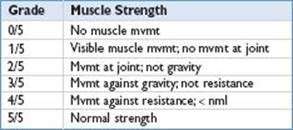
• Distinguishing upper motor neuron and lower motor neuron patterns of weakness


• Examples of neuromuscular conditions with primary LMN pattern of weakness

Duchenne/Becker Muscular Dystrophy (Pediatr Rev 2000;21:233; Pediatr Rev 2006;27:83)
• Epidemiology: Incidence 1 in 3,600–6,000  births; 1/3 cases 2/2 new mutations musc dystrophy; genetic d/o of skeletal muscle w/o 1° involve of CNS or PNS
• Pathology/pathophysiology
• X-linked recessive (Duchenne & Becker)
• Duchenne, Becker: Mutation in gene for dystrophin (deletion more common than duplication) → muscle damage & fibrosis
• Muscle bx w/ inflammation, degeneration/necrosis, & regeneration
• Clinical manifestations: Progressive weakness of prox muscles; legs before arms
• Duchenne: Asymptomatic at birth
• Develop proximal muscle weakness at 3–5 yo: Waddling gait, toe walking, frequent falls, difficulty running/jumping/climbing stairs
• As pelvic girdle weakness continues, develop Trendelenburg gait, lumbar lordosis, & “Gower sign” (stand from supine position “climbs up self”)
• Pseudohypertrophy of muscles, esp calves; feel rubbery & firm
• Wheelchair by 8–12 yo, then fixed skeletal deformities; scoliosis, equinovarus
• Declining resp fxn at 9–11 yo; progressive restrict defect, weak cough, ↑ risk PNA
• Weakness of arms in mid-teens; difficulty feeding & caring for themselves
• Cardiomyopathy in mid-teens; may be free of sx as wheelchair bound
• Also demonstrate nonprogressive cognitive & emotional disturbances; deficits in
verbal IQ, poor attention span, easily frustrated & distracted, OCD
• Usually die in 2nd–3rd decade from pulmonary or cardiac failure
• Becker: Similar clinically, but milder & slower progression; survive to adult
• Presents later in childhood; able to walk w/o assistance beyond 16 yo
• More severe cardiac dysfxn; 15% w/ cardiomyopathy <6 yo & 75% pts >40 yo
• Diagnostic studies
• Muscle enzymes: CK ↑ 50–100 × s nml; highest levels early when asymp; ↓ over time
• Mildly ↑ AST, ALT, & LDH; check GGT to verify that it is not of hepatic origin
• Gene mutation analysis
• 10–20% still need muscle bx for dx confirmation; needle or open bx; minimal histologic
• Δ if pt very young; when older, test for ↓ /absent dystrophin
• EMG changes nonspecific & not helpful in dx
• Treatment: Steroids preserve fxn & delay progression (QD prednisone or deflazacort)
• PT/OT, bracing, night splints, nutrition guidance when non-ambulatory
• Consider QD vit D & calcium for ↑ fracture risk from osteopenia of immobility
• Surgical tendon lengthening to prolong walking, scoliosis surgery
• Monitoring of pulmonary and cardiac functions
Guillain–Barré Syndrome (GBS) (Neurology 2011;76:294; Curr Treat Options Neurol 2011;13:590; Ped Rev 1997;18:10; Neurology 2003;61:736; Arch Dis Child Educ Pract Ed 2007;92:161)
• Definition and epidemiology: Annual incidence in pts >18 yo 0.5–2/100,000 pop
• Acute inflammatory demyelinating polyradiculoneuropathy (AIDP)
• Most common cause of acute generalized paralysis in all age groups
• Male-to-female ratio = 1.5:1
• Pathology/pathophysiology: Immune-mediated segmental demyelination of peripheral nerves w/ inflammatory cell infiltration
• Variants: (1) AIDP, (2) Miller-Fisher syndrome: Ophthalmoplegia, ataxia, &
areflexia, (3) acute motor axonal neuropathy: Less common but often more severe, (4) acute motor and sensory axonal neuropathy
• 50–70% w/ recent illness or vaccination w/i past 4 wk, URI or gastroenteritis; assoc w/ CMV, EBV, hepatitis, varicella, Mycoplasma pneumoniae and Campylobacter jejuni (most common preceding infxn; especially in Miller-Fisher variant)
• Clinical manifestations: Fever is atypical and suggests secondary infection
• 3 phases: Progression phase d to wks, plateau d to wks, recovery wks to mos
• Progressive, ascending, symmetric muscle weakness starting in legs, ↓ DTRs
• Sensory dysfxn: Pain or paresthesia (of back, extremities, or around mouth; may be in band distribution), ↓ position and vibratory sensation
• Ataxia out of proportion to muscle weakness; bowel and bladder dysfxn are rare
• CN involved (less common): Swallowing difficulty, facial weakness, ocular palsy
• Dysautonomia (less common): Tachy/bradyarrhythmias, hyper/hypotension
• Respiratory distress may progress to need for mechanical ventilation
• Diagnostic criteria/studies
• Hx; recent illness, onset and progression of sx, exposure to ticks, toxins or meds
• Exam w/ assessment of spinal tenderness and complete neuro exam w/ distribution and symmetry of strength, reflexes, and sensation
• Suggested workup: CBC, ESR, ANA, U/A, CSF protein and cells, EMG
• May also consider urine porphobilinogen, ALA, and heavy metal screen; quantitative immunoglobulins; Campylobacter jejuni Abs
• Required for dx: Progressive motor weakness >1 limb, areflexia or hyporeflexia
• Supportive of dx: Progression rapid but halts after 4 wk, relative symmetry, mild sensory deficits, CN involvement (facial weakness in 50%), autonomic dysfxn, initial absence of fever, recovery 2–4 wk after progression ceases
• Labs/studies supportive of dx:
• Albuminocytologic dissociation: ↑ CSF protein after 1 wk sx and CSF lymphs <10 per mm3; >50 cells/mm3 or PMN predom → alternative dx
• Nerve conduction studies w/ prolonged F waves, slowing or block (unless variant)
• Features that cast doubt on dx: Marked, persistent asymmetry, prolonged
progressive phase >4 wk (likely CIDP) or recurrent = CIDP, persistent bowel/bladder dysfxn or dysfxn present at onset, discrete sensory level, (suggests transverse
myelitis) CSF pleocytosis >50 wcc/mm3 (suggests infection)
• Features that exclude dx: Recent Hx of hexacarbon abuse, porphyria, recent diphtheria, Hx/evidence of lead intox, pure sensory syndrome
• Treatment: Symptomatic Rx and supportive care
• Measure FVC, negative inspiratory force and vital signs q6h until trend determined
• Admit ICU if any of following: Severe rapid progression, flaccid tetraparesis, bulbar palsy, vital capacity ≤20 mL/kg, autonomic cardiovasc instability
• Immunotherapy speeds recovery and lessens disability: Plasmapheresis (series of exchanges to total 250 mL/kg) or IVIg (2 g/kg divided over 2–4 d)
• Pain management: Opioids, NSAIDs, carbamazepine, amitriptyline
• Corticosteroids may worsen or slow recovery!
• Outcome/prognosis: 90–95% complete recovery in 3–12 mo; mortality <5%
• 5%–10% retain severe permanent disability
• Indicators of a poor prognosis: CN involvement, ventilatory dysfxn, rapid progressive tetraparesis, severe ↓ (<10% of nml) in compound muscle action potentials at 1st measurement, C. jejuni infection
• Deaths 2/2 cardiac arrhythmias, resp failure, dysautonomia, pulmonary embolism
Bell’s Palsy (J Child Neurol 2001;16:565; Clin Pediatr (Phila) 2010;49:411; Brain Dev 2011;33:644)
• Definition and epidemiology: Acute idiopathic lower motor neuron facial palsy
• Incidence 2.7/100,000 in children <10 yo; 10.1/100,000 in those 10–20 yo
• Pathology/pathophysiology
• Upper face innervated by fibers from both cerebral hemispheres while lower face innervated by corticobulbar fibers primarily from contralateral hemisphere
• Lower motor neuron dysfxn (as in Bell’s palsy) affects both upper and lower face, whereas upper motor neuron dysfxn affects lower face
• Portion of CN VII w/i temporal bone is most commonly affected
• Differential diagnosis
• Causes may be identified in up to 70% of children
• Most common cause of acute facial palsy in kids: AOM, Lyme (50% in endemic area)
• Infectious etiologies: HSV, Lyme, VZV (Ramsay Hunt), HIV, EBV, CMV, coxsackie, rubella, mumps, mycoplasma, TB
• If gradual onset, consider neoplasm (cholesteatoma, schwannoma, meningioma, glioma, lymphoma/leukemia, parotid tumor)
• Other: mastoiditis, trauma, GBS/MFS, toxins, vasculitis, sarcoidosis, HTN
• Clinical manifestations
• Rapid onset is usually unilateral upper & lower facial weakness w/ facial sagging
• Initial sx pain/tingling in ear canal. Typically aching is severe over mastoid
• No facial sensory loss, although may report a “numb” feeling
• May have disruption of taste on ant 2/3 tongue, hyperacusis, decreased tearing
• Inability to close eyelid w/ risk of corneal injury if not lubricated and patched
• Diagnostic studies: If typical presentation, no need for diagnostic testing; full neuro exam, otoscopy, BP check and CBC recommended
• Serologic testing for Lyme if endemic area
• If <2 yo or atypical features (slow onset) consider w/u for ddx above with serum studies, tox screen, LP, urgent neuro-imaging
• Consider neuroimaging if resolution does not begin within 1 mo
• Prognosis and treatment: ∼80–90% pts recover completely or satisfactorily from cosmetic perspective, many w/I 2 mo and most w/i 6 mo
• Evidence mixed if corticosteroids provide any benefit in children. Often prescribed in clinical practice (rx ideally begun w/i 72 hr onset symptoms)
• Evidence mixed if antivirals (acyclovir/valcyclovir) w/ any benefit
• Patch the eye and apply artificial tears several times a d
Myasthenia Gravis (MG) in Childhood (J Child Neurol 2009;24:584;
Pediatr Rev 1990;12:73; J Child Neurol 1999;14:41)
• Includes juvenile, congenital, and transient neonatal subtypes. Hallmark feature is fatigable muscle weakness; sensation intact; DTR nml
• Childhood forms comprise 10–15% of all MG cases in N. America; incidence ∼1.1/million
• Juvenile MG
• Pathophysiology: Presents <19 yo. Classic immune-mediated MG w/ Ab against postsynaptic acetylcholine receptor at NMJ. AChR Ab accounts for ∼80% cases; MuSK Ab much less frequent and often assoc w/ more severe sx. Prepubertal
children often seronegative. Thymoma/thymic hyperplasia identified in many cases
• Clinical manifestations: Ptosis & diplopia typically initial sx of both forms; pupillary function wnl
• Ocular MG: 1°ly ocular sx w/ +/− mild weakness limbs & face; may progress to total ophthalmoplegia or relapses/remissions; no resp distress or deficits in
swallowing or speaking; can progress to generalized sx (but less likely to progress if >1 yr w/ ocular sx only)
• Generalized myasthenia: Mod–severe limb & bulbar weakness; generalized weakness 1 yr after initial sx; dysphagia, facial diplegia, dysarthria, difficulty chewing, fatigable limb muscles; 1/3 w/ resp insufficiency → can lead to myasthenic crisis; permanent remission rare; ↑ incidence other autoimmune diseases
• Diagnosis
• Tensilon test (edrophonium, an AChEI): + test w/ transient improvement in weakness
• EMG – “decremental”; ↓ response w/ repetitive nerve stimulation (RNS)
• Test anti-AChR Ab in serum; if negative, consider anti-MuSK Ab
• Imaging for thymoma
• Differential diagnosis: Hypothyroidism, mitochondrial d/o, myopathy, GBS, ADEM, MS, botulism/other neurotoxins, Lambert–Eaton, tick paralysis, intracranial tumor
• Treatment
• AChEIs first-line therapy (neostigmine, pyridostigmine) Other: Steroids, steroid-sparing agents. IVIG/plasma exchange and respiratory support for myasthenic crisis (acute severe weakness)
• Consider thymectomy if thymoma present, or in anti-AChR + forms unresponsive to medications
• Congenital myasthenic syndromes: Associated w/ various genetic defects of NMJ jnctn. Not immune mediated, therefore not responsive to steroids, only some subtypes respond to AChEIs. Variable age of onset/dz severity. Usually presents in neonates/children, but can present in teens/adulthood
• Transient neonatal myasthenia: 2/2 to passive transfer of maternal Abs. Hypotonia & easy fatigability w/ feeding; weak cry, lack of facial expression; 15% w/ ocular findings; resp dysfxn uncommon; self-limited, worsens in first few d then usually resolves in few wks