
Tuberous Sclerosis
Diagnosis (Arch Neurol 2000;57:662; NEJM 2006;355:1345; J Child Neurol 2004;19:643)
• Need ≥2 major features or 1 major w/ 2 minor features. May emerge over time. Consider diagnosis if FamHx of TS and in infants w/ infantile spasms
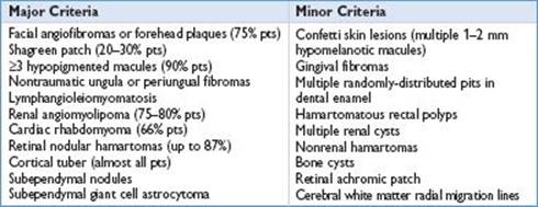
Epidemiology: Incidence is 1/5,000–10,000 live births (Neurol Clin 2003;21:983)
Pathophysiology (Arch Neurol 2000;57:662; N Engl J Med 2006;355:1345)
• Inheritance pattern: Autosomal dominant or sporadic (accounts for ⅔ of cases)
• Genes involved: TSC1 on chromosome 9, TSC2 on chromosome 16. Encode cell cycle regulation proteins. Variable penetrance. TSC1 mutations often w/ milder phenotype
• Screening (Arch Neurol 2000;57:662; N Engl J Med 2006;355:1345)
• Brain MRI yearly until 21 yo to evaluate for subependymal giant cell tumors
• Renal imaging at least q1–3yr for emergence/growth of renal angiomyolipomas
• Funduscopic exam to assess for retinal hamartomas
• Dermatologic exam w/ Wood lamp to assess for assoc skin findings. Rpt if needed
• Echo yearly to assess for growth of cardiac rhabdomyomas
• Chest CT if pulm sx develop to assess for lymphangiomyomatosis. Annual PFTs in patients with known lymphangiomyomatosis
• Neurodevelopmental testing
Complications (N Engl J Med 2006;355:1345)
• Neurologic: Epilepsy (in 70–80% of TS pts; infantile spasms strongly assoc w/ TS), mental retardation (affects ∼50% TS pts), neurobehavioral d/o/autism 2/2 cortical tubers. ↑ ICP, hydrocephalus 2/2 subependymal giant-cell tumors
• Renal: Sudden hemorrhage 2/2 renal angiomyolipomas. HTN/CRI 2/2 renal cysts
• Pulmonary: Pneumothorax/dyspnea due to lymphangiomyomatosis
• Cardiac: Cardiac failure in infancy (very rare), dysrhythmias due to rhabdomyomas
Neurofibromatosis Type 1
Diagnosis (JAMA 1997;278:51; Pediatrics 2000;105:608; Pediatrics 2008;121:633)
• Requires at least 2 of following, can emerge over time:
• ≥6 café-au-lait spots (1.5 cm ≥ postpuberty; 0.5 cm ≥ prepuberty). Most common feature in children, presents by 2 yo. Affects 99% of patients
• ≥2 neurofibromas, or 1 or more plexiform neurofibromas. Occur in brain and/or periphery, including SC tissue. Plexiform neurofibromas usually congenital, and can have multiorgan involvement. Neurofibromas occur in 48% of pts by 10 yo; ↑ to 84% by 20 yo. 60% of NF1 pts have brain neurofibromas
• Freckling in axilla or groin. 2nd most common feature in children. Commonly
presents 3–5 yo. Present in up to 90% of patients by 7 yo.
• Optic glioma (tumor of optic pathway). Presents in pts <6 yo. In up to 15% of pts
• 2 or more Lisch nodules (benign iris hamartomas that are best appreciated on
slit-lamp examination). Present in >70% of patients by 10 yo
• Distinctive bony lesion (dysplasia of the sphenoid bone or dysplasia/thinning of long bone cortex.) Often apparent by 1 yo. Present in ∼14% pts
• A 1st-degree relative w/ NF1
Epidemiology: Prevalence is 1/2,000–1/4,500 (Pediatrics 2000;105:608)
Pathophysiology (JAMA 1997;278:51; Pediatrics 2008;121:633)
• 50% AD, 50% sporadic; high rate of penetrance
• NF1 is a tumor suppressor gene on long arm of chromosome 17 band q11.2
Complications (Pediatrics 2000;105:608; Pediatrics 2008;121:633)
• Most pts w/ NF1 are mildly affected. Serious complications occur in ∼30% pts
• Growth abnormality, short stature, disfigurement 2/2 neurofibromas
• Vision loss, severe proptosis, hydrocephalus, precocious puberty 2/2 optic glioma, neurofibromas impinging on optic, hypothalamic, and pituitary tissues
• Malignancy: 5–10% of pts have malignant peripheral nerve sheath tumors, usually in adulthood. Less common malignancies include pheochromocytoma, rhabdomyosarcoma, leukemia, and CNS gliomas
• Gross organ damage due to plexiform neurofibromas
• HTN, ↑ stroke risk 2/2 vasc dysplasia; segmental vasculopathies, moyamoya
• Focal neurologic deficits, hydrocephalus, ↑ ICP 2/2 neurofibromas/gliomas
• Epilepsy (affects 5–7% pts)
• Learning disabilities in ∼60%. MR in ∼4% kids. Assoc w/ ADHD and autism
Screening (JAMA 1997;278:51; Pediatrics 2008;121:633; J Med Genet 2007;44:81)
• Routine imaging of the chest, abdomen, spine, and brain is not indicated
• Yearly ophtho exam until 7 yo to assess for optic glioma/associated vision defects
• Annual PE w/ BP check, neurodevelopmental assessment, skin exam to assess for progression of neurofibromas & underlying plexiform neurofibromas, skeletal exam to assess for scoliosis, vertebral angulation, limb abnormalities, tibial dysplasia, head circum to assess for hydrocephalus, and pubertal development to assess for precocious puberty
Neurofibromatosis Type 2
• Autosomal dominant. Less common than NF1
• Diagnostic criteria: Bilateral vestibular schwannomas (VS) or FHx NF2 (1st-degree relative) and unilateral VS or any 2 of following: Meningioma, glioma, schwannoma, juvenile posterior subcapsular lenticular opacities/juvenile cortical cataract
Sturge–Weber
• Congenital vascular capillary disorder, nonheritable. Characterized by port wine
stain (facial capillary angioma) typically in distribution of trigeminal nerve 1st and 2nd divisions. Assoc w/ ipsilateral leptomeningeal angioma w/ risk of venous thrombosis and/or surrounding brain ischemia. Complications include szr, MR, & glaucoma
Von Hippel–Lindau
• Rare AD condition assoc w/ multiple neoplasms: RCC, pheochromocytomas,
hemangioblastomas (often retinal, intracranial, or intraspinal), endolymphatic sac tumors, pancreatic tumors (neuroendocrine and serous cystadenomas), and papillary cystadenomas of the epididymis. Screening in VHL pts includes yearly ophtho exams, serum catecholamines, and MRI of brain/spine
Ataxia Telangiectasia
• Rare AR condition assoc w/ progressive cerebellar ataxia, immune def, abnl eye mvmt/impaired tracking, & oculocutaneous telangiectasias