
Gastric Aspirates
Checked by oral or NGT before feeding to see if feeds tolerated and digested
• Issues to consider
• If bilious emesis → malrotation until proven otherwise
• Prob w/ regimen: Interval btw feeds too short or delayed gastric emptying
• Ddx: Constipation, bowel obstruct, NEC, meconium plug or ileus, Hirschsprung dz, malrotation, volvulus, ileus, factitious (tube in duodenum or jejunum), pyloric stenosis, infxn, bleeding disorder (DIC, congenital coagulopathy), stress ulcer, meds
• Evaluation
• Assess volume and characteristics (bloody, bilious, undigested formula/milk) of aspirate, vitals, PE (distension, visible bowel loops, visible erythema)
• Labs: CBC, Bld cx (?sepsis/bleeding), stool guaiac, blood gas (if ? met acidosis)
• Radiology: KUB; check gas pattern, pneumatosis intestinalis or portal venous air, ileus, obstruction
• Left lateral decubitus film: Check for air – fluid levels (obstruction) and for layering of air above liver (free air, perforation)
Necrotizing Enterocolitis (NeoReviews 2001;2:e103; NeoReviews 2001;2:e110)
• Definition: Death of intestinal tissue in neonate 2/2 combo of vascular, mucosal, metabolic, infectious, and other unidentified insults to relatively immature gut
• Epidemiology
• Risk factors: Prematurity, asphyxia, acute cardiopulm dz, enteral feeds, polycythemia and hyperviscosity syndromes, exchange transfusions, enteric pathogenic microorganisms (E. coli, Klebs, Enterobacter, Pseudomonas, Salmonella, etc.), umbilical A-lines
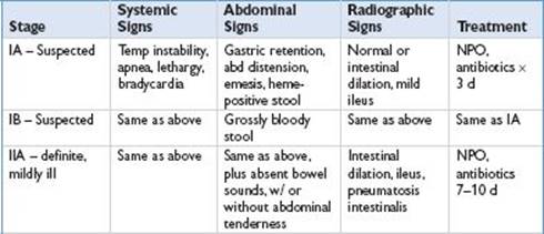
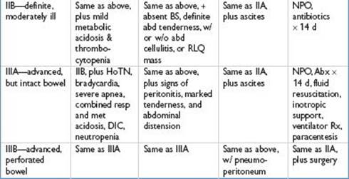
• Clinical presentation: Modified Bell staging criteria for NEC
• Diagnostic studies
• Early may resemble sepsis; but classically w/ triad of feeding intolerance, abd distension, and guaiac + stool
• Labs: CBC/diff, (Plts <50,000/μL), bld cx, guaiac, ABGs, electrolytes
• Radiology: Serial KUBs (assess for abn bowel gas patterns, ileus, portal venous or intramural gas [pneumatosis intestinalis], fixed sentinel loop of bowel). Lat decubitus (assess for pneumoperitoneum, indicates perforation)
• Management
• Initial NEC protocol: NPO, NG tube for decompression
• Abx coverage (ampicillin, gentamicin or cefotaxime; consider anaerobic Rx w/ clindamycin or Flagyl if pneumatosis or pneumoperitoneum seen)
• F/u I/O, remove K from IVF if anuric, septic w/u, x-rays q6–8h in acutely ill
• Surgical management: Resection of nonviable viscera with re-exploration later
Tracheal Esophageal Fistula and Esophageal Atresia (Am Fam Physician 1999;54:4:910)
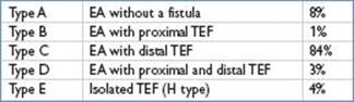
• Definition: Abn communication btw esophagus and trachea; assoc w/ esophageal atresia (EA), where proximal and distal portions of esophagus do not communicate
• Pathophysiology
• 1° morbidity pulmonary. Esophageal obstruct w/ inability to clear secretions, drooling, aspiration/regurg of food/pharyngeal contents → pneumonia
• W/ crying, straining, coughing, air enters stomach → abd distension, dilated loops of bowel, elevated diaphragm → basilar atelectasis and resp distress
• Clinical presentation
• W/ esophageal atresia: Polyhydramnios in utero; excess salivation, choking, coughing, cyanosis; diff swallowing excess fluid need freq suctioning
• ∼½ of pts w/ EA/TEF have other assoc congenital anomalies (i.e., VACTERL), cardiac anomalies (VSD or tetralogy of Fallot), other GI anomalies
• Diagnostic studies: Inability to pass NGT & resistance 10–12 cm from nares
• Imaging: Barium swallow may identify fistula, need caution and use nonionic contrast or dilute barium; +/− distal gas clue to presence and location of TEF; bronchoscopy is the most accurate procedure
• Management: Surgical correction is required
• Presurgical management
• Discontinue oral feedings, avoid bag-mask ventilation, and start IV fluids
• Elevate head to limit risk of aspiration and NGT in pouch w/ continuous suction
• Oxygen therapy if needed and mechanical ventilation if respiratory failure
• Evaluation for other congenital anomalies, especially VACTERL
• Repair delayed in VLBW infants and those w/ major concomitant anomalies
• Surgical complications
• Stricture 2/2 gastric acid erosion of shortened esophagus, leak of contents at anastomosis, fistula recurrence, tracheomalacia, abn swallow & esoph dysmotility, GERD
Gastroschisis
• Definition: Centrally located full-thickness abd wall defect (a) w/o protective covering over extruded intestine, and (b) w/ intact umbilical cord. Typically, liver and spleen reside w/i peritoneal cavity. No syndromic association
• Pathophysiology
• Unprotected intestine exposed to irritating amniotic fluid in utero → edematous, indurated, foreshortened appearance; w/ delay of peristaltic activity and effective absorption by several wk
• Diagnosis: Predominantly by prenatal U/S; differential dx: Ruptured omphalocele
• Management: Delivery at center equipped to provide definitive care
• Debate if C-section vs. vag delivery ↑ mortality/morbidity w/ gastroschisis
• Temperature regulation: Intestinal surface area exposed to environment puts infant at risk for hypothermia. Rx w/ protective covering: Saline-soaked gauze dressing and covering abdomen in plastic bag up to chest
• Nasogastric decompression; antibiotic coverage; TPN
• Surgical correction: Complete reduction of herniated intestine w/ 1° closure of wall or placement of unreduced intestine in prosthetic silo w/ staged reduction over 7–14 d
Omphalocele
• Definition: Herniation of abdominal contents into base of umbilical cord
• Has protective covering enclosing malpositioned abd contents. Umbilical cord comes together over apex of sac to form a normal appearing cord
• 24–40% assoc w/ other congenital anomalies including Beckwith – Wiedemann, trisomies, chromosomal abnormalities, CDH and various cardiac defects
• Clinical presentation: Vary in size; giant omphaloceles may contain liver and spleen
• Peritoneal cavity underdeveloped as growth proceeded w/o solid organ in position
• Diagnostic studies: Typically picked up by prenatal ultrasound
• Requires further evaluation for other anomalies
• Management: If ruptured sack, treat similar to gastroschisis w/ surgical correction
• Intact sack: Less urgent surgery required, and may require period of time for daily dressing changes to dessicate/toughen sac
Hirschsprung Disease (Pediatr Rev 2006;27:e56)
• Definition: Congenital absence of ganglion cells of the myenteric and submucosal plexus of the intestines resulting in poor/dysfxnl intestinal motility
• Begins at internal anal sphincter and variably extends proximally, may be patchy
• Short-segment Hirschsprung: (75–85%) distal to splenic flexure
• Long-segment Hirschsprung: (20%) aganglionic section extends past splenic flex
• Total intestinal aganglionosis: Rare form involving all small and large bowels
• Epidemiology: Incidence: Highest in African-Americans and Asians, ∼4:1  to
to 
• Pathophysiology
• Craniocaudal migration failure of ganglion cells precursors during embryogenesis
• Earlier arrest of migration results in longer segment involvement
• Clinical manifestations
• Delay in passage of meconium (>48 hr) always concerning for Hirschsprung
• Can p/w infreq BMs, abdominal distension, vomiting, diarrhea, refusal to feed
• Exam w/ contracted anal sphincter, no stool in vault on rectal exam but explosive stool release w/ w/d of finger, abd distension
• Isolated defect (70%) but 10× more common in pts w/ Trisomy 21
• Diagnostic studies: Gold std is rectal bx w/ staining for ganglion cells
• Barium enema w/ transition zone btw contracted distal and dilated proximal bowel is very suggestive; but absence of transition does not r/o
• Anorectal manometry to check reflex relaxation of anal sphincter (dx if no reflex)
• Useful for controversial ultrashort segment dz w/ <5 cm involvement, (unclear whether this condition really exists)
• Management: Resection of aganglionic section (Am J Surg 2000;180:382)
• Multi surgical approaches; most w/ 1° endorectal pull through (1-stage procedure)
• Age when done & if staged approach used (w/ diverting colo in btw) varies
• Complications
• Even w/ surgical Rx, 30% w/ cont constipation or never develop fecal continence
• Major mortality/morbidity w/ Hirschsprung 2/2 enterocolitis (occurs in ∼20% in one series) (Am J Surg 2000;180:382)
• Hirschsprung assoc enterocolitis: Mostly pts <2 yo p/w abd distension, explosive watery stool, fever, and hypovolemic shock