
ASSESSMENT PRINCIPLE #1
A complete and detailed clinical and radiographic assessment of the child’s foot is required before treatment is initiated.
It is hard to further clarify or justify this principle. How to do it is the focus of this chapter.
HISTORY
ASSESSMENT PRINCIPLE #2
Clinical evaluation of the child’s foot begins with a clinical evaluation of the child.
Although the foot deformity or malformation is the reason for the requested evaluation by you, children with these conditions often have underlying neuromuscular, genetic, or chromosome disorders as well as other deformities and/or malformations of the lower extremities and spine. These must be recognized and factored into the decision-making process to ensure that the most appropriate of the possible nonoperative and operative interventions is chosen (see Basic Principle #14, Chapter 2).
Idiopathic congenital clubfoot, congenital vertical talus, flatfoot, metatarsus adductus, skewfoot, and positional calcaneovalgus deformity are often seen in normal children. These deformities can also be seen in children with underlying neuromuscular, genetic, or chromosome disorders. By way of contrast, almost all cavus foot deformities are the result of an underlying neuromuscular disorder, though congenital idiopathic cavus exists.
Foot deformities in children with neuromuscular, genetic, and chromosome disorders have appearances similar to those in normal children, but the natural history and response to treatment are often quite different. Therefore, differentiation is important.
Underlying conditions that are associated with congenital foot deformities and mal-deformations include myelomeningocele, lipomeningocele, arthrogryposis, sacral agenesis, fibular and tibial hemimelia, Apert syndrome, congenital hemiatrophy, myotonic dystrophy, Down syndrome, Ehlers–Danlos and Marfan syndromes, and a whole host of other chromosome abnormalities.
ASSESSMENT PRINCIPLE #3
Congenital and developmental deformities should be differentiated (see Basic Principle #12, Chapter 2).
Ask if the deformity was present at birth. Congenital deformities are rarely progressive in their natural history, yet rarely regressive. Tendons and joint capsules are usually co-contracted. For example, in a clubfoot (congenital talipes equinocavovarus) in an older child that does not correct with nonoperative management, posterior ankle capsulotomy is often required in addition to tendo-Achilles lengthening.
Developmental deformities, by definition, are progressive in their natural history, though the rate of progression is variable. Contracture of tendons precedes contracture of joint capsules. In a developmental equinocavovarus foot deformity in an older child, an tendo-Achilles lengthening is usually sufficient to correct the equinus deformity.
ASSESSMENT PRINCIPLE #4
Static and progressive foot deformities should be differentiated, and the rate of progression established, if possible.
Ask if the deformity has changed noticeably over time and, if so, by how much over what interval. As stated in Assessment Principle #3, most congenital foot deformities are static, rather than progressive, in nature. Muscle imbalance is the underlying problem in many acquired foot deformities. The muscle imbalance can be static, as in children with myelomeningocele, lipomeningocele, and postinfectious poliomyelitis; or it can be progressive, as in children with Charcot–Marie–Tooth disease, muscular dystrophy, spinal cord tumors, tethered cord, and diastematomyelia. Whether the muscle imbalance is static or progressive, the deformity is likely to progress. Unfortunately, the rate of progression is rarely predictable for either static or progressive muscle imbalances. Progression will increase the complexity of reconstruction.
ASSESSMENT PRINCIPLE #5
It is often more challenging to ascertain the history of pain and/or dysfunction that is related to a foot deformity in a child than in an adult, but it is worth the effort.
Otherwise, it is like practicing veterinary medicine. Reasons for children to be poor historians include too young, “too adolescent,” intellectually challenged, neurologically impaired. The importance of an accurate assessment of the pain and dysfunction is that there are many clinically and radiographically apparent normal anatomic variations of the child’s foot. If the pain location, severity, and temporal and activity-related patterns do not match the known pain pattern of a particular deformity/condition, the two might not be related. Do not go for the low-hanging fruit.
ASSESSMENT PRINCIPLE #6
Assessment of pain must be specific—ask where, when, what level/severity, what associations.
There are many anatomic variations of the foot, including a host of accessory ossicles, which could be the source of pain or merely incidental findings. It is easy, for example, to ascribe reported foot pain to a tarsal coalition or an accessory navicular that is identified on an x-ray. However, since most anatomic variations including tarsal coalitions and accessory naviculars do not hurt, it is important to know the exact site(s) of pain (see Assessment Principle #15, this chapter), as well as the activities that incite and relieve the pain. Severity of the pain should be quantified. Visual analog pain scales have been shown to be reliable in even very young children. The pain location, pattern, and severity must all match those of the presumed diagnosis. Chronic pain in a nonphysiologic distribution that occurs continuously during all waking hours and is reported to be of an exaggerated severity suggests chronic regional pain syndrome, a.k.a. reflex sympathic dystrophy, reflex neurovascular dystrophy, and pain amplification syndrome.
PHYSICAL EXAMINATION
ASSESSMENT PRINCIPLE #7
Physical evaluation of the child’s foot begins with a physical evaluation of the child (see Basic Principle #14, Chapter 2).
This includes a careful examination of the hips and spine in a newborn. Visual gait analysis, torsional profile analysis, and angular alignment assessment are used for older children and adolescents.
Visual gait analysis is carried out by watching the child walk, run, toe walk, heel walk, squat and stand, and hop on each foot. These observations are used to evaluate symmetry, strength, coordination, and comfort.
The child’s torsional profile must be ascertained. The foot progression angle, which is assessed while the child walks at a normal pace in a long hallway, is the summation of all segmental rotational alignments/deformities in the lower extremities. The segmental rotational alignment values are determined with the child prone on an examination table. The degrees of internal and external hip rotation reflect femoral torsion. Utilization of the thigh–foot angle (TFA) for assessment of tibial torsion is predicated on the absence of a foot deformity in the limb being tested. Determination of the transmalleolar axis (TMA) is required to assess tibial torsion when there is hindfoot/subtalar joint deformity and/or equinus deformity. Assessment of the TMA is less reliable than assessment of the TFA.
The importance of accurate assessment of lower limb torsion is highlighted in children with flatfoot deformity, whether idiopathic or associated with cerebral palsy or tarsal coalition. There is rarely coincident pathologic tibial torsion in these conditions. The external rotation of the foot in relation to the limb exists almost entirely in the subtalar joint. A flatfoot will create an out-turned, or positive value, TFA because of eversion/external rotation of the subtalar joint (up and out) (see Basic Principles #6 and 7, Chapter 2). If the clinical TFA equals the radiographic standing anteroposterior (AP) talus–1st metatarsal (MT) angle (see Assessment Principle #18, this chapter). All of the external rotation is in the subtalar joint (foot) and none in the tibia. If the TFA is greater than the standing AP talus–1st MT angle, the difference is the degree of external tibial torsion.
In contrast, developmental cavovarus foot deformities are usually associated with external tibial torsion which is exposed after the foot deformity is corrected. A cavovarus deformity will internally rotate the foot in relation to the limb because of inversion/internal rotation of the subtalar joint (down and in) (see Basic Principles #6 and 7, Chapter 2). The thigh–foot angle is neutral to slightly internally rotated before the foot surgery and outwardly rotated afterward, reflecting the external tibial torsion that was already present. Families need to be apprised of this fact before the foot surgery is performed, or they will assume that the foot deformity was overcorrected (see Management Principle #10, Chapter 4). The change in TFA after correction of a cavovarus deformity will equal the preoperative AP talus–1st MT angle (see Assessment Principle #18, this chapter).
Equinus deformity will also make it challenging to determine tibial torsion using the TFA, because the planar axis of the foot is not parallel with the planar axis of the femur. The TMA is necessary to determine tibial torsion in this situation as well.
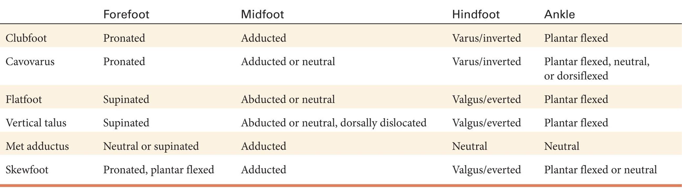
|
TABLE 3-1 |
Deformity-specific segmented deformities of the foot and ankle |
Exaggerated genu varum and genu valgum will cause the foot to bear weight unusually because of the altered angular relationship between the tibia and the ground. This can create an apparent foot deformity when none exists.
ASSESSMENT PRINCIPLE #8
Assessment of each of the segmental deformities of the foot and ankle is imperative before planning treatment, as a plan needs to be established to correct each one (Table 3-1, Figure 3-1).
The segments are:
1. Forefoot—pronated or supinated; plantar flexed (equinus) or dorsiflexed
a. Recall that alignment (and deformity) is defined as the relationship between a more distal anatomic part and the next more proximal anatomic part. Therefore, pronation or supination refers to the alignment of the forefoot in relation to the midfoot/hindfoot, not the tibia/leg. This has been a source of confusion for many who believe the forefoot in a flatfoot is neutrally aligned (in relation to the tibia) when, in fact, it is supinated—in relation to the mid/hindfoot (Figure 3-2).
2. Midfoot—abducted or adducted (see Metatarsus adductus, Chapter 5)
3. Hindfoot—varus/inverted or valgus/everted (see Figures 2-4 and 2-7, Chapter 2)
4. Ankle—varus or valgus (see Figure 3-12, this chapter); plantar flexed (equinus) or dorsiflexed (calcaneus)
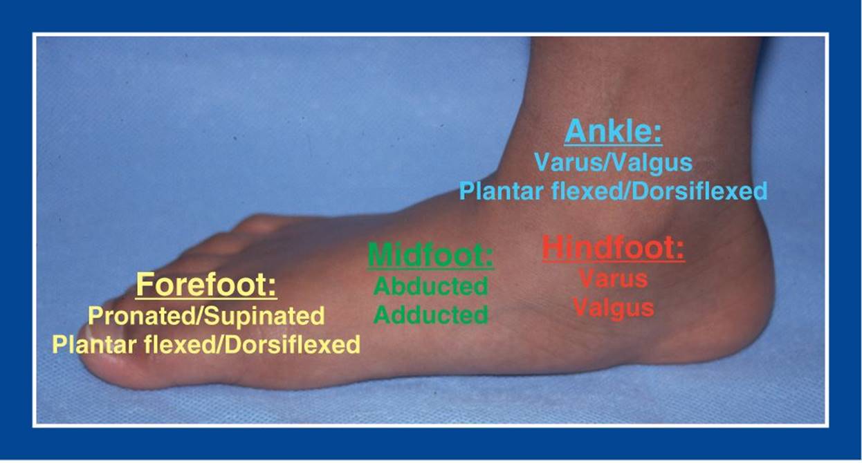
Figure 3-1. Identify each of the segmental deformities of the foot and ankle before initiating treatment.
ASSESSMENT PRINCIPLE #9
Each segment of the foot should be evaluated for shape/deformity, flexibility, and skin integrity. Documentation should be specific.
Accurate assessment of the shape of each segment of the foot is the first step. For a cavovarus foot deformity, the segmental deformities are pronation of the forefoot, adduction of the midfoot, varus of the hindfoot, and possibly equinus of the ankle (Table 3-1). Equally important is the flexibility of each segment. The first segment to lose flexibility is the forefoot. Loss of flexibility of the hindfoot, which is assessed by the Coleman block test, eventually follows (Figure 3-3).
I have found that the block test, as described by Coleman, is uncomfortable and awkward to perform and, therefore, unreliable. With the entire lateral column of the foot on the block, it is tempting for the child to balance the foot on the block, rather than allowing the forefoot to pronate off the block. Price and Mubarak have independently proposed alternate methods for the clinical assessment of hindfoot flexibility in a cavovarus foot. However, neither is performed in weight-bearing. A more comfortable, reliable, and accurate way to assess weight-bearing hindfoot flexibility in a cavovarus foot is to perform a modified Coleman block test in which a 2.5-cm block is placed under the lateral 2–3 MT heads. The heel remains on the ground and the medial MT heads seek the ground as the forefoot pronates off the block (Figure 3-4).
Skin integrity should be assessed, as it can identify unsafe foot pressures which is especially important in children with insensate skin. In the cavovarus foot, exaggerated pressures are seen at the base of the 5th MT and under the 1st and 5th MT heads (Figure 3-5).
The segmental deformities of a flatfoot include supination of the forefoot, abduction or straight alignment of the midfoot, valgus of the hindfoot, and equinus of the ankle (Table 3-1). Equally important is the flexibility of each segment. Flexibility of the hindfoot is assessed in a different manner than that used for a cavovarus foot. There is not a reliable “reverse” Coleman block test. Instead, toe standing (Figure 3-6) and the Jack toe raise test are utilized to assess hindfoot flexibility (Figure 3-7).
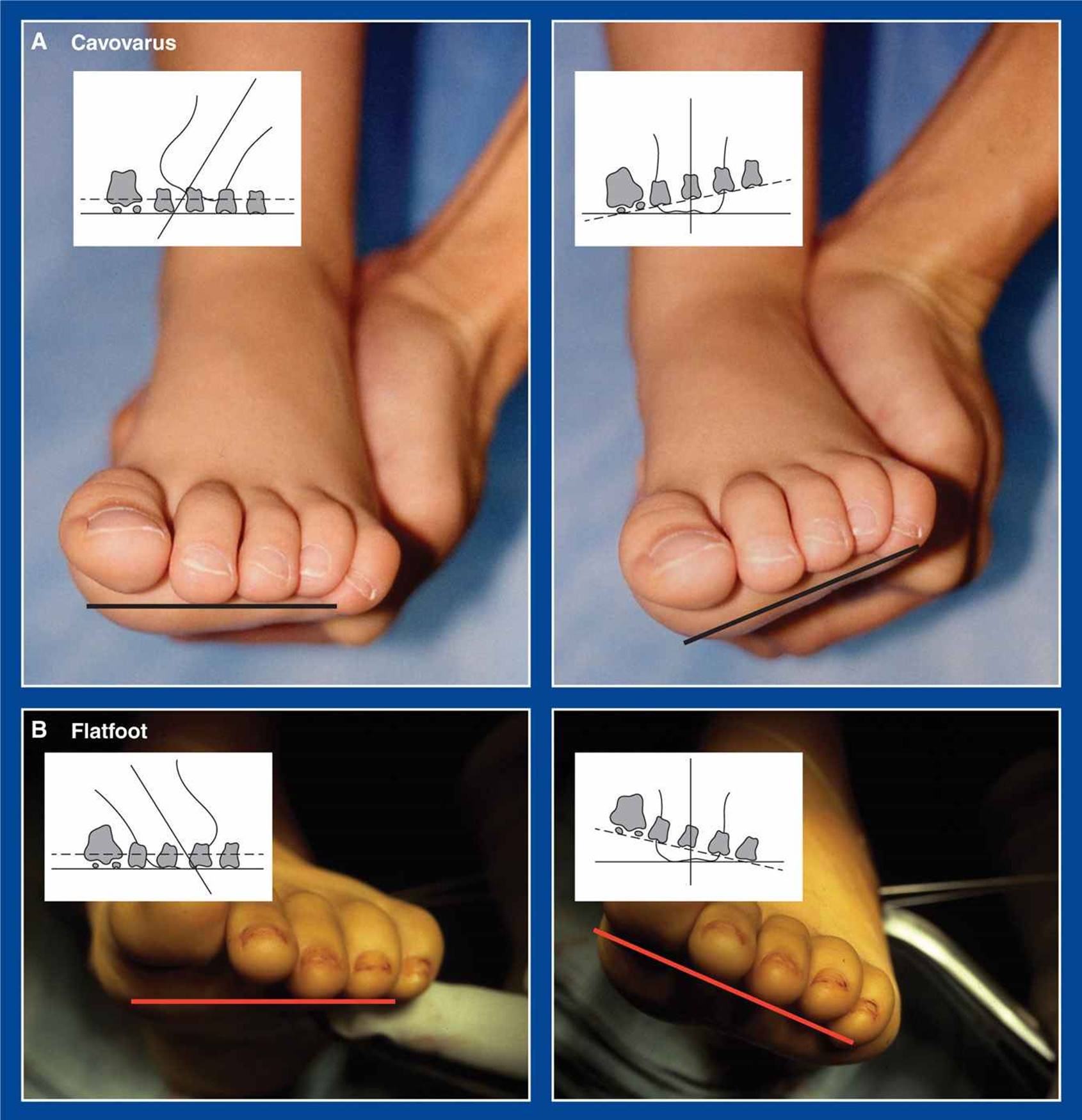
Figure 3-2. A. Pronation of the forefoot in a cavovarus foot is unappreciated until the hindfoot varus is corrected to neutral. B. Supination of the forefoot in a flatfoot is unappreciated until the hindfoot valgus is corrected to neutral.
Evidence of exaggerated skin pressures in a flatfoot are identified under the medial midfoot. The skin in this area is rarely stressed except when a flatfoot is associated with contracture of the gastrocnemius or the entire triceps surae (tendo-Achilles) (Figure 3-8).
The clubfoot should be assessed for shape and flexibility using one or both of the two most commonly used classification systems, those of Pirani and Dimeglio.
ASSESSMENT PRINCIPLE #10
The accurate assessment of subtalar motion is an inexact science, but you can better at it by practicing.
There are no studies documenting the accuracy of assessment of subtalar motion. It is particularly challenging in very small feet and fat feet. The ankle should be held in neutral dorsiflexion. The dome of the talus is biconical in shape, narrower posteriorly than anteriorly. Dorsiflexion of the foot engages the widest portion of the talar dome in the ankle mortis, thereby creating bony stability as well as tightening the collateral ligaments to eliminate false inversion/eversion motion at that joint. The calcaneus is held in a cupped hand and moved in the axis of the subtalar joint, “down and in” and “up and out” (Figures 2-7, 2-8, and 3-9).

Figure 3-3. The Colman block test is used to assess flexibility of the hindfoot in a cavovarus foot with rigid forefoot pronation. Early in the course of development of the cavovarus deformity, the hindfoot varus (seen in A) remains flexible despite rigid forefoot pronation. It corrects to valgus (seen in B) with a block of wood under the lateral forefoot which allows the forefoot to pronate freely over the edge of the block. At that stage, correction of the forefoot deformity alone will result in spontaneous correction of the hindfoot. In time, the hindfoot varus deformity becomes rigid. Correction of the hindfoot deformity must then be combined with correction of the forefoot deformity. (From Coleman SS, Chestnut WJ. A simple test for hindfoot flexibility in the cavovarus foot. Clin Orthop Relat Res. 1977;123:60–62, with permission.)
The other hand is used to note the motions at the midfoot and forefoot. It should not be used to attempt to move the subtalar joint, because hypermobility of Chopart joints (talonavicular and calcaneocuboid) can give a false impression of subtalar joint motion when none exists (Figure 3-10).
In my experience, this hypermobility of Chopart joints often develops in feet with solid talocalcaneal tarsal coalitions. It gives a false impression that a rigid flatfoot is flexible, not only when subtalar joint motion is incorrectly assessed manually, but also when it is assessed with toe standing (Figure 3-11).
The best way to improve your skills for assessing subtalar joint motion is to practice in the OR during a foot deformity correction operation while observing your technique and the resultant motions of the subtalar joint under mini-fluoroscopy.
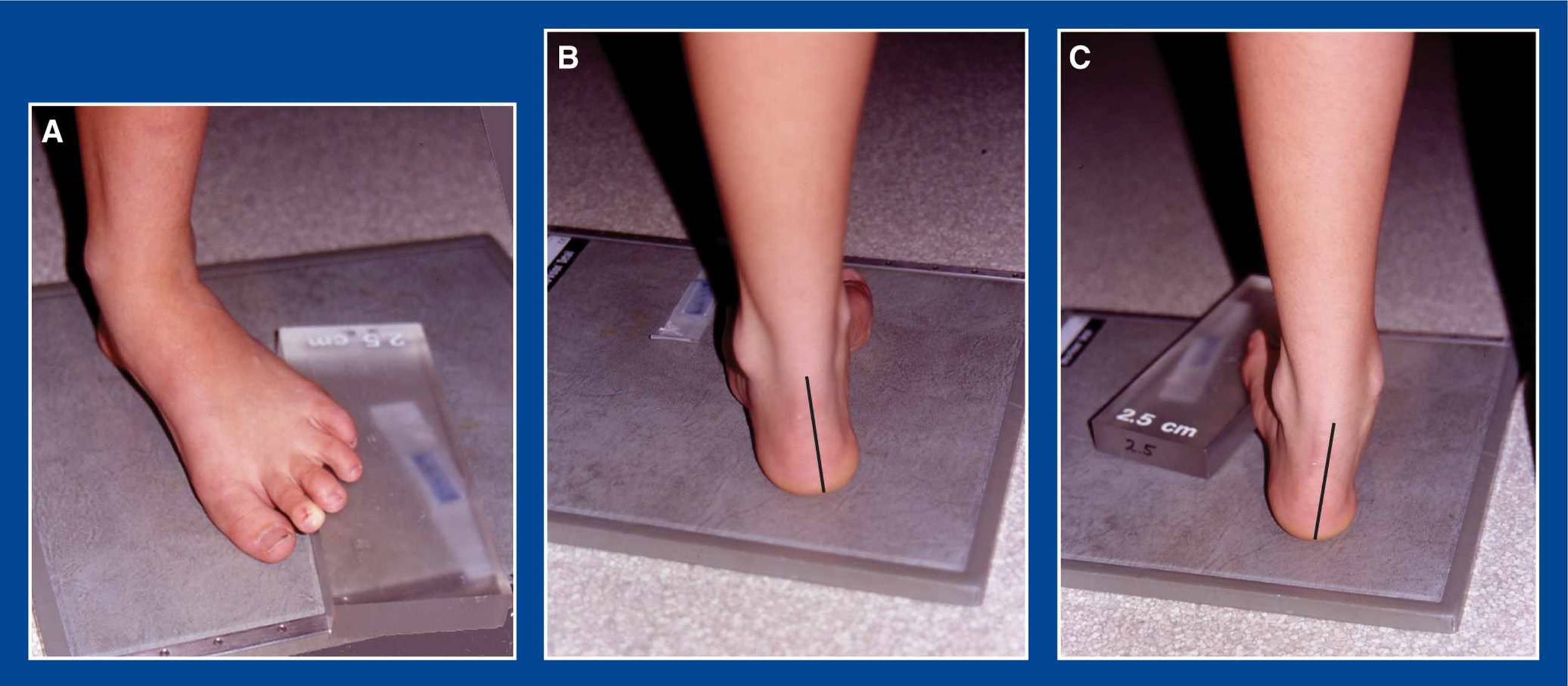
Figure 3-4. Modified Coleman block test. A. A 2.5-cm block of Plexiglas (or wood) is placed under the lateral 2–3 MT heads while keeping the heel on the ground. B. The posterior view with no block. The hindfoot is in varus. C. With the block under the lateral 2–3 MT heads, the hindfoot varus has converted to valgus, indicating flexibility of the subtalar joint. This can be confirmed radiographically (see Assessment Principle #19, this chapter).
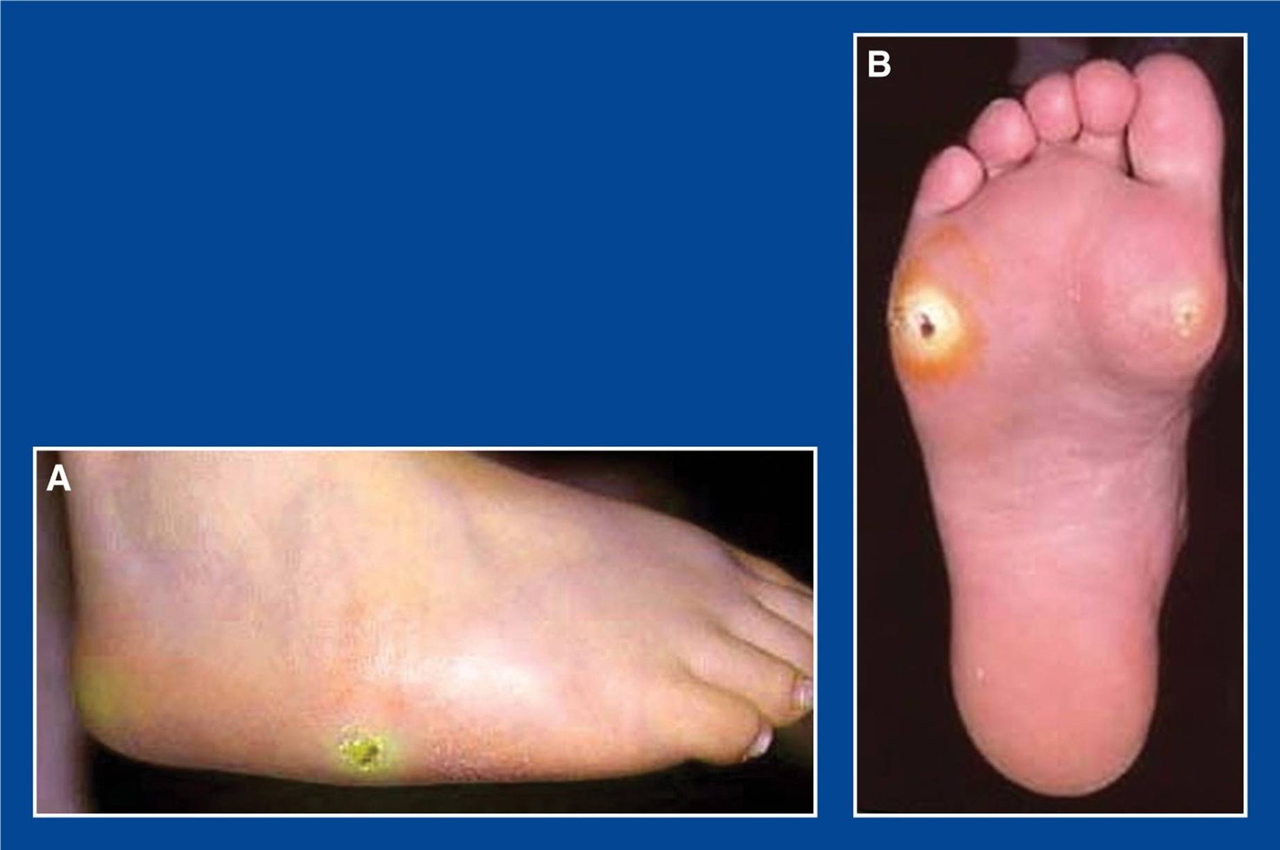
Figure 3-5. Exaggerated and unsafe skin pressures in the cavovarus feet of children with myelomeningocele. A. A cavovarus foot with hemorrhagic callus following healing of a neurotrophic ulcer. B. Deep neurotrophic ulcer with large surrounding area of thick callus formation under the 5th MT head and a recently healed ulcer under the 1st MT head.
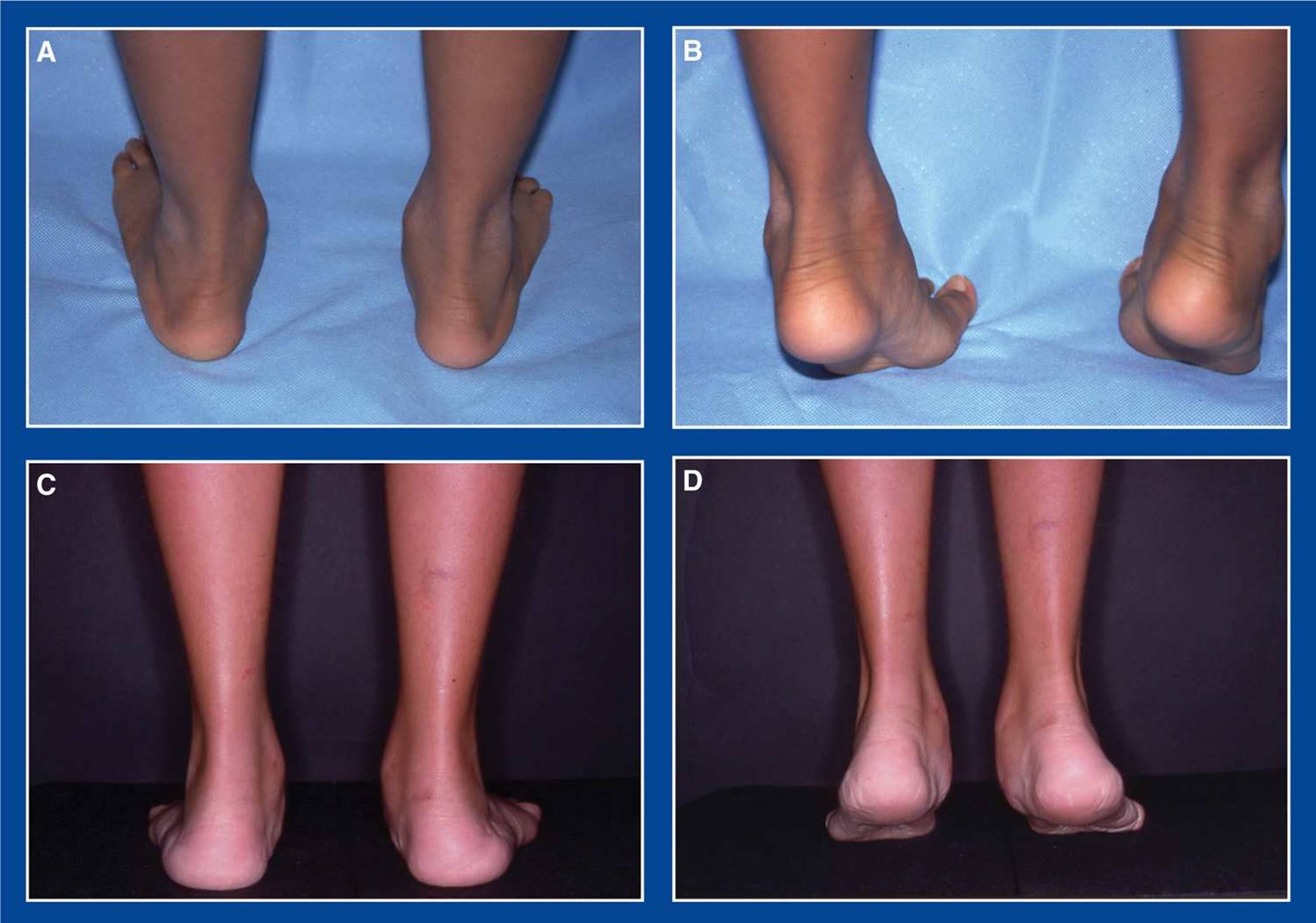
Figure 3-6. A. Flexible flatfeet with hindfoot valgus, forefoot supination, and the “too many toes” sign (toes seen lateral to the hindfoot when viewed from behind). B. With toe standing, the valgus hindfoot of a flexible flatfoot converts to varus, the arch elevates with reversal of the forefoot supination to pronation, and the toes appear medial to the hindfoot. C. Rigid flatfeet with the same segmental deformities as the flexible flatfoot, i.e., hindfoot valgus, forefoot supination, and the “too many toes” sign. D. With toe standing, nothing changes except that the heels elevate off the ground.
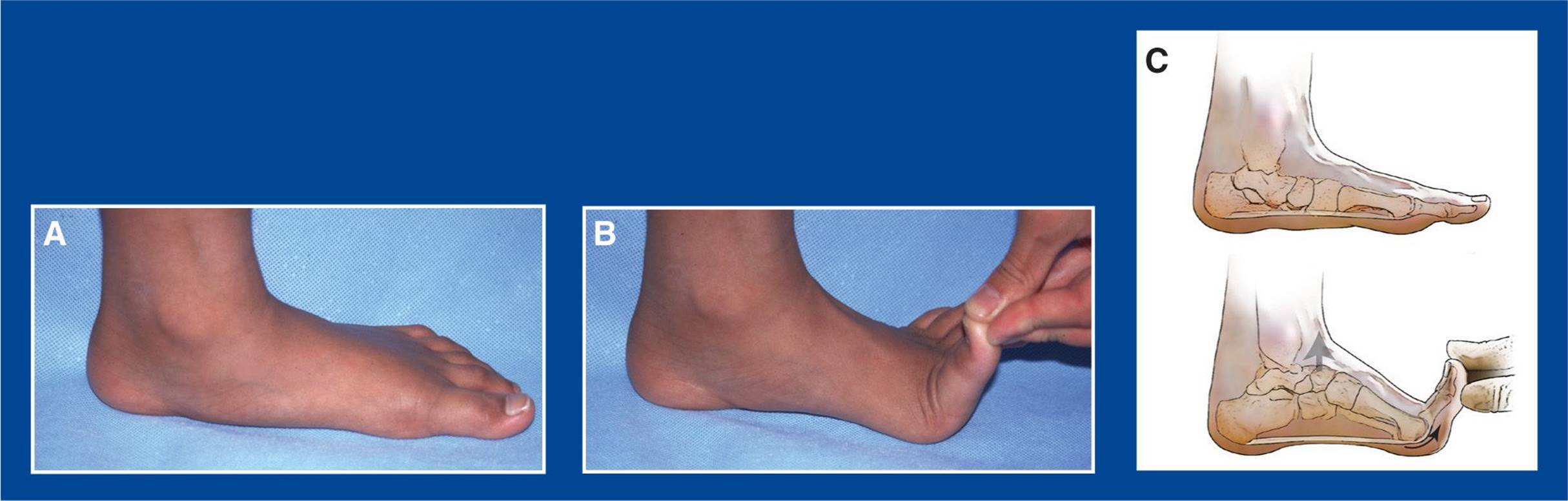
Figure 3-7. The Jack toe raise test, like toe standing, demonstrates hindfoot/subtalar joint flexibility in a flexible flatfoot (A) by means of the “windlass action” of the plantar fascia. The plantar fascia originates on the plantar aspect of the calcaneus and inserts into the plantar aspect of the toes through multiple interconnections. Great toe dorsiflexion (B) pulls the plantar fascia distally under the pulley of the head of the 1st MT. Since the plantar fascia is of fixed length, the great toe can only fully dorsiflex if the calcaneus is pulled distally toward the MT heads, thereby shortening the foot, elevating the longitudinal arch, and inverting the subtalar joint (C).
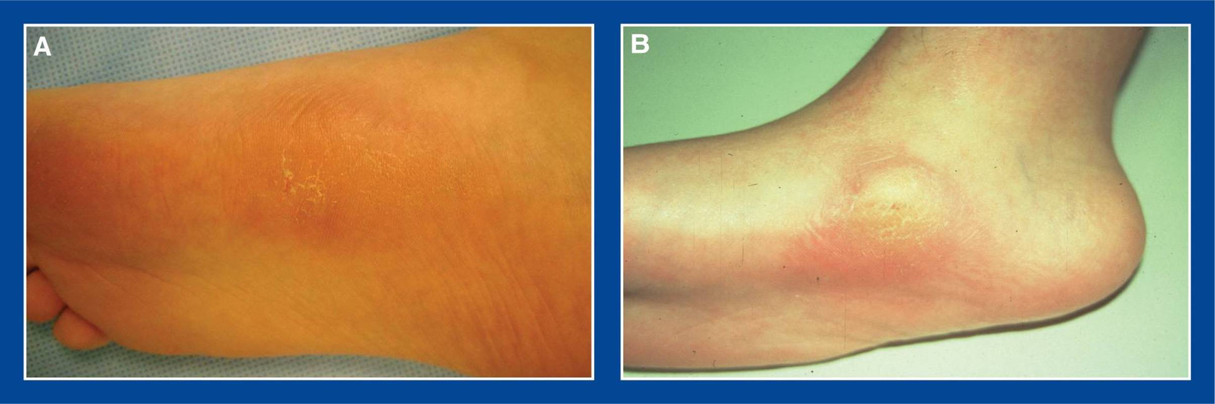
Figure 3-8. Contracture of the gastrocnemius or the entire triceps surae prevents the talus from dorsiflexing in the ankle joint. The calcaneus can dorsiflex past the plantar flexed talus by taking advantage of subtalar joint eversion—dorsiflexion, external rotation, and pronation of the calcaneus/acetabulum pedis. The talus remains rigidly plantar flexed while the navicular and the rest of the acetabulum pedis move “up and out,” thereby concentrating all the weight-bearing stresses under the talar head (A & B). Since the plantar flexion of the talus is unyielding, firm or rigid arch supports will increase skin pressure and pain at that site.
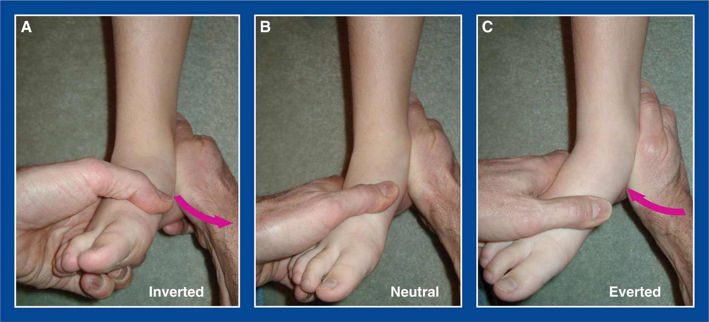
Figure 3-9. Attempt to move the subtalar joint “down and in” and “up and out” with a cupped hand on the heel, while maintaining the ankle at neutral dorsiflexion. Do not attempt to move the hindfoot with the hand on the forefoot because there can be excessive motion through Chopart’s joints (talonavicular and calcaneocuboid) that gives the false impression of subtalar motion. Only use the hand on the forefoot to stabilize the foot and to detect false motions. A. Inverted. B. Neutral. C. Everted.
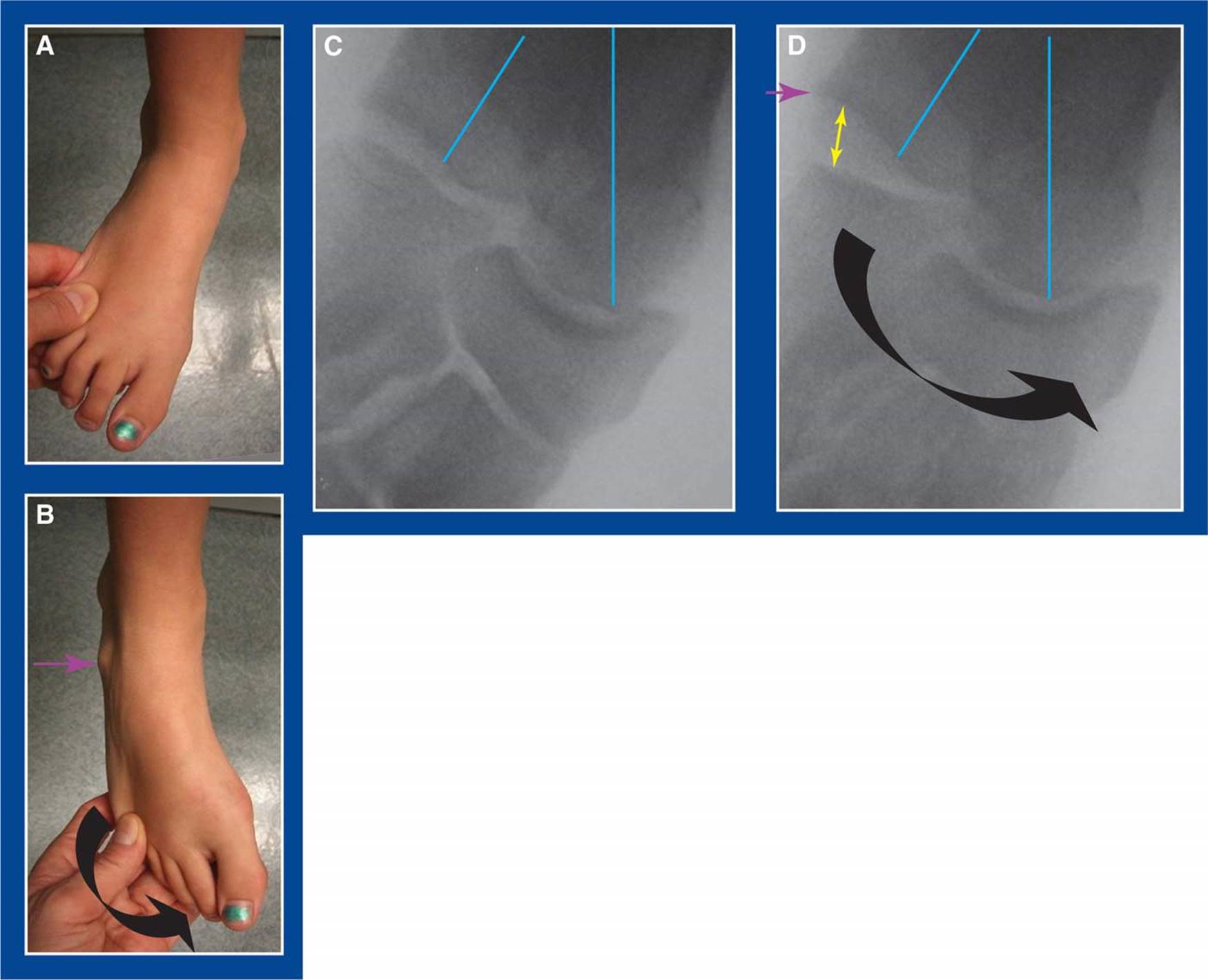
Figure 3-10. A, B. Foot with a large, solid talocalcaneal tarsal coalition and no motion possible between the talus and calcaneus, but apparent subtalar motion when examined incorrectly. The purple arrow points to a bony prominence that is noted with the forefoot “inverted” (actually adducted), but not apparent with the foot in its normal everted position. C. AP x-ray of the foot in its normal everted position. The dark blue lines are the axes of the talus and the calcaneus. D. The curved arrow shows the direction that the forefoot/midfoot was moved. The navicular has rotated into better axial alignment with the talus suggesting inversion of the subtalar joint, but there is no change in the relationship between the talus and calcaneus (see dark blue lines). Instead, the apparent inversion took place because of acquired hypermobility at the calcaneocuboid joint. Normally a nonmobile joint, the calcaneocuboid joint opened up like a book (yellow double-headed arrow). The bony prominence (at the tip of the purple arrow) is the anterior end of the calcaneus that has been exposed because of the plantar–medial movement of the cuboid, along with the navicular, at Chopart joints.
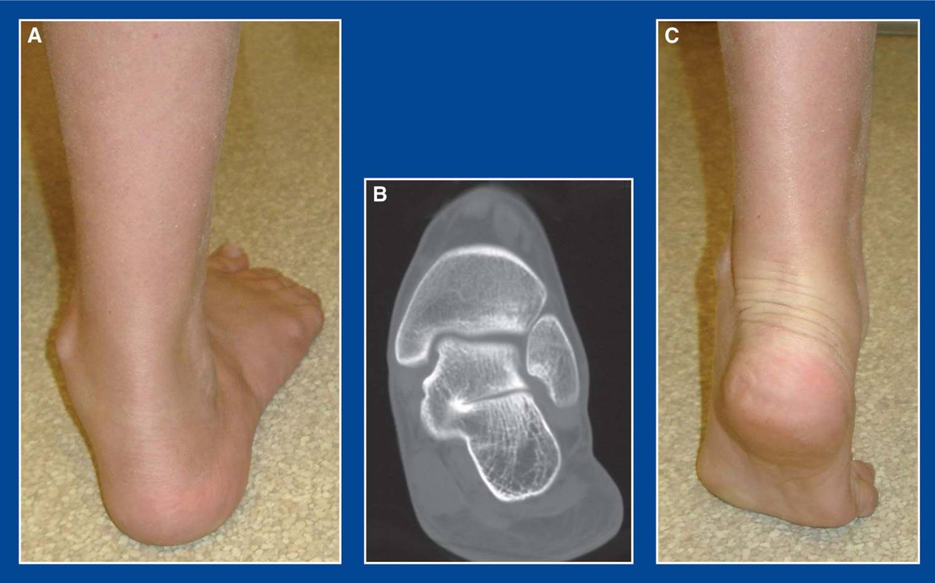
Figure 3-11. The same foot as in Figure 3-10. A. Valgus hindfoot with the “too many toes” sign. B. Coronal CT scan cut shows large osseous middle facet coalition, narrow posterior facet, and excessive valgus deformity. C. With toe standing, the hindfoot valgus deformity “corrects” to apparent varus, the arch elevates, and there are less toes seen laterally. This is a physiologic adaptation that can only happen because of acquired hypermobility at Chopart joints, specifically at the calcaneocuboid joint. The reason for the clinical appearance of hindfoot varus when the calcaneus is in rigid valgus alignment under the talus is unknown.
ASSESSMENT PRINCIPLE #11
An ankle joint deformity may coexist with a foot deformity, or it may be an isolated deformity. It must be differentiated.
The ankle joint is in valgus orientation to the anatomic axis of the tibia in all normal newborns. In otherwise normal children, the distal fibula and lateral distal tibia grow relatively faster than the medial distal tibia until about age 3 to 4 years. At that point, the ankle joint/tibial plafond becomes perpendicular to the tibia. It maintains that anatomic alignment through skeletal maturity (Figure 3-12).
That spontaneous change from physiologic neonatal ankle valgus to neutral alignment does not occur in children with myelomeningocele, lipomeningocele, early-onset poliomyelitis, other early-onset flaccid paralytic conditions, and approximately 66% of limbs with a clubfoot. The clinical assessment of ankle joint alignment and the differentiation from subtalar joint alignment are helpful in older children, particularly in those with the stated underlying conditions. In spastic conditions, such as cerebral palsy, normal spontaneous correction of neonatal ankle valgus to neutral occurs.
The lateral malleolus is longer/taller than the medial malleolus at all ages and in all underlying conditions (except fibular hemimelia). Therefore, with a valgus ankle joint, the distal tip of the lateral malleolus and that of the medial malleolus are in a transverse plane that is often perpendicular to the tibia (Figure 3-12A). When the ankle joint has assumed its adult alignment perpendicular to the tibia, the distal tip of the lateral malleolus is closer to the floor and further from the knee than the medial malleolus (Figure 3-12B). This assessment of the relative heights of the malleoli is helpful in the clinical determination of ankle alignment. It is particularly helpful in the clinical determination of the site of hindfoot valgus deformity, which can exist in the ankle joint, the subtalar joint, or in both joints. There may also be pathologic valgus in the ankle and varus in the subtalar joint, varus in both joints, or varus in the ankle and valgus in the subtalar joint. Radiographs of the ankle joint will confirm the specific anatomy (see Assessment Principle #21, Figure 3-27, this chapter).
The ankle joint can also have a procurvatum or recurvatum deformity. These are almost always acquired deformities. A flat-top deformity of the talus can occur following both nonoperative and operative treatment of clubfoot deformity, and results in a true or “functional” procurvatum deformity of the ankle (see Anterior ankle impingement, Figure 5-1, Chapter 5). Iatrogenic posterior distal tibial physeal arrest following clubfoot surgery can cause a true procurvatum deformity (see Anterior ankle impingement, Figure 5-3, Chapter 5).

Figure 3-12. Clinical assessment of the relative heights of the lateral and medial malleoli relative to the floor can provide a clue as to the alignment of the ankle joint (varus or valgus). Yellow line connects the distal tips of the lateral and medial malleoli. Black line represents the plane of the ankle joint. A. The ankle joint is in valgus alignment relative to the tibia from birth until age 3 to 4 years, resulting in malleoli that are at the same transverse level. Those neonatal relationships persist in many paralytic conditions and a large percentage of clubfeet. B.The ankle joint is perpendicular to the tibia after the age of 3 to 4 years. The lateral malleolus is further from the knee and closer to the ground than the medial malleolus thereafter.
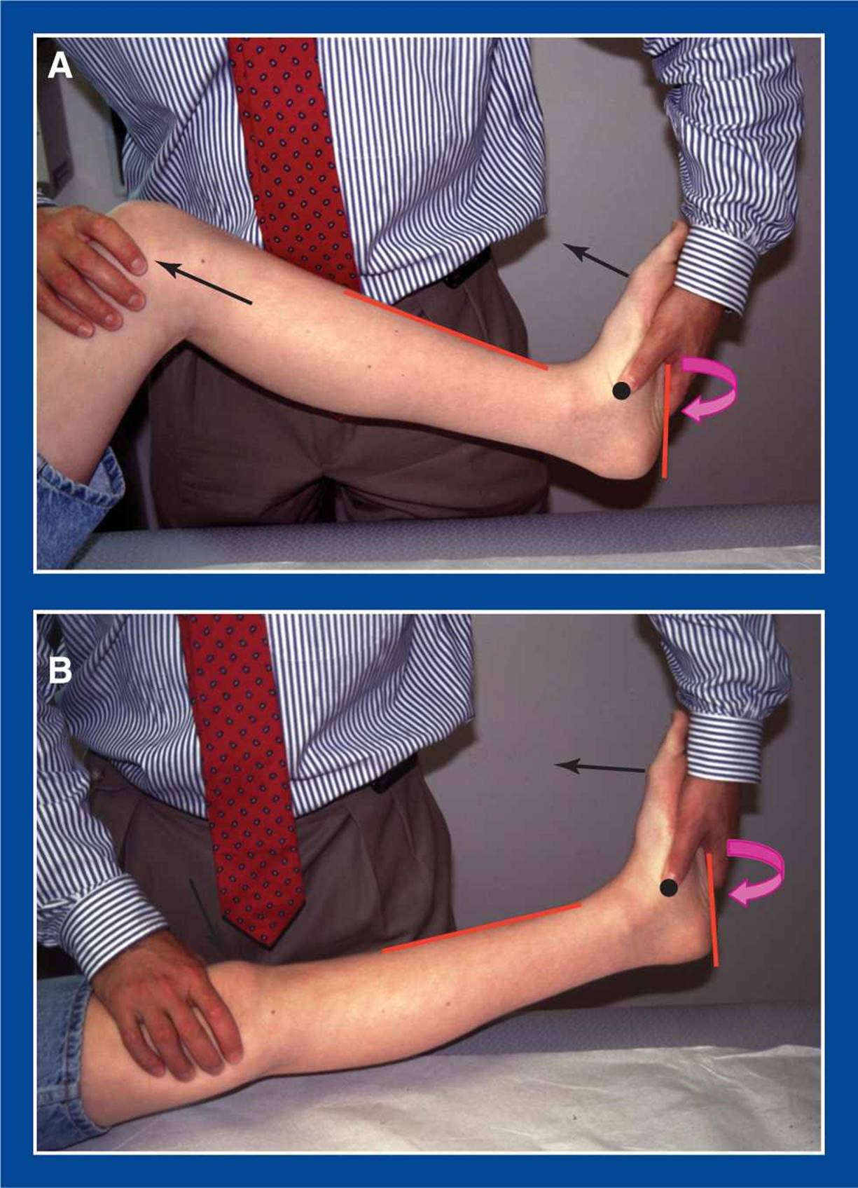
Figure 3-13. The Silfverskiold test. A. Testing the soleus and, effectively, the entire triceps surae/tendo-Achilles: 1. Flex the knee to relax the gastrocnemius (black arrow at knee). 2. Ensure that the talonavicular joint is in neutral alignment. If the subtalar joint is everted, as in a flatfoot, invert (“lock”—see Basic Principle #7, Chapter 2) the subtalar joint to neutral, and confirm anatomic subtalar joint alignment with a thumb over the plantar–medial aspect of the talonavicular joint (black dot). If the subtalar joint is inverted, as in a cavovarus foot, evert the subtalar joint to neutral. 3. Maximally dorsiflex the ankle joint (black arrow above foot) and record the angle between the plantar–lateral border of the foot (red line), which is the true proxy for the foot, and the anterior border of the tibial shaft (red line). Do not use the plantar–medial border of the foot as the reference line because supination or pronation deformity of the forefoot will give a false impression of ankle joint position. Ankle dorsiflexion greater than or equal to 10° is normal, as in this example.B. Testing the gastrocnemius: 1. While maintaining subtalar neutral, extend the knee to tighten the proximal end of the gastrocnemius. 2. The ankle will lose some dorsiflexion in most cases. 3. Record the angle between the plantar–lateral border of the foot and the anterior border of the tibial shaft. In this case, the ankle lacks about 5° of dorsiflexion from neutral, indicating contracture of the gastrocnemius.
ASSESSMENT PRINCIPLE #12
The presence of a gastrocnemius or an tendo-Achilles contracture must be identified and differentiated from each other.
Many foot deformities do not cause pain or functional disability unless they are accompanied by a contracture of the heel cord (the gastrocnemius alone or the entire triceps surae/tendo-Achilles). The ankle joint should have at least 10° of dorsiflexion with the knee extended and the subtalar joint in neutral alignment (“locked”—see Basic Principle #7, Chapter 2). The Silfverskiold test should be used to determine whether there is a contracture of the heel cord and, if so, whether the contracture is of the gastrocnemius alone or the tendo-Achilles. This will ensure that the proper tendon is lengthened if surgery is indicated, thereby avoiding under or overlengthening. The Silfverskiold test must be mastered (Figure 3-13).
The flatfoot presents a special challenge when determining contracture of the heel cord. The reason is that both the ankle joint and the subtalar joint dorsiflex and plantar flex (see Basic Principles #6 and 7, Chapter 2). The goal is to assess dorsiflexion at the ankle joint, i.e., the upward movement of the talus relative to the tibia. To do so, the subtalar joint must be anatomically aligned, or locked (see Basic Principle #7, Chapter 2), and stabilized by means of inversion to prevent subtalar dorsiflexion from being attributed to the ankle joint (Figure 3-13).
The cavus foot presents a different challenge to the assessment of a possible heel cord contracture. Cavus means plantar flexion of the forefoot on the hindfoot, i.e., equinus of the forefoot. Therefore, assessment of ankle equinus can only be performed by isolating the hindfoot. The forefoot should be obscured from your vision with your hand so that only the hindfoot can be seen (Figure 3-14).
ASSESSMENT PRINCIPLE #13
A detailed evaluation of strength, sensation, reflexes, and vascularity is required.
This is particularly true for the cavovarus foot, but is important for all foot deformities. Do not rely on EMG findings or on someone else to do it.
ASSESSMENT PRINCIPLE #14
The foot must be assessed clinically in weight-bearing, not just on the examination table.
Do this first to learn about the true deformities and functions/dysfunctions of the foot. The foot deformity will look very different when weight-bearing and non–weight-bearing. A flatfoot looks better than it truly is when it is not bearing weight (Figure 3-15).
And a cavovarus foot looks worse than it truly is when non–weight-bearing. Pain and/or disability are usually, if not always, experienced when weight-bearing. Observation of the weight-bearing foot helps understand the pattern of pain and disability.

Figure 3-14. Assessing hindfoot dorsiflexion in a cavovarus foot. A. Evert the hindfoot to neutral (if possible), dorsiflex the foot, and extend the knee. Obscure the forefoot from your vision and assess hindfoot dorsiflexion. The vertical green line represents the axis of the tibia. The black line represents the inclination of the hindfoot. There appears to be ankle dorsiflexion above neutral, though it is somewhat limited in degree. B. With the forefoot exposed, the plantar aspect of the foot is represented by the red line. Using this line, there is an apparent significant lack of dorsiflexion of the foot at the ankle. In fact, there is lack of dorsiflexion of the forefoot (yellow line) in relation to the hindfoot (black line), i.e., cavus. One’s eye is drawn to the position of the MT heads relative to the tibia which falsely gives the impression of equinus of the entire foot at the ankle. This foot needs correction of the cavus deformity alone. Inappropriate lengthening of the tendo-Achilles would convert cavovarus to calcaneocavus (see Management Principle #23, Figure 4-19, Chapter 4).
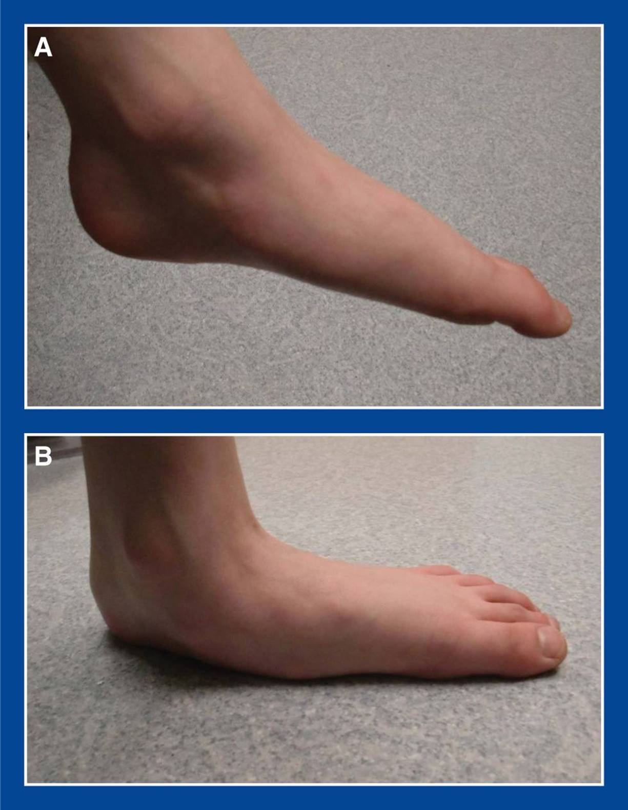
Figure 3-15. A. A severe flexible flatfoot seen dangling in space, with the child seated on the exam table with the leg hanging down. B. The same foot in full weight-bearing.
ASSESSMENT PRINCIPLE #15
If pain is a complaint, the child should be asked to point to the exact location(s).
By having the child identify the point(s) of maximal tenderness, you can start your physical examination away from that site(s) and learn about the surrounding area(s) before creating pain that might limit the rest of the examination. You can also quickly determine if your working diagnosis (based on the history) is valid even before you touch the foot (Figure 3-16).
ASSESSMENT PRINCIPLE #16
Signs and symptoms must match the presumed pathology, so ensure that you have enough information before focusing on a radiographic finding.
There are many common anatomic foot variations, such as tarsal coalitions and accessory naviculars, that do not cause pain or functional disability in the majority of affected individuals. Therefore, it is important to ensure that the signs and symptoms match those associated with the radiographic finding. If they do not, the two are unrelated and a more thorough investigation is required.
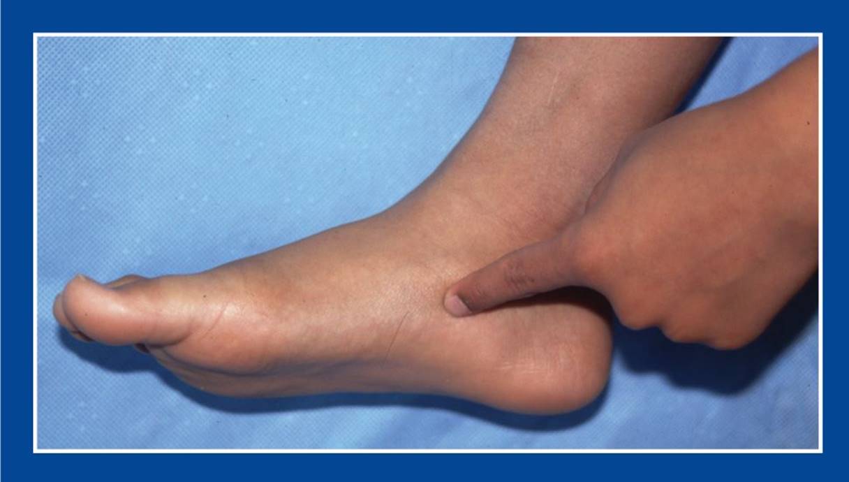
Figure 3-16. Exact identification by the child of the site(s) of pain is important.
RADIOGRAPHS AND OTHER IMAGING
ASSESSMENT PRINCIPLE #17
All radiographs for the assessment of foot deformities should be btained in weight-bearing, or simulated weight-bearing if the former is not possible because of extreme youth or the child’s inability to stand.
This is the radiographic version of Assessment Principle #14. The appropriate clinical assessment of foot deformities is performed in weight-bearing. Radiographs must, therefore, be obtained in weight-bearing to correlate the anatomic alignment of the bones and joints with the outward appearance of the foot. Recall that a flatfoot looks better than it truly is when it is not bearing weight (Figure 3-17).
And a cavovarus foot looks worse than it truly is when non–weight-bearing. Specialized views, such as oblique views, can be taken non–weight-bearing because they are used to identify anatomic abnormalities other than bone and joint alignment. The standard radiographic views for assessing foot deformities are standing AP, lateral, and (medial or standard) oblique; additional views include lateral oblique and Harris axial views.

Figure 3-17. A. Partial weight-bearing lateral foot x-ray. Normal-appearing foot with straight talus–1st MT line. The white line indicates the plantar cortex of the medial cuneiform. The green line is the calcaneal pitch. The yellow arrow shows dorsal positioning of the head/neck of the talus in relation to the dorsal cortex of the navicular, a subtle sign of partial weight-bearing. B. Full weight-bearing lateral x-ray of the same foot. Note the plantar sag at the talonavicular joint with the foot-CORA in the talar head, the lowering of the calcaneal pitch, the plantar position of the plantar cortex of the medial cuneiform, and the level alignment of the dorsal cortices of the talus and navicular. These are all characteristics of a flatfoot.
ASSESSMENT PRINCIPLE #18
The foot-CORA (center of rotation of angulation) method should be used pre-, intra-, and postoperatively for the most objective evaluation of foot deformities and malformations.
For over a century, radiographs have been used to add objectivity to the clinical assessment of bones and joints and their alignment. The mechanical axis is the most basic radiographic measurement used to assess overall lower extremity alignment. A normal mechanical axis is one in which there is a straight line/linear relationship between the centers of the hip, knee and ankle. It is intuitive that, in a limb with a mechanical axis deviation (i.e., the centers of the three joints are not on the same line), one or more angular deformities exist at some point(s) between the hip and the ankle. It is perhaps less intuitive that, in a limb with a normal mechanical axis, two or more opposite direction angular deformities can exist. These intervening bone deformities can create joint mal-orientation, a feature of limb alignment that is as important to normalize as is the mechanical axis. Normalization of both features is important for the health and longevity of the joints.
Normative data exist for the shape of each of the long bones of the extremities. Those data were derived by quantifying the angle between the shaft of each bone and either the adjacent articular surface or the unique configuration of the end of that particular bone. An assumption is that the shafts of all long bones are straight, except for the femur in the sagittal plane.
Assessment of the site(s) of deformity within a bone is best done using Paley’s CORA method. A line is drawn through the longitudinal axis of each straight segment of the shaft. These lines are related to each other as well as to the adjacent joint orientation lines. The site at which a normal and an abnormal, or an abnormal and an abnormal, segmental axis line intersect is a CORA. The CORA represents a static, fixed, structural deformity that exists within a bone. A CORA can exist in the epiphysis, the physis, the metaphysis, or the diaphysis. Using the CORA assessment principles, the site(s) of deformity can be determined and used as a guide for deformity correction. The CORA method can also be used to assess the success of deformity correction.
There are several justifications for a unique CORA method for assessment of foot deformities. The bones of the midfoot are (1) small; (2) irregularly shaped, without clearly definable axes; (3) hard to see/visualize and measure on radiographs, in part because they have overlapping shadows; (4) not ossified or not fully ossified in young children; (5) often not amenable to drawing axis lines because the ossification centers are spherical (note: the axis of a sphere is a dot, not a line); and (6) truly deformed in only a few conditions, including metatarsus adductus, metatarsus primus varus, skewfoot (only the forefoot deformity), and only the cavus component of cavovarus deformity.
Unlike the midfoot bones, the oval-shaped ossification centers of the hindfoot bones (talus and calcaneus) (1) are present at birth and (2) roughly represent the true shape of those bones, even in infancy, so that axis lines can be drawn fairly reliably. However, these two bones are more often malaligned than deformed.
Ossification of the MT bones represents the true shape of those bones, even in infancy. Axis lines can be drawn very reliably. Interestingly though, the MTs, like the hindfoot bones, are more often malaligned than deformed.
These features of the foot bones make it unreliable or impossible to apply the CORA method that is used for the long bones of the extremities to the assessment of foot deformities, particularly in children.
Normative, static radiographic measurements for adult foot alignment exist. They relate the axis of one long bone with another, such as the talus to the 1st MT, the talus to the calcaneus, and the calcaneus to the 4th MT. The talus is the stable structure around which the acetabulum pedis rotates on the fixed oblique axis of the subtalar joint (see Basic Principles #6 and 7, Chapter 2). The axis of the talus can be used as a linear proxy for the sagittal plane alignment of the ankle joint because the axis of the talus is perpendicular to the axis of dorsiflexion/plantar flexion of the ankle joint in the coronal plane. The 1st MT is the distal–medial extension of the calcaneus/acetabulum pedis block that rotates around the talus. The axis of the 1st MT can be determined with more accuracy and reliability than that of the calcaneus, so it can be used as a proxy for the calcaneus when relating the axis of the calcaneus/acetabulum pedis block to the axis of the talus. This is true unless there is a second deformity distal to the acetabulum pedis, as there is in a skewfoot (see below).
Deformities of the foot and ankle are typically due to exaggerated malalignments of the bones of the subtalar joint complex (varus/inversion and valgus/eversion) and the ankle joint (plantar flexion/equinus and dorsiflexion/calcaneus), rather than deformities within bones; however both may exist. Deformity correction in the foot and ankle most often involves realignment of the bones in the subtalar and ankle joints, rather than osteotomies of bones, though both may be necessary.
I have developed a modified CORA method, the “foot-CORA,” to assess the sites of deformity in feet and ankles to more accurately characterize the deformities and to help ensure that they are corrected at those sites, if at all possible. The basis of the method is the assessment of the relationship between the axis of the talus and the axis of the 1st MT in the transverse (AP) and sagittal (lateral) planes and, to a lesser extent, the relationship between the axis of the talus and the axis of the tibia in the sagittal (lateral) plane.
The normal AP talus–1st MT angle ranges from 12° (abducted) to −10° (adducted), with an average value of 4° (abducted) (Figure 3-18).
I have observed that, on the standing AP radiograph of a foot with normal alignment, the axis of the talus and the axis of the 1st MT are either parallel and narrowly translated from each other or they intersect in the head/neck of the talus. In a foot with isolated valgus/eversion or varus/inversion malalignment of the hindfoot, the axes of those bones consistently intersect in the head of the talus or as far anterior as the talonavicular joint. The point of intersection of the axis lines can be considered a CORA. In contrast to a conventional CORA, this is a CORA between bones, rather than within a bone. In a foot with valgus/eversion deformity of the hindfoot, there is exaggerated abduction of the 1st MT axis in relation to the talar axis at the foot-CORA in the talar head (Figure 3-19).

Figure 3-18. A. The axis of the talus and that of the 1st MT are parallel and narrowly translated from each other on this standing AP radiograph of a normal foot. B. The axis of the talus and that of the 1st MT intersect in the head/neck of the talus and are abducted less than 12° from each other on this standing AP radiograph of a normal foot.
In a foot with varus/inversion deformity of the hindfoot, there is exaggerated adduction of the 1st MT axis in relation to the talar axis at the foot-CORA in the talar head (Figure 3-20).
In contrast to the CORA in a long bone, an osteotomy is never performed at the subtalar foot-CORA. Instead, soft tissue procedures and/or osteotomies are preformed around the subtalar joint to align the axes of the talus and the 1st MT at the foot-CORA (Figures 3-19 and 3-20). The talus–1st MT angle can be used to quantify the degree of eversion and inversion deformity before and after correction.
It should also be acknowledged that there are some foot deformities in which the foot-CORA is within a bone. The two most common examples are metatarsus adductus and cavus, i.e. the forefoot plantar flexion deformity in a cavovarus foot. In both cases, the deformity is within the medial cuneiform (Figures 3-21 and 3-22).
The skewfoot, as well as some other unique deformities and malformations, presents a special challenge to the basic foot-CORA method that is resolved by the introduction of a new and unique axis line, the “tarsal line.” It is a summary line, or proxy, for the overall alignment of the midfoot bones, which is otherwise difficult to assess due to the challenges noted above (the bones are small, irregular in shape, and delayed in ossification). Utilization of the tarsal line is particularly helpful when there are two opposite direction deformities between the talus and the 1st MT, as classically seen in skewfoot deformities. The tarsal line is drawn from the point of intersection of the axis of the talus with the subchondral bone of the head of the talus to the point of intersection of the axis of the 1st MT with the subchondral bone at the base of the 1st MT. The tarsal line and the axis of the 1st MT are collinear when there is no deformity of the forefoot on the midfoot (adduction or abduction) in the frontal plane. The tarsal line and the axis of the talus are collinear when there is no deformity of the subtalar joint (i.e., no inversion or eversion) (Figure 3-23).

Figure 3-19. The foot-CORA for flatfoot. A. The foot-CORA for valgus/eversion deformity of the hindfoot is in the head of the talus on the AP x-ray. B. The eversion deformity has been corrected at the foot-CORA with a calcaneal lengthening osteotomy (gray trapezoid-shaped graft highlighted) (see Calcaneal Lengthening Osteotomy, Chapter 8). The axes of the talus and the 1st MT became aligned without actually doing anything to either of the named bones. The osteotomy was performed in a different bone, the calcaneus. C. The foot-CORA on the lateral x-ray is also in the head of the talus. D. The axes of the talus and the 1st MT became aligned in this plane as well following the calcaneal lengthening osteotomy. The allograft in the calcaneus appears to be healed, but not yet remodeled, in this early postoperative x-ray. Within a year after surgery, it was difficult to identify the graft on x-ray.
ASSESSMENT PRINCIPLE #19
Hindfoot flexibility in a cavovarus foot deformity should be assessed objectively with the radiographic equivalent of the modified Coleman block test.
The modified Coleman block test, and the justification for the modification, was described in Assessment Principle #9, Figure 3-4, this chapter. Objective assessment of the flexibility of the hindfoot in a cavovarus foot deformity can be documented with standing AP radiographs of the foot both off and on the block. The normal AP talus–1st MT angle ranges from 12° (abducted) to −10° (adducted) (see Assessment Principle #18, Figure 3-18, this chapter). Varus/inversion deformity of the hindfoot is characterized by exaggerated adduction of the talus–1st MT angle. Correction of the AP talus–1st MT angle to the normal range when standing on the block indicates flexibility of the subtalar joint (see Assessment Principle #18, Figure 3-20, this chapter), whereas incomplete correction indicates inflexibility (Figure 3-24).

Figure 3-20. The foot-CORA for cavovarus. A. The foot-CORA for pure varus/inversion deformity of the hindfoot is in the head of the talus on the AP x-ray. In this example, there is mild associated metatarsus adductus which moves the foot-CORA slightly anteriorly to the talonavicular joint. B. The inversion deformity is flexible and has been corrected at the foot-CORA with the modified Coleman block test, as confirmed radiographically (see Assessment Principle #19, this chapter). The axes of the talus and the 1st MT became colinear without actually doing anything to either of the named bones. The same outcome follows plantar–medial soft tissue release of the subtalar joint (see Superficial Plantar-Medial Release and Deep Plantar-Medial Release, Chapter 7).

Figure 3-21. Metatarsus adductus. A. The talonavicular joint (and, by association, the subtalar joint) is well aligned. The axes of the talus and the 1st MT intersect in the medial cuneiform, indicating that to be the foot-CORA (orange stripes represent the interosseous ligaments). B.A medially-based opening wedge osteotomy of the medial cuneiform, along with a closing wedge osteotomy of the cuboid, has been performed. The foot-CORA has been improved significantly. The osteotomy began approximately half way from distal to proximal along the medial border of the medial cuneiform and angled slightly distal to end adjacent to the 2nd MT/middle cuneiform joint. Having created the osteotomy adjacent to that joint, the fragments have more mobility than if the osteotomy had ended more proximally adjacent to the medial cortex of the middle cuneiform. The interosseous ligaments maintained the appropriate amount of control of the fragments (see Medial Cuneiform Osteotomies, Chapter 8).

Figure 3-22. Cavovarus sagittal plane deformity. A. The foot-CORA on the lateral x-ray of a cavovarus foot is in the body of the medial cuneiform. This location indicates that the apex of the midfoot cavus deformity (not the hindfoot inversion/varus deformity) is within the medial cuneiform. Also note the exaggerated plantar flexion of the 1st MT in relation to the 5th MT (purple line). B. The axes of the talus and the 1st MT became aligned following a plantar-based opening wedge osteotomy in the medial cuneiform (see Medial Cuneiform [Dorsiflexion] Plantar-based Opening Wedge Osteotomy, Chapter 8). The angle between the 1st and 5th MTs became more parallel, the normal relationship.

Figure 3-23. The foot-CORA method for skewfoot. A. The axis of the talus and the axis of the 1st MT are parallel, though severely translated, creating an angle between them of 0°. That would indicate no deformity, though significant deformities exist within this foot. The “tarsal line” helps to resolve this puzzle by creating a second foot-CORA. The talus–tarsal angle is abducted (+) at the posterior foot-CORA and the tarsal–1st MT angle is equivalently adducted (−) at the anterior foot-CORA in this foot. The opposite direction angles are added to each other when determining the summary talus–1st MT angle (0° in this example). The yellow line would represent the axis of the 1st MT if no midfoot/forefoot adduction deformity existed. In that situation, the talus–tarsal angle would be equivalent to the talus–1st MT angle. B. The axes of the talus and the 1st MT intersect in the body of the talus (yellow dot) far from the talar head, which is the foot-CORA in a pure flatfoot deformity. This is the summary foot-CORA and is indicative of a second deformity (second foot-CORA) between the talus and the 1st MT. The talus–tarsal angle represents the true subtalar deformity, which is much more exaggerated than the talus–1st MT angle suggests. The yellow line would represent the axis of the 1st MT if no midfoot/forefoot plantar flexion/cavus deformity existed. In that situation, the talus–tarsal angle would be equivalent to the talus–1st MT angle.

Figure 3-24. Radiographic modified Coleman block test. A. The patient stands with the lateral (4th and 5th) MT heads on a 2.5-cm block for an AP x-ray. B. Standing AP x-ray off the block. The talus–1st MT angle is adducted with the foot-CORA in the talar head, indicating hindfoot varus/inversion. C.Standing AP x-ray on the block (purple rectangle). The talus–1st MT angle corrects fully, indicating a flexible subtalar joint. D. Standing AP x-ray off the block. The talus–1st MT angle is adducted with the foot-CORA in the talar head, indicating hindfoot varus/inversion. E. Standing AP x-ray on the block (purple rectangle). The talus–1st MT angle corrects only partially, indicating inadequate flexibility of the subtalar joint.
ASSESSMENT PRINCIPLE #20
There is usually a projectional artifact on the lateral radiograph of a foot with a varus/inverted or valgus/everted hindfoot deformity.
When a foot is C-shaped due to inversion or eversion of the hindfoot, the lateral x-ray beam cannot simultaneously pass perpendicular to the forefoot and the hindfoot. Therefore, order specifically positioned views to see each segment in a true lateral projection. The radiology technicians can easily visualize the forefoot and will generally aim the x-ray beam perpendicular to the MTs. That creates a rotational projectional artifact of the hindfoot in varus/inversion and valgus/eversion hindfoot deformities. Recall that one component of inversion is internal rotation of the subtalar joint/acetabulum pedis in relation to the talus/ankle which means, conversely, external rotation of the hindfoot in relation to the forefoot (Figure 3-25).
Also recall that one component of eversion is external rotation of the subtalar joint/acetabulum pedis in relation to the talus/ankle which means, conversely, internal rotation of the hindfoot in relation to the forefoot (Figure 3-26).
Finally, be aware that the best way to assess proper hindfoot positioning for a lateral radiograph is to note the relationship between the distal fibula and tibia. The posterior cortex of the distal fibula metaphysis and the posterior ossification margin of the distal tibial epiphysis are colinear in a true lateral x-ray of the hindfoot/ankle. It is unreliable to use the shape of the dome of the talus as a means to determine a true lateral projection because the ossification of the dome is not particularly dome-shaped in young children. Furthermore, there are many instances in which the dome had been crushed, devascularized, or otherwise injured, thereby, flattening its dome shape. And, as has just been discussed, flattening of the dome can be a projectional artifact. Therefore, use the distal fibula to tibia relationships to determine if the projection is a true lateral of the hindfoot/ankle.

Figure 3-25. A. Standard positioning of a cavovarus foot: The radiology technician typically aligns the forefoot parallel with the plate (because the axis of the hindfoot is difficult to appreciate) and the beam perpendicular to the forefoot and plate. A true lateral image of the forefoot is obtained. Inversion of the subtalar joint includes internal rotation of the subtalar joint/acetabulum pedis in relation to the talus/ankle. That equates to external rotation of the ankle in relation to the forefoot—note positions of the malleoli (purple lateral malleolus and green medial malleolus)—and the radiographic appearance of an AP or mortis view of the ankle on the “lateral” x-ray of the foot. B. Proper positioning for assessment of the hindfoot: To see a true lateral image of the hindfoot/ankle, the technician must turn the forefoot toward the beam until the hindfoot is parallel with the plate (purple curved arrow). The forefoot image will look odd, but the hindfoot will appear as it should, with the posterior cortex of the distal fibula metaphysis in line with the posterior ossification margin of the distal tibial epiphysis. C. False appearance of a flat-top talus is seen in the standard positioning view. It is actually the normal talar dome appearance of a mortis view. D. The true talar dome appearance is seen when the foot is positioned properly.
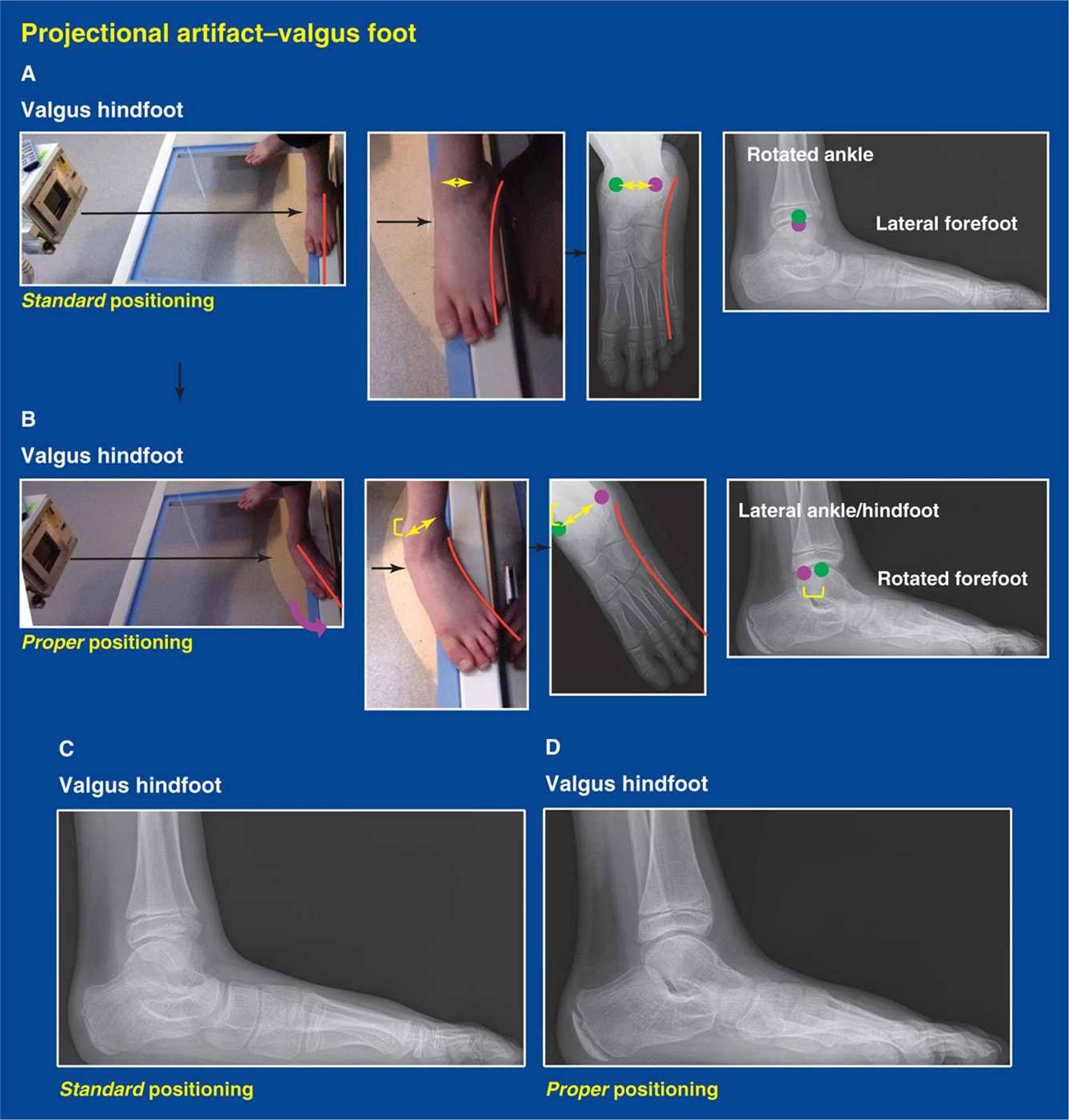
Figure 3-26. A. Standard positioning of a flatfoot: The radiology technician typically aligns the forefoot parallel with the plate (because the axis of the hindfoot is difficult to appreciate) and the beam perpendicular to the forefoot and plate. A true lateral image of the forefoot is obtained. Eversion of the subtalar joint includes external rotation of the subtalar joint/acetabulum pedis in relation to the talus/ankle. That equates to internal rotation of the ankle in relation to the forefoot—note positions of the malleoli (purple lateral malleolus and green medial malleolus). The lateral malleolus projects half way between the anterior and posterior cortices of the tibia. B. Proper positioning for assessment of the hindfoot: To see a true lateral image of the hindfoot/ankle, the technician must turn the forefoot away from the beam (purple curved arrow) until the hindfoot is parallel with the plate. The forefoot image will look odd, but the hindfoot will appear as it should, with the posterior cortex of the distal fibula metaphysis in line with the posterior ossification margin of the distal tibial epiphysis. C. Odd-shaped talus is seen in the standard positioning view. D. The true talus and talar dome appearances are seen when the foot is positioned properly.

Figure 3-27. A. The ankle joint (blue line) is in valgus alignment at birth and gradually corrects to neutral; except it persists in many limbs with clubfoot and in those affected by paralytic conditions such as myelomeningocele, lipomeningocele, and poliomyelitis. The yellow line represents the distal tips of the medial and lateral malleoli, which are at approximately the same transverse level when the ankle joint is in valgus alignment. B. The ankle joint gradually becomes perpendicular to the tibia (blue line) and the lateral malleolus grows distal to the medial malleolus (yellow line) by age 3 to 4 years in normal limbs.
ASSESSMENT PRINCIPLE #21
Do not forget about ankle radiographs.
Ankle radiographs (standing AP, lateral, mortis) are not a standard part of every assessment of a foot deformity or malformation, but should be ordered if clinically indicated. The ankle joint is in valgus alignment at birth (see Assessment Principle #11, this chapter). The distal fibula and lateral distal tibia grow relatively faster than the medial distal tibia until approximately age 3 to 4 years, at which point the ankle joint is perpendicular to the tibial shaft. Neonatal ankle valgus deformity persists in children with paralytic conditions (such as myelomeningocele, lipomeningocele, and poliomyelitis) and in many children with clubfeet for unknown reasons. The ankle joint undergoes its normal conversion to neutral alignment in children with cerebral palsy (Figure 3-27).
ASSESSMENT PRINCIPLE #22
A CT scan in all three orthogonal planes and with 3D reconstruction is the best imaging modality for more detailed assessment of complex foot deformities and malformations. It is the imaging modality of choice to assess tarsal coalitions.
For most deformities and malformations, plain radiographs provide sufficient information to corroborate the physical examination findings. CT scans show the shapes of bones and the alignment of joints in three dimensions, the exact information needed to assess the more complex deformities and malformations, particularly those that have been operated on previously. MRI scans are best at the assessment of soft tissue pathology, which is not the intent of structural assessment. The exorbitant cost of an MRI (even in comparison with a CT scan) makes it fiscally irresponsible to obtain this study without careful consideration of the indications and the information desired, considerations that apply to all imaging studies. CT scans use ionizing radiation, but at a distance far from the most radiation sensitive parts of the body.
Importantly, the CT scan is the definitive imaging study for the diagnosis and management of talocalcaneal tarsal coalitions because the generally accepted criteria for choosing the appropriate treatment modality are based on CT scan findings (see Talocalcaneal Tarsal Coalition, TCTC, Figure 5-2, Chapter 5) (Figure 3-28).
ASSESSMENT PRINCIPLE #23
An MRI is rarely helpful or indicated for assessment of foot deformities and malformations, except in special circumstances.
Radiographs and CT scans are useful in assessing bone and joint abnormalities, specifically deformities and malformations. MRI scans are useful in assessing soft tissue abnormalities, but not as useful in assessing deformities and malformations. The exorbitant cost of an MRI of the foot might be justified in the assessment of a complex deformity or malformation in a very young child who has minimal ossification of the tarsal bones (Figure 3-29).
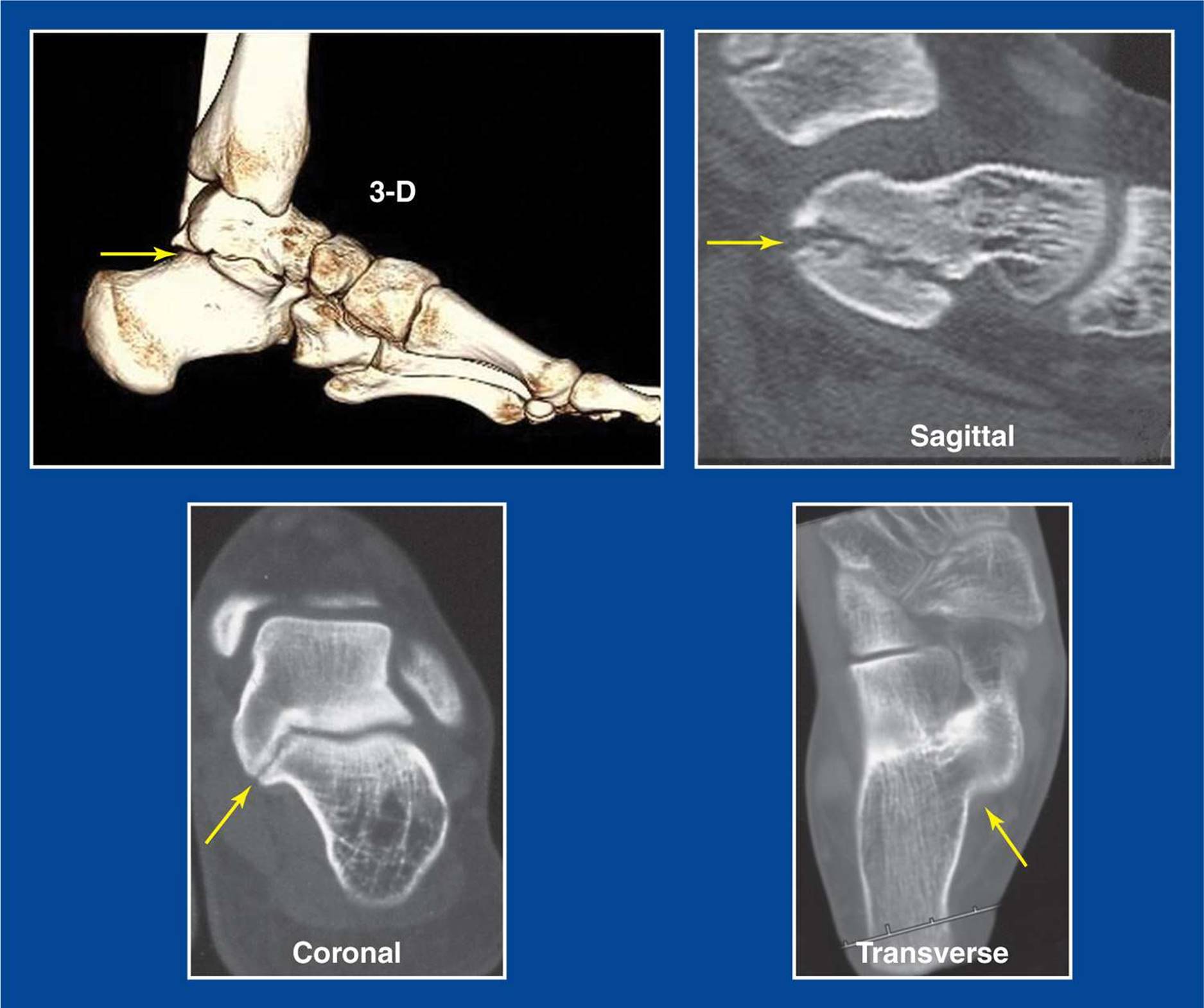
Figure 3-28. Collage of CT scan images of a foot with a middle facet talocalcaneal tarsal coalition (yellow arrow). The formerly healthy joint is narrow, sclerotic, irregular, and down-sloping.

Figure 3-29. A. Lateral x-ray of a surgically treated clubfoot in a 3-year-old with a taller than expected midfoot and suspicion for dorsal subluxation/overcorrection at the talonavicular joint. B. MRI confirms dorsal subluxation/overcorrection at the talonavicular joint.
MRIs are the study of choice for soft tissue tumors and infections (Figure 3-30).
ASSESSMENT PRINCIPLE #24
A bone scan is a good and relatively inexpensive way to identify a specific site(s) of inflammation/pain, and is excellent at diagnosing complex regional pain syndrome.
There are many anatomic variations of the foot that, in many/most cases, do not cause pain. These include tarsal coalition, accessory navicular, os trigonum, and a host of rare accessory ossicles. It is imperative to ensure that a radiographically identified anatomic variation is the cause of the pain and not merely an incidental finding (see Assessment Principles #6, 15, and 16, this chapter). A bone scan can be used to confirm if the anatomic variation is the source of the pain when the signs and symptoms are not characteristic for the anatomic variation that has been identified on physical examination, plain radiographs, and/or CT scan.
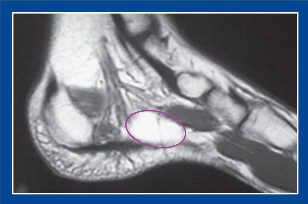
Figure 3-30. MRI reveals a lipoma (purple oval) in the tarsal tunnel in the abductor hallucis muscle that was compressing the medial plantar tibial nerve, creating pain and numbness in the distribution of the nerve.

Figure 3-31. Bone scan demonstrating complex regional pain syndrome, a.k.a. reflex sympathetic or neurovascular dystrophy, pain amplification syndrome. A. AP image of the feet shows stocking–glove distribution decreased tracer uptake in the entire left foot and ankle. To the casual observer, and without benefit of the clinical history or visualization of the more proximal parts of the limbs, one might diagnose right foot and ankle pathology with generalized increased uptake. B. Lateral image of the feet confirms stocking–glove distribution decreased tracer uptake in the entire left foot and ankle. C. AP image of the knees shows essentially symmetric tracer uptake in the growth plates of the two limbs. D. AP image shows right-to-left symmetry of the proximal tibias/fibulas, but marked asymmetry at the ankles/feet with decreased tracer uptake on the left side.
The bone scan should be ordered with magnified views and SPECT images in multiple projections. Both lower extremities must be seen for comparison, especially if complex regional pain syndrome is being considered, in which case there is most often a stocking–glove distribution decrease in uptake in the affected foot and ankle (Figure 3-31).
It is true that there is a theoretic risk of excessive radiation exposure to the gonads until the technetium is expelled from the urinary bladder, especially in females. But bone scans should be used infrequently and only for the rare indications stated. The alternative is to use an MRI scan, with its exorbitant cost and lack of specificity, to find the true site(s) of pain. The significance of “bone edema,” which is frequently identified on MRI scans, is unknown.