
MANAGEMENT PRINCIPLE #1
The decision (to operate) is more important than the incision (i.e., the surgical technique).
And the decision to operate on a foot deformity or malformation is based on (1) the known natural history of the condition, (2) the symptomatic and/or functional responses to nonoperative treatment (where appropriate), and (3) the reported risks and complications of surgery. A “well executed” operation for the right indication is far better for the patient than the “most skillfully executed operation in the history of surgery” for the wrong indication. The best surgeon is not necessarily the most skillful, but the one who knows when to operate. Of course, it is nice to make the best decisions and be technically excellent. We all strive for that combination of knowledge and skills.
MANAGEMENT PRINCIPLE #2
A less-than-ideal surgical or nonsurgical outcome can be due to a poor technique, a poor technician, or both.
This principle assumes that the patient satisfies reasonable indications for the technique in question. A surgical or nonsurgical (e.g., Ponseti) technique is developed and, hopefully, tested by the originator before it is presented to the medical community. There is perhaps no technique that is so simple or foolproof that mere knowledge of the concept allows another surgeon to perform the procedure as well as the originator. And for some/many techniques, attention to all of the fine details of the procedure is critical for success. Failure to perform the procedure as described by the originator might result in a good outcome, but a poor outcome cannot automatically be attributed to the technique. It can, perhaps, be considered a poor technique only if other surgeons skillfully follow the fine details of the procedure (as published and without modifications) and fail to achieve outcomes comparable to those achieved by the originator. Before abandoning or modifying a procedure that has been shown by others to be effective, make sure to perform it as described by the originator. Personal observation of, or tutoring by, an expert might be required, depending on the complexity and uniqueness of the technique. Though it is possible that the technique, as described by the originator, can be successfully performed only by the originator, such procedures should be extremely rare.
Admittedly, detailed descriptions for many of the procedures that are commonly and uncommonly performed are not published or otherwise accessible. I have included my time-tested techniques for many soft tissue and bony foot procedures in Chapters 7 and 8 of this text. Some are original to me, but most are my interpretation of the originals that often have not been well described in the literature. Some of the articles describing the original techniques can also be found in the bibliography in Chapter 9.
MANAGEMENT PRINCIPLE #3
You cannot un-operate on anyone.
Foot deformities and malformations are never lethal. Nonoperative treatment might prolong the temporary pain and disability, but might eliminate both, thereby avoiding the reported risks and complications of surgery.
MANAGEMENT PRINCIPLE #4
The (surgical) treatment could be worse than the condition itself.
No operation is without potential risks and complications that are unacceptable if the natural history of the condition or the response to nonoperative treatment provides favorable outcomes with little to no long-term disability. Nonoperative treatment corrects a high percentage of many congenital deformities (clubfoot, congenital vertical talus, and metatarsus adductus) and/or resolves pain and functional disability in a high percentage of certain other conditions (tarsal coalition, juvenile hallux valgus, and accessory navicular). Natural history trumps all treatment modalities. Many anatomic variations correct spontaneously through normal growth and development (flexible flatfoot, metatarsus adductus, and position calcaneovalgus) or persist without resulting in pain or functional disability (flexible flatfoot, metatarsus adductus).
MANAGEMENT PRINCIPLE #5
Modalities that correct deformities: (1) natural history, (2) physical stretching, (3) serial casting, and (4) surgery.
The natural history of congenital metatarsus adductus and positional calcaneovalgus is spontaneous correction in almost all cases (see Basic Principles #3 and 4, Chapter 2). Though perhaps better classified as an anatomic variation rather than a deformity, physiologic flexible flatfoot changes to an arched foot in most cases through its natural history.
Physical stretching exercises will increase the rate of dorsiflexion deformity correction for positional calcaneovalgus and will correct ankle plantar flexion deformity in many mild cases of congenital and acquired tendo-Achilles contracture. The technique for heel cord stretching in children with flatfoot/short tendo-Achilles must be performed in a specific manner to ensure that the proper structure, the tendo-Achilles, is stretched and that the proper joint, the ankle joint, achieves the increase in dorsiflexion. The reason was explained in Basic Principle #6 and illustrated in Figure 2-7 in Chapter 2. Dorsiflexion of the acetabulum pedis/calcaneus in relation to the talus, as seen in flatfoot deformity, is a component of eversion of the subtalar joint. Unless the subtalar joint is inverted to neutral and “locked” (see Basic Principle #7, Figure 2-9, Chapter 2), dorsiflexion stress will likely increase dorsiflexion/eversion through the subtalar joint rather than dorsiflexion in the ankle joint (Figure 4-1).
Serial casting can fully correct most cases of rigid congenital metatarsus adductus. It can fully correct most cases of congenital clubfoot and congenital vertical talus with the addition of minor surgery (Achilles tenotomy). And serial casting can correct some of the cases of congenital and acquired heel cord contracture that do not fully correct with physical stretching exercises. Serial casting can, at a minimum, partially correct foot deformities in children who are at even fairly advanced ages, so as to decrease the extent of required surgery.
Surgery is the final common pathway for foot deformities that do not correct spontaneously or respond fully to nonoperative treatment. Surgery involves soft tissue releases and/or plications, osteotomies, and, rarely, arthrodeses. Tendon transfers do not correct structural deformities.

Figure 4-1. Heel cord stretching for a flexible flatfoot with a short tendo-Achilles must be performed with the subtalar joint inverted (A) and the knee extended (B). Recall that one component of eversion is dorsiflexion of the acetabulum pedis around the talus. If the subtalar joint is not inverted/”locked” (see Basic Principle #7, Figure 2-9, Chapter 2), it will merely further evert/dorsiflex, thereby stretching the medial soft tissues of the subtalar joint rather than the heel cord across the ankle joint. The knee must be extended to ensure that the gastrocnemius is also stretched at its proximal end as it crosses that joint. (Arch Safe™ Rubber biplanar wedge courtesy of Prasad Gourineni, MD with permission.)
As a corollary, natural history is the only modality that results in permanent deformity correction. There is a risk of deformity recurrence following all treatment modalities.
A commonly held belief by some health care professionals and most grandparents is that special “orthopedic shoes” and orthotics correct foot deformities in children. There is no scientific evidence to support that belief. The myth has been perpetuated because those devices have been credited with the deformity correction that has, in fact, occurred as a result of the natural history of the condition.
MANAGEMENT PRINCIPLE #6
Modalities that correct dynamic deformities: (1) focal injection of tone-reducing medication into muscles and (2) muscle-balancing tendon surgery.
Dynamic deformities are flexible; i.e., the joints can be passively moved through a full range of motion. They are due to muscle imbalance from underlying neuromuscular disorders in which there may be spasticity or weakness. Injection of botulinum toxin (BOTOX) into a spastic muscle has been shown to temporarily paralyze and weaken it, resulting in improved muscle balance across a joint. Although the effect is not permanent, it can be repeated. This is an appropriate treatment modality for a young child with spastic muscles in whom a delay in surgery until the child is older will often improve the results of muscle-balancing surgery.
Tendon lengthening/weakening and tendon transfer techniques are more permanent solutions to muscle imbalance, but they are not entirely reliable, predictable, or definitive. The main problem with a dynamic deformity is that it is the result of the problem (an underlying neuromuscular disorder) and not the primary problem (see Basic Principle #12, Chapter 2). After tendon surgery, the child still has the underlying nerve or muscle disorder. Therefore, recurrence of deformity and overcorrection are real possibilities (see Management Principle #10, this chapter).
MANAGEMENT PRINCIPLE #7
Modalities that maintain deformity correction: (1) focal injection of tone-reducing medication into muscles, (2) special shoes/braces, (3) orthotics, (4) physical stretching, and (5) balanced muscles.
Recurrence of a corrected deformity is common in many congenital and acquired deformities of the child’s foot and ankle. In deformities caused by an underlying progressive neuromuscular disorder, recurrence is even more likely. Recurrence of a deformity is also common in children with underlying collagen disorders such as arthrogryposis and, at the other end of the spectrum, the ligament laxity syndromes. There are fewer recurrences in some deformities that are corrected later in childhood. However, delaying treatment is not always an acceptable option. The bottom line is that, unlike in adult foot surgery, maintenance of deformity correction in children and adolescents is a very important component of the overall treatment plan. It must be given consideration equal to the deformity correction itself and monitored long term.
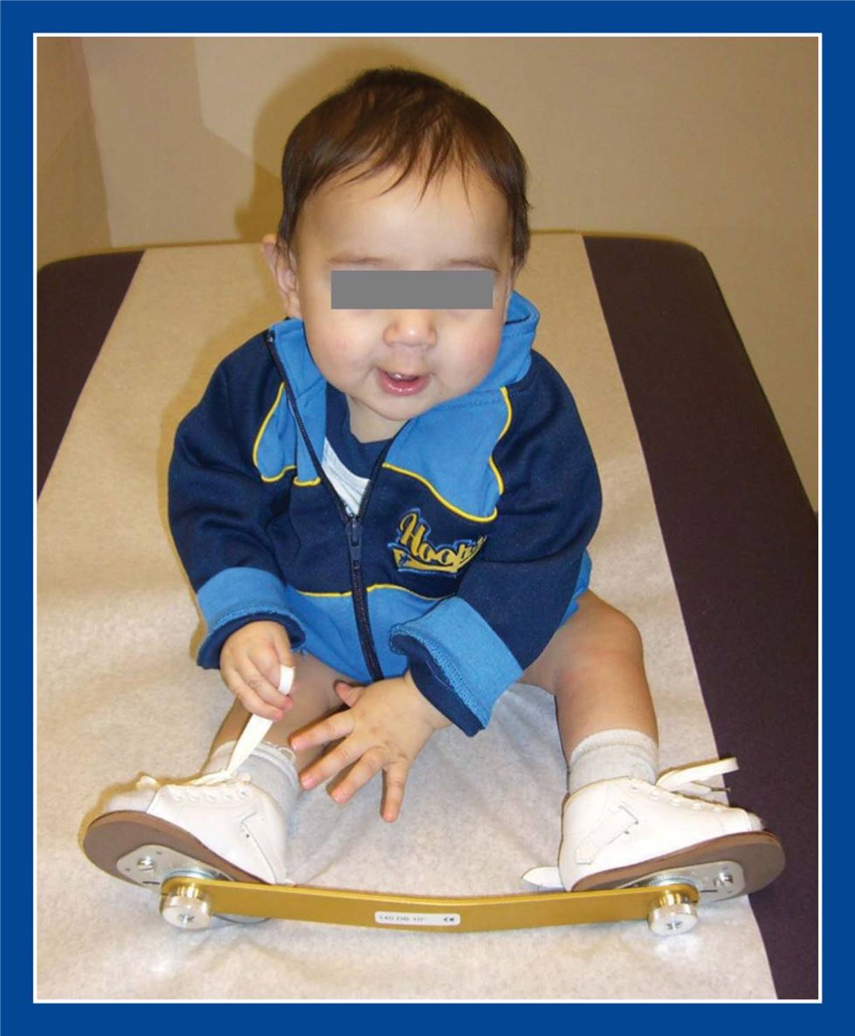
Figure 4-2. Foot-abduction brace worn at night for several years after clubfoot deformity correction using the Ponseti method.
Focal injection of tone-reducing medication into muscles can correct dynamic deformities and reduce the risk or rate of recurrence, but they do not guarantee maintenance of deformity correction because their effect is not permanent (see Management Principle #6, this chapter).
Special shoes/braces and orthotics do not correct deformities, but they are often helpful in maintaining deformity correction, even if worn only at night (Figure 4-2).
Daily stretching exercises are also an important modality for maintenance of deformity correction (Figure 4-3). The modified technique for heel cord stretching must be used for maintaining, as well as for achieving, correction in flexible flatfoot with short tendo-Achilles (Figure 4-1).
Surgically balanced muscles can maintain deformity correction, but achieving balance is an art and may not be achievable (see Management Principle #22-4, this chapter). Maintaining muscle balance is particularly challenging in progressive neuromuscular disorders (see Management Principle #6, this chapter).
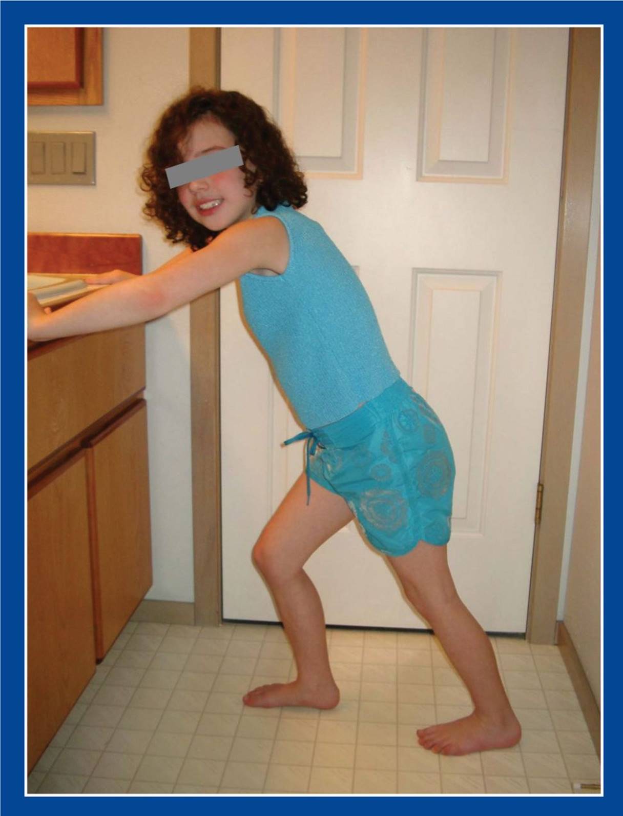
Figure 4-3. A few minutes of tendo-Achilles stretching can help maintain deformity correction after heel cord lengthening. A physical therapist is not required, except perhaps to teach the technique(s). Requesting that the stretching be performed immediately before or after brushing teeth (twice per day) could be the link that ensures compliance.
MANAGEMENT PRINCIPLE #8
Treatment (nonoperative and/or operative) is indicated for:
1. Congenital deformities and malformations that are known, or expected, to cause pain and/or functional disability unless corrected.
These include congenital clubfoot, congenital vertical talus, rigid metatarsus adductus, rigid skewfoot, polydactyly, macrodactyly (Figure 4-4). They are treated well before they become symptomatic.
2. Developmental and acquired deformities and malformations that are creating pain and/or functional disability.
These include cavovarus foot, flexible flatfoot with short tendo-Achilles, idiopathic equinus, tarsal coalition, accessory navicular, spastic and paralytic foot deformities, iatrogenic deformities (Figure 4-5).
For both pain and functional disability, the treatment is disease-specific and can be nonoperative and/or operative.
MANAGEMENT PRINCIPLE #9
Surgical treatment is indicated for:
1. Congenital deformities and malformations that do not, or cannot, correct with nonoperative treatment and are known to cause pain and/or functional disability unless corrected.
These include congenital clubfoot and vertical talus that do not respond to nonoperative (Ponseti and reverse Ponseti) management, macrodactyly, longitudinal epiphyseal bracket of the 1st metatarsal, polydactyly.
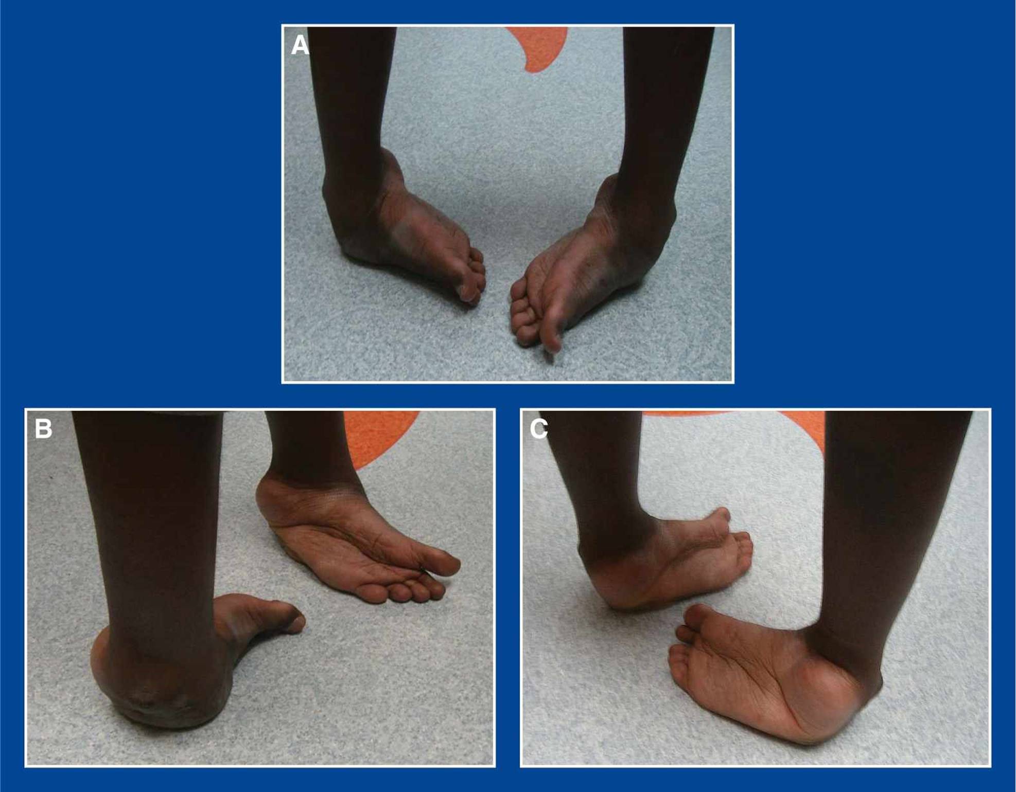
Figure 4-4. A. Front. B. Side. C. Back views of untreated congenital clubfeet in an 18-year-old male. They are unshoeable and are painful when walking on hard surfaces. Clubfoot never corrects without treatment. The natural history is well known. Early treatment can avoid disability in this common condition.
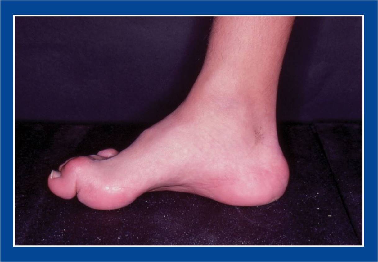
Figure 4-5. Cavovarus foot with claw toes in a boy with Charcot–Marie–Tooth disease. His feet were normally shaped, strong, and comfortable until 3 to 4 years prior to this photo. He presented with the obvious deformities along with instability, frequent ankle sprains, and weight-bearing pain under the 1st metatarsal head and the base of the 5th metatarsal on both feet. The natural history has been playing out for the last 3 to 4 years. There is no reason to believe that the pathologic changes will reverse or slow down. Therefore, there is no reason to delay treatment.
2. Progressive cavovarus foot deformities that are associated with pain and/or functional disability.
3. Other developmental, persistent, and recurrent deformities that do not adequately respond to prolonged attempts at nonoperative treatment designed to correct the deformity, maintain deformity correction, relieve pain, and diminish or relieve functional disability.
These include skewfoot, recurrent and overcorrected congenital clubfoot and vertical talus, idiopathic equinus, flexible flatfoot with short tendo-Achilles, tarsal coalition, accessory navicular, juvenile hallux valgus, spastic and paralytic foot deformities.
MANAGEMENT PRINCIPLE #10
Provide clear, accurate, and reasonable expectations to the patient and family of the short- and long-term outcomes of nonoperative and operative management.
Foot deformities and malformations are rarely “cured,” i.e., made normal. But long-term comfort and function can be anticipated for many or most of them. Deformities attributable to neuromuscular disorders are the result of the problems and not the primary problems. Recurrence of deformity and the need for future treatment can be anticipated in many of these cases. Normal growth and development of a foot with a primary deformity can have an anticipated or unanticipated effect on the long-term outcome of the intervention. Share your predictions about future comfort and function and about the need for future treatment with the patient and family. That way there should be few surprises down the line.
MANAGEMENT PRINCIPLE #11
A surgical plan for each of the segmental deformities and muscle imbalances needs to be established before proceeding with surgery.
This means creating a list of the multiple related and unrelated procedures that are to be performed either during a single operative session or sequentially in cases of staged procedures. Some deformities are not evident until others are corrected. This needs to be anticipated before the start of the operation, based on one’s knowledge and understanding of deformities, with a surgical plan ready for each additional deformity that might be identified intraoperatively. Be prepared, rather than surprised.
MANAGEMENT PRINCIPLE #12
Correct deformity at the site of the deformity. If that is not possible, use compensatory bone and soft tissue procedures.
That means:
1. Perform a calcaneal lengthening osteotomy (CLO) (see Chapter 8) rather than posterior calcaneal medial displacement osteotomy (see Chapter 8) for valgus/eversion deformity of the hindfoot. The former procedure (CLO) corrects all components of subtalar joint eversion at the site of deformity, whereas the latter procedure creates a compensatory deformity to correct valgus alignment of the hindfoot.
2. Perform a plantar–medial soft tissue release of the subtalar joint (see Chapter 7) rather than posterior calcaneal lateral displacement osteotomy (see Chapter 8) for varus/inversion deformity of the hindfoot. The former procedure corrects subtalar joint inversion at the site of deformity, whereas the latter procedure creates a compensatory deformity to correct varus alignment of the hindfoot.
3. Perform a medial cuneiform opening wedge osteotomy (see Chapter 8) rather than 1st metatarsal osteotomy (see Chapter 8) for cavus deformity (plantar flexion deformity of the 1st ray). The foot-CORA (center of rotation of angulation) for cavus (see Assessment Principle #18, Chapter 3) is in the medial cuneiform.
4. Perform a medial cuneiform opening wedge osteotomy (see Chapter 8) and cuboid closing wedge osteotomy (see Chapter 8) rather than metatarsal osteotomies or tarsometatarsal capsulotomies for metatarsus adductus. The foot-CORA for metatarsus adductus (see Assessment Principle #18, Chapter 3) is in the medial cuneiform.
When Willie Sutton was asked why he robbed banks, he said: “…because that’s where the money is!” Go where the money is!
MANAGEMENT PRINCIPLE #13
Preserve joint motion (particularly subtalar joint motion) in the feet of children and adolescents by utilizing soft tissue releases/plications and osteotomies instead of arthrodeses.
Arthrodesis of the subtalar joint results in debilitating stress transfer to adjacent joints, particularly the ankle joint, leading to premature degenerative arthritis. Arthrodesis also has a detrimental effect on future growth and development of the foot. The subtalar joint is the shock absorber of the foot and, in fact, the entire lower extremity. Preserve its function at all costs (Figure 4-6).
MANAGEMENT PRINCIPLE #14
Use biologic, rather than technologic, interventions; i.e., rearrange and/or reshape anatomic parts rather than replace or interfere with them.
The overall reported short term complication rate of subtalar arthroereisis (“pseudoarthrodesis”) with synthetic implants is 3.5% to 30%, with more recent reports of 3.5% to 11%. However, the actual rates are much higher if one includes the inappropriate implantation of these devices into normal physiologic flexible flatfeet, a practice employed by some health care providers. Complications can be categorized as surgeon error, problems with biomaterials, biologic problems, and inappropriate implantations. Long term outcome studies have not been reported.
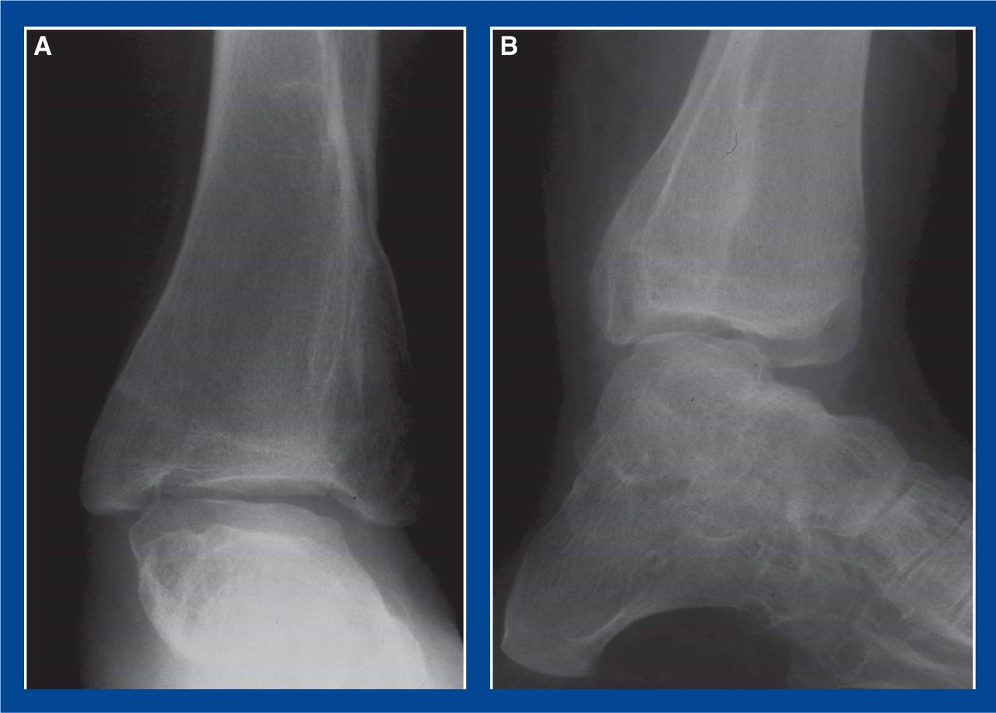
Figure 4-6. Degenerative arthrosis of the ankle joint several years after triple arthrodesis. A. AP x-ray. B. Lateral x-ray.
MANAGEMENT PRINCIPLE #15
Correct deformities and balance muscle forces.
1. Deformity correction will not correct muscle imbalance.
Deformity correction without muscle balancing can result in recurrent deformity. If muscle imbalance created the deformity, as is usually the case in cavovarus foot deformities, persistence of the muscle imbalance will recreate the deformity despite adequate initial deformity correction.
2. Tendon transfers will not correct structural deformities.
Muscle balancing without deformity correction will create a balanced deformity. That is not the goal (see Management Principle #22-2, this Chapter).
MANAGEMENT PRINCIPLE #16
Principles of cavovarus deformity correction:
1. Release the plantar–medial soft tissues to realign the subtalar joint.
The default position of the subtalar joint is valgus/everted (see Basic Principle #9, Chapter 2); release of the plantar–medial soft tissues (see Chapter 7) will result in partial or complete correction of varus/inversion. The subtalar joint is inverted in a cavovarus foot deformity, just as it is in a clubfoot. On the basis of the segmental deformities, one could consider an (equino-)cavovarus foot deformity an “acquired” clubfoot. One would never consider performing compensatory osteotomies or arthrodeses before attempting subtalar joint release and alignment in a clubfoot. The same approach should be used for a cavovarus deformity.
2. Perform osteotomies to correct residual bone deformities.
Depending on the severity and rigidity of the subtalar joint inversion deformity, plantar–medial soft tissue release might not be sufficient to realign the subtalar joint. In those cases, one or more hindfoot osteotomies (see Chapter 8) are required to correct the residual varus deformity. They should not, however, be used primarily in place of the plantar–medial soft tissue release.
Furthermore, alignment of the hindfoot does not correct the forefoot pronation deformity, which is a separate deformity that requires its own treatment (see Basic Principle #5, Chapter 2), specifically a dorsiflexion osteotomy of the medial cuneiform (see Chapter 8).
3. Reserve arthrodesis of the subtalar joint as a salvage procedure.
Most cavovarus deformities can be corrected with a combination of soft tissue releases and osteotomies. Arthrodesis of the subtalar joint can and should be avoided in children and adolescents (see Management Principle #13, this chapter) unless there is advanced arthritis in that joint, a rare finding in children and adolescents.
MANAGEMENT PRINCIPLE #17
Principles of planovalgus deformity correction:
1. Perform osteotomies to correct bone deformities and/or align the subtalar joint.
The default position of the subtalar joint is valgus/everted (see Basic Principle #9, Chapter 2). Therefore, release of the lateral soft tissues will result in no change in the eversion deformity, and plication of the plantar–medial soft tissues will not maintain deformity correction. The calcaneal lengthening osteotomy (see Chapter 8) corrects all components of valgus/eversion deformity of the subtalar joint at the site of deformity. The posterior calcaneus medial displacement osteotomy (see Chapter 8) corrects valgus alignment of the hindfoot without correcting the other components of eversion deformity. Specifically, it does not correct the dorsiflexion and external rotation malalignment at the talonavicular joint. The posterior calcaneus medial displacement osteotomy, when combined with other procedures, has a role in the correction of some specific planovalgus deformities.
Alignment of the hindfoot does not correct the forefoot supination deformity, which is a separate deformity that requires its own treatment (see Basic Principle #5, Chapter 2), specifically a plantar flexion osteotomy of the medial cuneiform (see Chapter 8).
2. Plicate soft tissues to further stabilize the subtalar joint.
Following correction of the eversion deformity of the subtalar joint with the CLO, the plantar–medial talonavicular joint capsule and the posterior tibialis tendon are lax. They should be tightened by means of a plantar–medial plication (see Chapter 7) to take up the redundancy in the capsule and to reset the muscle tension.
3. Reserve arthrodesis of the subtalar joint as a salvage procedure.
Most planovalgus deformities can be corrected with a combination of osteotomies and soft tissue plications. Arthrodesis of the subtalar joint can and should be avoided in children and adolescents (see Management Principle #13, Chapter 4) unless there is advanced arthritis in that joint, a rare finding in children and adolescents.
MANAGEMENT PRINCIPLE #18
The calcaneocuboid joint is the most distal site at which the lateral column of the foot can be shortened or lengthened to realign the talonavicular joint/acetabulum pedis in a foot with a varus/inverted or a valgus/everted hindfoot deformity. The body of the cuboid is too far distal.
The talonavicular and calcaneocuboid joints are collectively known as Chopart joints. The talonavicular joint is the anteromedial extent of the acetabulum pedis. As such, the navicular, along with the rest of the acetabulum pedis, rotates around the axis of the subtalar joint, i.e., “down and in” for inversion and “up and out” for eversion (see Basic Principles #6 and 7, Chapter 2). The calcaneocuboid joint, on the other hand, is a fairly nonmobile joint withinthe acetabulum pedis, analogous to the transverse limb of the triradiate cartilage within the acetabulum in the ilium (see Basic Principle #7, Figure 2-11, Chapter 2). The body of the cuboid, on the other hand, is distal to Chopart joints and the acetabulum pedis.
Plantar–medial soft tissue release of a varus/inverted hindfoot will produce partial-to-complete eversion of the subtalar joint with realignment of the talonavicular joint. In a long-standing deformity, full correction and realignment might not be possible because secondary bone deformity, manifest as a long lateral column of the foot, has developed. In such a case, there is residual inversion following a deep plantar-medial release (see Chapter 7). The long lateral column of the foot can be shortened to pull the navicular dorsolaterally to align with the talar head. Three procedures are effective in accomplishing this: the Evans calcaneocuboid joint resection/arthrodesis, the Lichtblau anterior calcaneus resection, and an anterior calcaneus lateral closing wedge osteotomy (see Chapter 8 for a description of each procedure). They are most commonly employed to treat resistant, residual, or recurrent hindfoot varus in clubfoot deformities in older children. A closing wedge osteotomy of the cuboid (see Chapter 8) is too far distal to affect the relationship between the navicular and the head of the talus. Its primary action is to help correct metatarsus adductus, particularly when combined with a medial opening wedge osteotomy of the medial cuneiform (see Chapter 8) (Figure 4-7).
In contrast to a foot with a varus/inverted hindfoot deformity, the lateral column of a foot with a valgus/everted hindfoot deformity is too short. The CLO (see Chapter 8) corrects valgus/eversion deformity of the hindfoot at the site of deformity and realigns the entire subtalar joint complex, including the talonavicular joint. An opening wedge osteotomy of the cuboid is too far anterior to affect bone relationships within the subtalar joint complex. Its primary action is to help correct metatarsus abductus (if, in fact, there exists such a deformity), particularly when combined with a medial closing wedge osteotomy of the medial cuneiform (Figure 4-8).
MANAGEMENT PRINCIPLE #19
When considering a dorsiflexion or plantar flexion osteotomy of the medial cuneiform for the correction of forefoot pronation or supination, one should also consider the alignment in the transverse plane (adduction or abduction).
The medial cuneiform has been recognized for some time as being the ideal site for correcting metatarsus adductus (see Chapter 5) with a medially-based opening wedge osteotomy, often combined with a closing wedge osteotomy of the cuboid. It is the foot-CORA for that deformity (see Assessment Principle #18, Figure 3-21, Chapter 3). Less well recognized or acknowledged is the fact that the medial cuneiform is the foot-CORA for forefoot pronation (i.e. cavus) and supination (see Assessment Principle #18, Figure 3-22, Chapter 3). The base of the 1st MT is not the site of deformity (foot-CORA) for any forefoot or midfoot deformity.
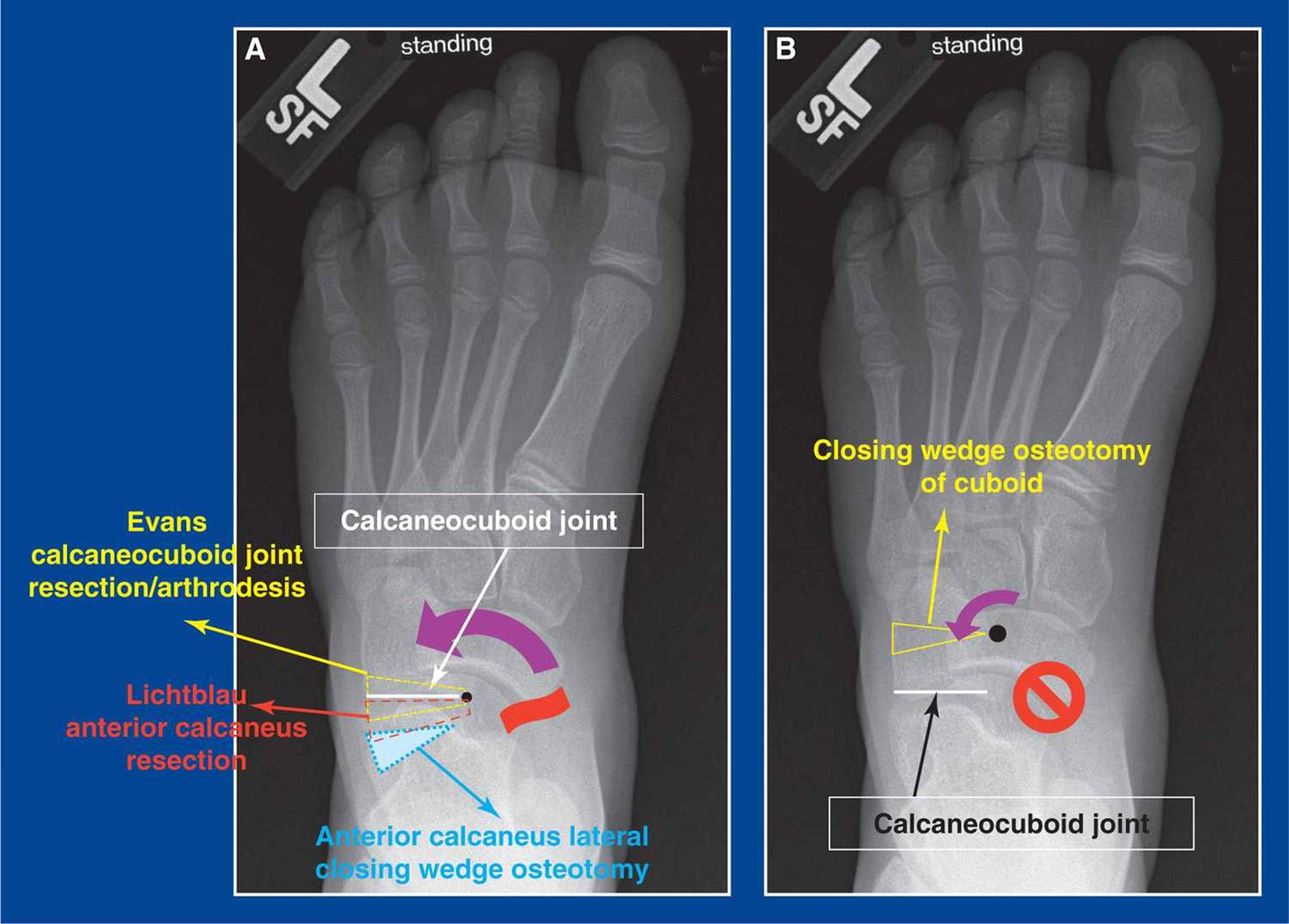
Figure 4-7. Lateral column shortening in a cavovarus foot. A. Following a deep plantar-medial release (wavy red line) (see Chapter 7), there may be residual inversion of the subtalar joint. The Evans calcaneocuboid resection/arthrodesis, the Lichtblau anterior calcaneus resection, and the anterior calcaneus lateral closing wedge osteotomy (see Chapter 8 for a description of each procedure) are all capable of shortening the lateral column of the foot and, thereby, correcting residual inversion of the subtalar joint with realignment of the navicular on the head of the talus (curved purple arrow). The black dot in the head of the talus represents the foot-CORA of the subtalar joint (see Assessment Principle #18, Chapter 3) around which the acetabulum pedis rotates following each of these three osteotomies. B. A closing wedge osteotomy of the cuboid (seeChapter 8) does not affect bone relationships in the subtalar or talonavicular joints  . Its foot-CORA (black dot) is the medial cortex of the cuboid. In this foot, a closing wedge osteotomy of the cuboid would not realign the navicular on the head of the talus, but merely create a compensatory abduction deformity (curved purple arrow) distal to the true deformity in the subtalar joint (see Management Principle #12, this chapter).
Osteotomies in the medial cuneiform can, in fact, be used to correct forefoot pronation and supination, midfoot adduction and abduction, as well as combinations of those deformities (see Medial Cuneiform Osteotomies, Chapter 8). The medial cuneiform is, therefore, the workhorse of the medial column of the foot.
When treating pronation (plantar flexion of the 1st ray) and supination (dorsiflexion of the 1st ray) deformities of the forefoot, it is important to recognizing the alignment of the midfoot, i.e., adduction or abduction. Knowledge of this second plane alignment can help determine whether an opening or closing wedge osteotomy should be used to correct not only the rotational deformity (pronation or supination), but also the angular deformity (adduction or abduction), or at least avoid exaggerating the deformity in that second plane.
The medial cuneiform is bordered laterally by two bones (the base of the second metatarsal and the middle cuneiform) and a joint (the second metatarsal–middle cuneiform joint) with interosseous ligaments along its entire border. The medial border is merely covered by soft tissues (skin, fat, and the anterior tibialis tendon). These features of the local anatomy of the medial cuneiform create four biplanar osteotomy scenarios (Figure 4-9).
1. A medial cuneiform dorsiflexion plantar-based opening wedge osteotomy (MC-PF-OWO) will always additionally create slight abduction, because the lateral ligaments create a tether on the two bone fragments that is not created medially. This would be best for forefoot pronation in cavovarus and skewfoot deformities. The base of the wedge is positioned plantar–medially in a skewfoot.
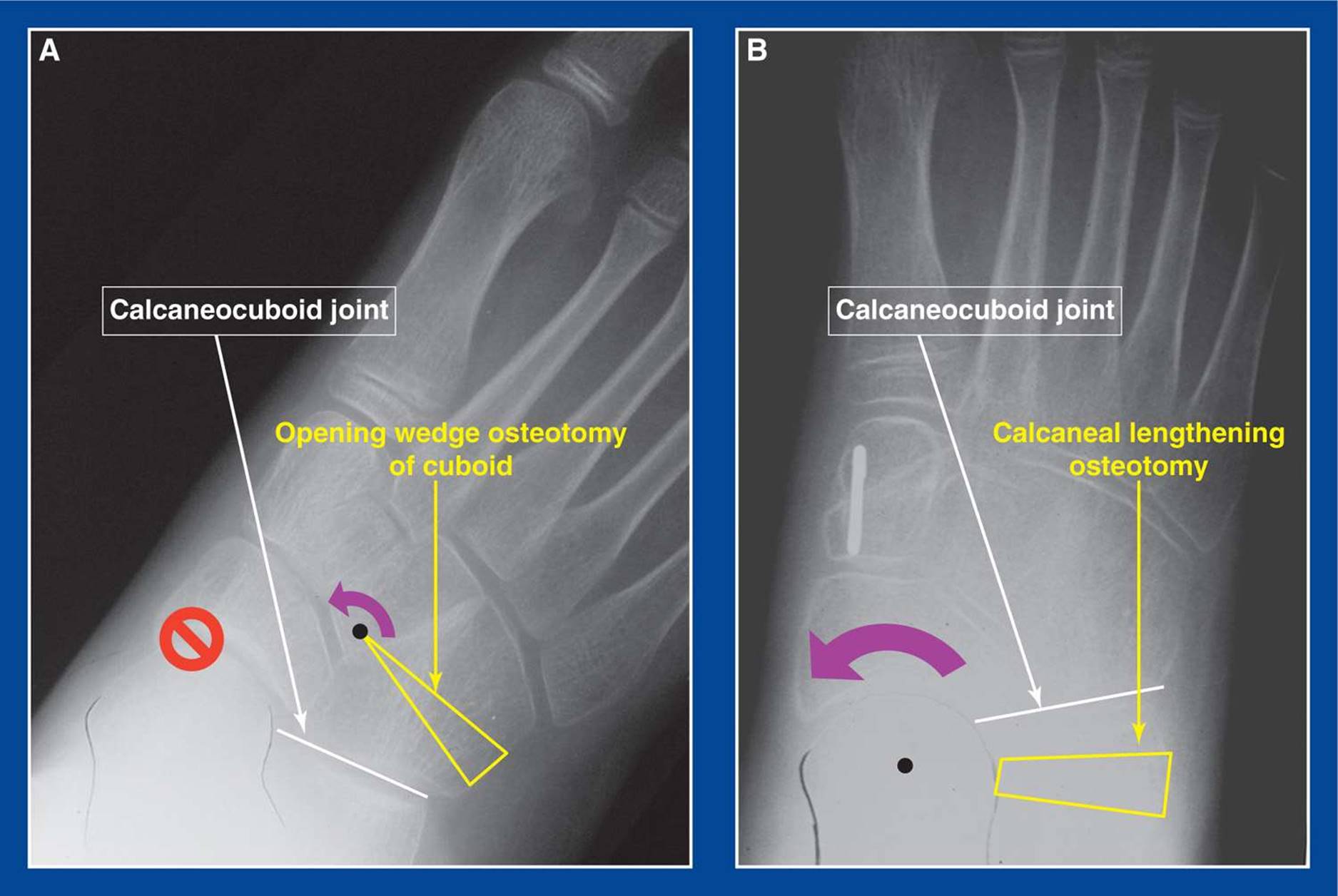
Figure 4-8. Lateral column lengthening in a flatfoot. A. An opening wedge osteotomy of the cuboid does not affect the relationship between the navicular and the talus or correct eversion deformity of the subtalar joint. Its foot-CORA (black dot) is the medial cortex of the cuboid. It merely creates a compensatory adductus deformity (curved purple arrow) anterior to the true deformity in the subtalar joint. B. The calcaneal lengthening osteotomy (see Chapter 8) lengthens the lateral column of the foot and, thereby, corrects all components of eversion deformity of the subtalar joint with realignment of the navicular on the head of the talus. The black dot in the head of the talus represents the foot-CORA of the subtalar joint (see Assessment Principle #18, Chapter 3) around which the acetabulum pedis rotates following a CLO (curved purple arrow). This can also be accomplished by a distraction arthrodesis of the calcaneocuboid joint, which is unnecessary in children and adolescents, but preferred by some surgeons for the correction of the painful adult flatfoot.
2. A medial cuneiform plantar flexion plantar-based closing wedge osteotomy (MC-PF-CWO) will always additionally create slight adduction, because the lateral ligaments create a tether on the two bone fragments that is not created medially. This may be best for forefoot supination with no midfoot adduction deformity in flatfoot and dorsal bunion deformities.
3. A medial cuneiform plantar flexion dorsally-based opening wedge osteotomy (MC-PF-OWO) will always additionally create slight abduction, because the lateral ligaments create a tether on the two bone fragments that is not created medially. This would be best for forefoot supination with mild-to-severe midfoot adduction in flatfoot, skewfoot, and dorsal bunion deformities. It should not be used for typical flatfoot with neutral to slight abduction deformity of the midfoot. The additional abduction is undesirable.
4. A medial cuneiform dorsiflexion dorsally-based closing wedge osteotomy (MC-DF-CWO) will always additionally create slight adduction, because the lateral ligaments create a tether on the two bone fragments that is not created medially. This may be best for forefoot pronation (cavus) with midfoot abduction, a combination rarely seen, except perhaps as an iatrogenic deformity. It should not be used for typical cavovarus with neutral to slight adduction deformity of the midfoot. The additional adduction is undesirable.
MANAGEMENT PRINCIPLE #20
Principles for distal tibia and fibula deformity correction osteotomies (see Distal Tibia and Fibula Varus, Valgus, Flexion, Extension, Rotational Osteotomies, Chapter 8):
1. The fibula must be cut in conjunction with all distal tibial deformity correcting osteotomies. The reasons are based on geometry and the CORA principles (Figures 4-10 and 4-11).
When correcting angular and/or rotational deformities of the tibia and fibula, the goal is to align the central axes of the proximal and distal tibial fragments, thereby centering the ankle directly under the knee. This means that the central axes of the proximal and distal fibula fragments can never be aligned. Therefore, without an osteotomy, the fibula will resist tibial deformity correction.
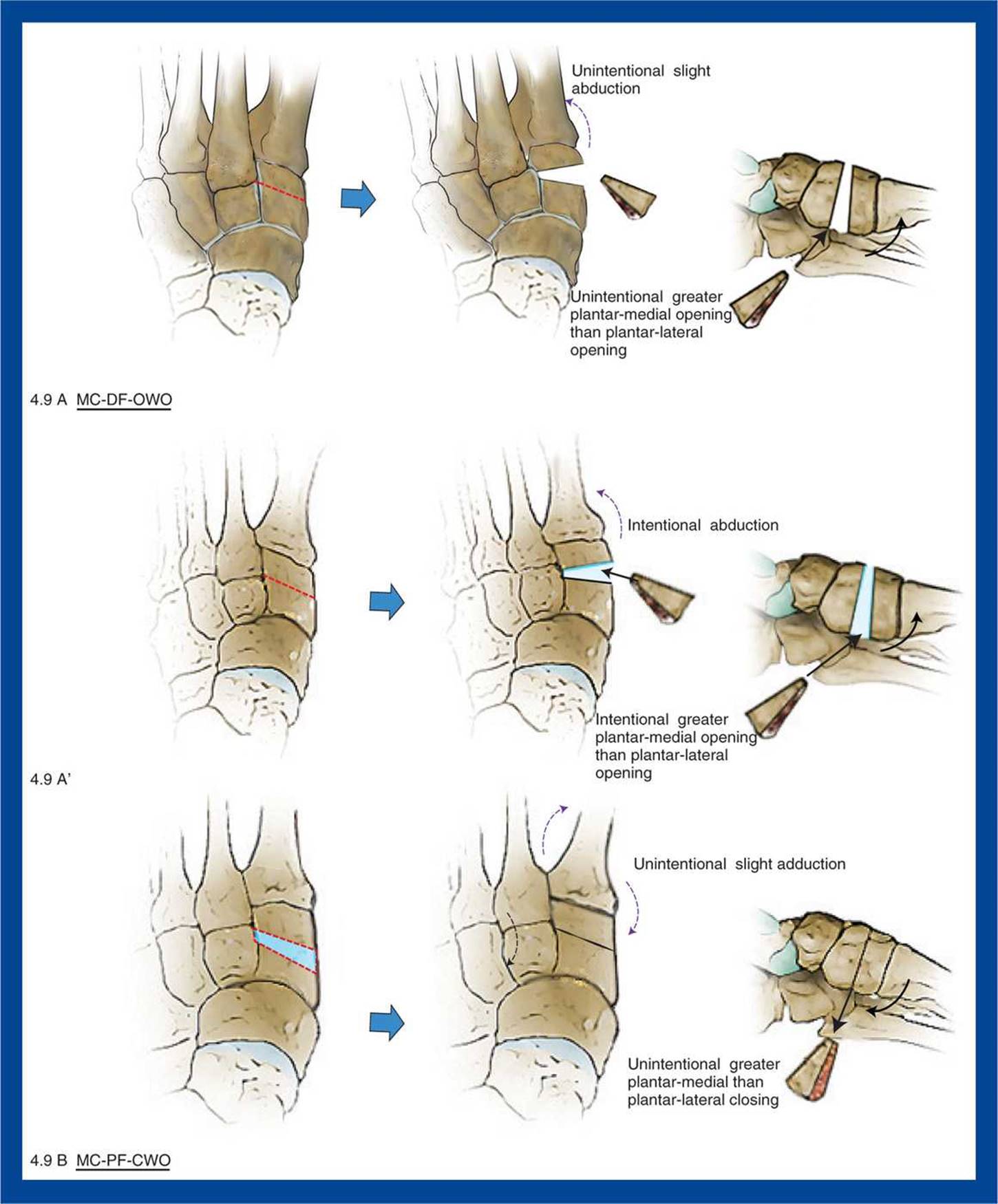
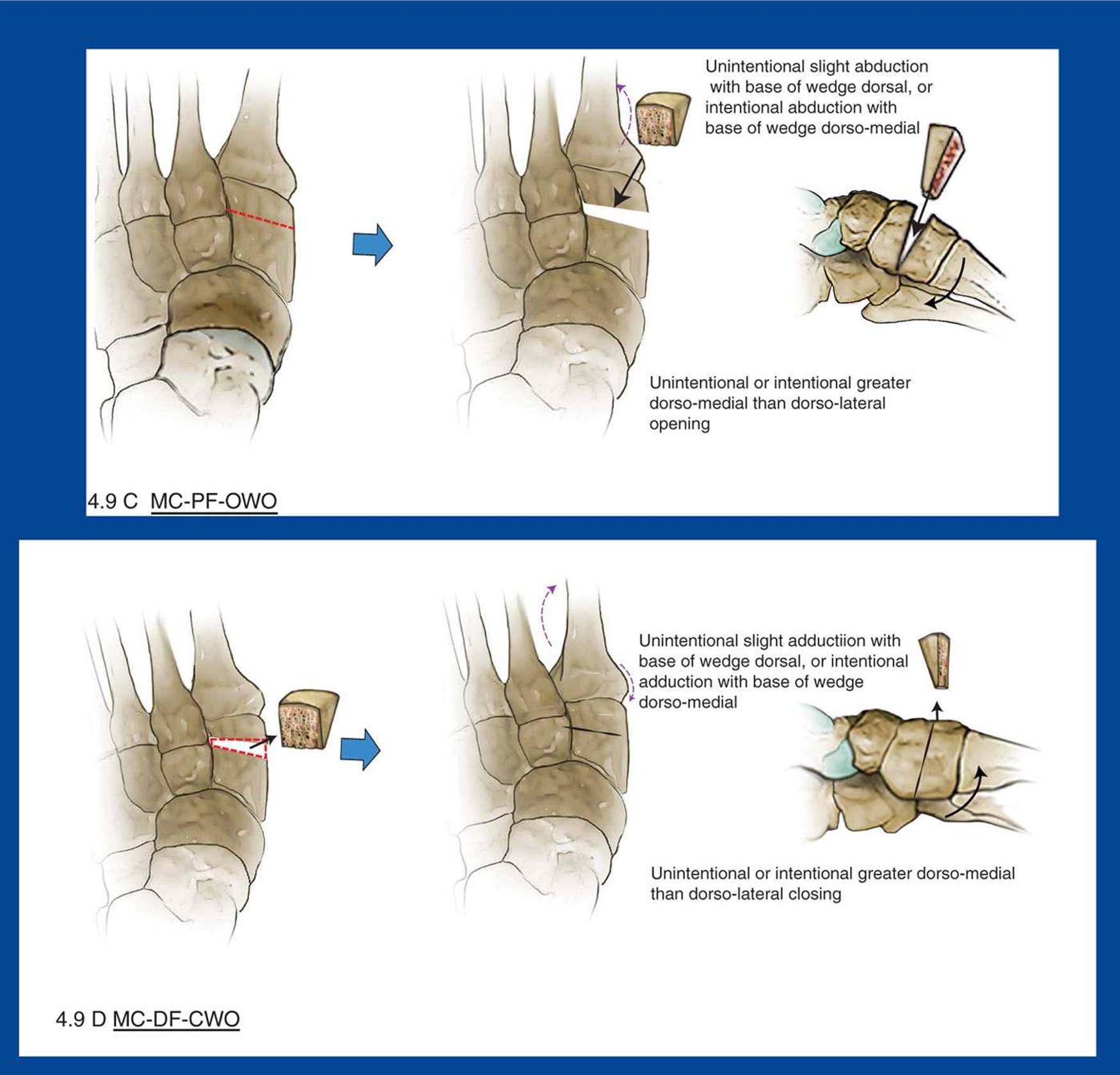
Figure 4-9. Medical cunieform dorsiflexion and plantar flexion osteotomies. Curved purple arrows on AP images show unintentional and intentional changes that occur in the frontal plane (adduction or abduction) when these osteotomies are performed (see text). Curved black arrows on the lateral images show the intentional dorsiflexion and plantar flexion changes that occur. A. Medial cuneiform (dorsi-flexion) plantar-based opening wedge osteotomy (MC-DF-OWO). This is best for a cavovarus deformity, as it corrects forefoot pronation (plantar flexion of the 1st ray) and adds some unintentional, yet acceptable, midfoot abduction to the abduction/eversion that is being achieved in the hindfoot with the plantar-medial soft tissue release. This frontal plane deviation is due to the tethering effect of the bones and ligaments on the lateral side of the medial cunieform fragments, an effect that is observed with all medical cunieform osteotomies despite attempts to create pure sagittal plane correction. A’. Consideration of both planes and tha lateral tethering effects are also useful for a skewfoot withadduction/pronation deformities of the forefoot/midfoot. Intentional plantar-medial alignment of the base of the wedge will correct both deformities simultaneously. B. Medial cuneiform (plantar flexion) plantar-based closing wedge osteotomy (MC-PF-CWO). This is best for a flatfoot, as it corrects forefoot supination (dorsiflexion of the 1st ray) and adds some unintentional, yet acceptable, midfoot adduction to the adduction/inversion that is being achieved in the hindfoot with the calcaneal lengthening osteotomy. It is also useful for a dorsal bunion with no midfoot angular deformity. C. Medial cuneiform (plantar flexion) dorsal-based opening wedge osteotomy (MC-PF-OWO). This is best for: a skewfoot withadduction/supination deformities (align the base of the wedge dorsomedially); a dorsal bunion with midfoot adduction; and possibly a flatfoot with forefoot supination and mild midfoot adduction (if neutral or abducted, the additional abduction might be undesirable). D.Medial cuneiform (dorsi-flexion) dorsal-based closing wedge osteotomy (MC-DF-CWO). This is best for forefoot pronation (cavus) and midfoot abduction, a combination rarely seen, except perhaps as an iatrogenic deformity. If used for a typical cavus deformity with neutral or slight adduction deformity, the unintentional additional adduction might be undesirable.
More specifically, for angular deformity correction, the tibial osteotomy is rarely performed at the CORA, which in children is usually the growth plate. Therefore, translation of the fragments is required and, geometrically, the fibula must translate even further than the tibia.
Furthermore, the lateral tibial cortex is never the apex or base of the angular deformity. It is the intended apex or base of the deformity correction. The lateral cortex of the fibula is the apex or base of the deformity. Without a fibula osteotomy, the tibial osteotomy surfaces will not meet.
2. Consider the intended direction of movement of the distal tibial fragment to determine the proper plane for the fibula osteotomy (Figures 4-12 and 4-13).
The fibula should be cut obliquely to create broad surfaces for rapid healing because, as discussed above, the ends will not be in exact or direct contact and fixation will not be used. The plane of obliquity should be designed to allow the fragments to move in the intended direction(s) without obstructing that movement. For varus or valgus tibial deformity correction, make an oblique coronal plane fibula osteotomy. For rotational deformity correction as well as flexion or extension tibial deformity correction, make an oblique sagittal plane fibula osteotomy.

Figure 4-10. A. For angular deformity correction at the ankle in children, the tibial osteotomy is rarely performed at the CORA, which is usually at the growth plate, as in this case. Therefore, translation (small yellow arrow) in addition to angulation (curved yellow arrows) is required. Without translation, the axis of the distal fragment (oblique blue line) would be parallel with (dotted vertical blue line) the axis of the shaft fragment (solid vertical blue line), but the axes would not be colinear, as they should be. The lateral cortex of the tibia is the apex of the deformity correction, whereas the lateral cortex of the fibula is the apex of the deformity. B. A fibula osteotomy is required to enable both angulation and lateral translation of the distal fragments, and it must be made in the proper plane, in this example, the oblique coronal plane (1). The axes of the fragments have become anatomically aligned (solid blue line). If the osteotomy were made in the oblique sagittal plane from distal/lateral to proximal/medial (2), the fragments would abut each other and prevent angular deformity correction. If the osteotomy were made in the oblique sagittal plane from proximal/lateral to distal/medial (3), the fragments would separate and lose contact with each other, thereby possibly delaying healing.
3. Achieve control of the distal tibial fragment before the osteotomy is performed, if at all possible (Figure 4-14).
After the osteotomy is performed, it is difficult (or even impossible) to appreciate the complex three-dimensional shape and alignment of the distal tibial fragment. Fixation on the “anticipated” distal fragment will make it easy to move it to the intended new location after the osteotomy is completed. It is always easy to see the shaft fragment.
4. Cut the tibia perpendicular to the shaft for a pure rotational osteotomy (Figure 4-15).
If the tibia is not cut perpendicular to the axis of the shaft, rotation will result in undesired flexion, extension, varus, valgus, or combinations of these deformities.
5. For closing wedge angular deformity correction osteotomies, make the first tibial cut parallel with the ankle (while you can still see parallel), and make the second tibial cut perpendicular to the shaft on the shaft fragment(Figure 4-16).
If the second tibial cut is not perpendicular to the axis of the shaft, any desired (or undesired) change in rotation will result in undesired flexion, extension, varus, valgus, or combinations of these deformities.
MANAGEMENT PRINCIPLE #21
Iliac crest is the ideal bone graft material for foot deformity correction surgery in children and adolescents. Allograft has advantages over autograft (Figure 4-17).
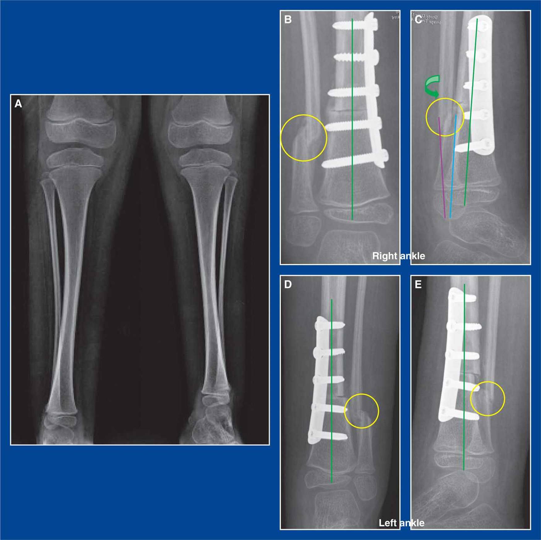
Figure 4-11. A. Bilateral severe external tibia and fibula rotational deformities in a child with myelomeningocele. B. For pure rotational deformity correction at the ankle in children, the fibula must be cut, because the central axis of rotational deformity correction is that of the tibia (green line). Two adjacent solid objects cannot rotate on the axis of one of them without the other resisting rotation. C. If a fibula osteotomy is not performed, the fibula will restrict rotation of the tibial fragments and the medial articular surface of the lateral malleolus (purple line) will flex or extend in relation to the lateral articular surface of the talus, thereby, creating incongruity. An oblique sagittal plane osteotomy of the fibula will enable the adjacent articular surfaces of the lateral malleolus and the talus to remain congruous and the axes of the distal fragments to remain parallel (blue and green lines) when the distal fragments are rotated around the central axis of the tibia (green line). D. and E. Forty-five degrees of rotational correction was achieved in this example. The obliquity of the fibula osteotomy ensured maintenance of some contact between the fragments (yellow circles) which, combined with the subperiosteal exposure of the fibula, ensured rapid healing in this extreme case of rotational deformity correction. An oblique coronal plane osteotomy of the fibula would have either created an obstruction to rotation or led to separation of the fibula fragments.
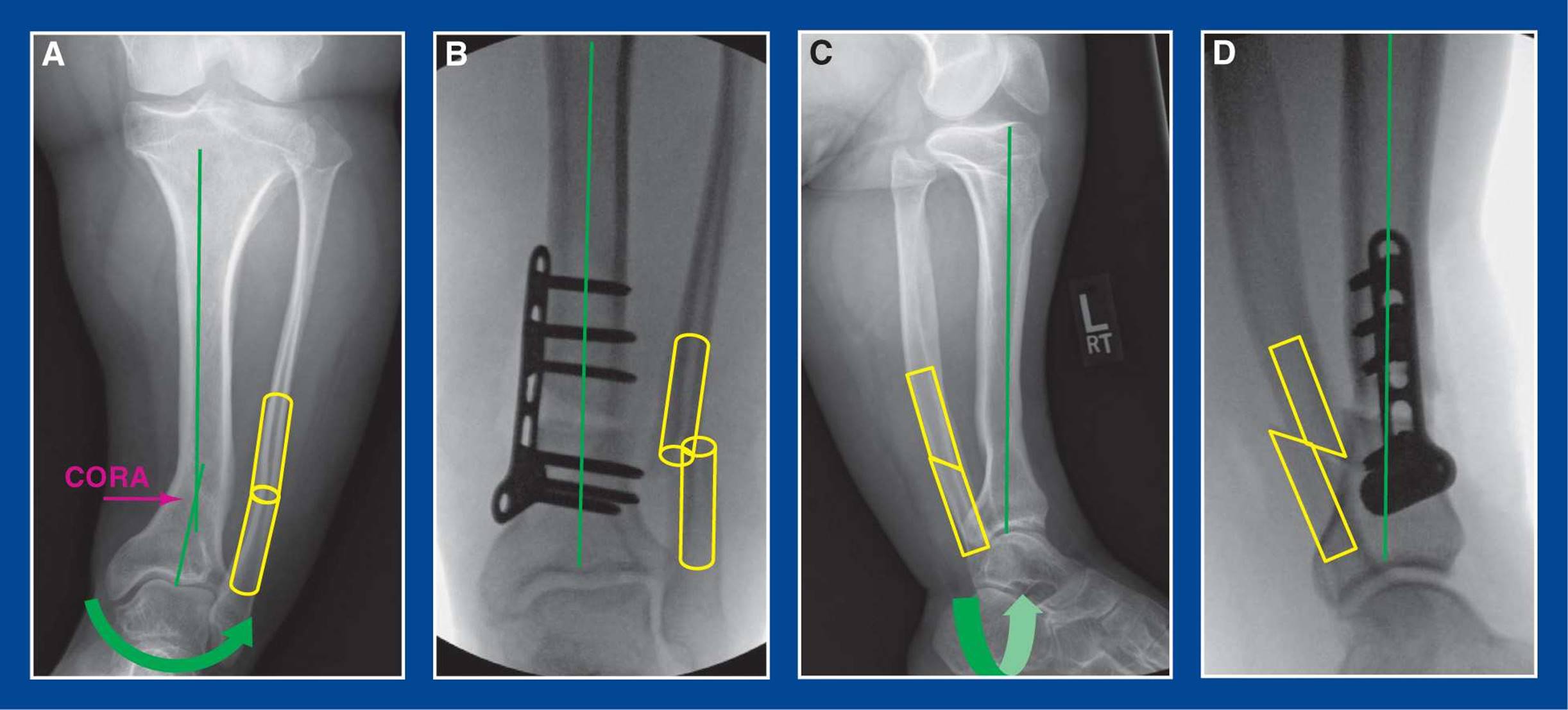
Figure 4-12. For varus and valgus correcting osteotomies, the fibula osteotomy should be made in the oblique coronal plane.A. This AP x-ray of the leg of an adolescent with achondroplasia shows the CORA in her tibia, which became the site of her varus correcting osteotomy. The fibula osteotomy was performed in the oblique coronal plane. B. The fibula osteotomy enabled frontal (coronal) plane deformity correction of the tibia at the CORA (straight green line). The fibula fragments slid past each other. C. The lateral x-ray before deformity correction with the site and direction of the fibula osteotomy indicated. D. The fibula osteotomy enabled coronal plane deformity correction of the tibia at the CORA (with no change in the straight green line). The fibula fragments slid past each other.

Figure 4-13. For rotational osteotomies, the fibula osteotomy should be made in the oblique sagittal plane (also see Figure 4-11). A. This AP x-ray of the ankle of a child with myelomeningocele shows the oblique sagittal plane of the fibula osteotomy. B. The lateral x-ray after rotational deformity correction shows the anterior displacement of the distal fibula fragment that enabled the tibial fragments to rotate on their common central axes.
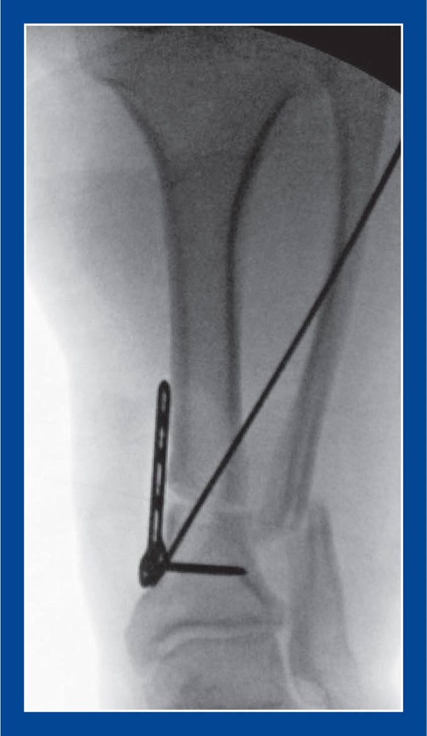
Figure 4-14. For this varus deformity correction, the plate was bent and the screws were inserted distally before the osteotomy was performed to ensure proper alignment on, and good control of, the distal fragment while the alignment of the distal fragment could still be determined. The plate and screws were then removed, the plate was straightened, the osteotomy was performed, and the plate and screws were reattached. The plate could have been attached anteriorly, thereby obviating the need to pre-bend it. For correction of a valgus deformity, pre-bending of the plate would not have been necessary regardless of where it was positioned.
The thick cortices of corticocancellous iliac crest bone grafts provide immediate structural support, and the abundant cancellous bone provides rapid early healing. We have shown that there is no difference between freeze dried iliac crest allograft and iliac crest autograft in the rate of healing, the quality of healing, and complications, based on allograft obtained from a reliable and reputable bone bank. The costs are comparable, i.e., the charge for the allograft and the surgical fee for obtaining autograft. The use of allograft obviates the time needed to obtain autograft and the need for an additional surgical site, one that is reported to be associated with significant pain. Finally, autograft is only bicortical in children and young adolescents. Allograft is tricortical, thereby making it more structurally sound and able to withstand forceful impaction into the osteotomy site.
MANAGEMENT PRINCIPLE #22
Principles of tendon transfers:
The best muscle balance across a joint exists when all of the muscles that cross the joint have normal strength. The next best muscle balance scenario exists when all of the muscles that cross a joint are equally weak or absent. The third, and worst, scenario exists when there are both strong and weak muscles across a joint, as these muscle imbalances create deformities. This last scenario is typically seen in foot deformities of neuromuscular origin. It is important to improve muscle balance at the time of deformity correction; otherwise the deformities will recur. Muscle/tendon balancing is part science and part art. Attention to the following principles will improve surgical outcomes.
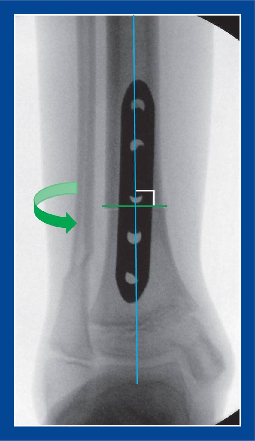
Figure 4-15. For a pure rotational osteotomy, the plate is attached distally with two screws, then removed; the osteotomy is performed perpendicular to the axis of the shaft; the plate is reattached to the distal fragment; it is then attached to the shaft with three screws after the rotational deformity has been corrected. If the osteotomy is not perpendicular to the axis of the shaft, rotation of the distal fragment will result in undesired flexion, extension, varus, valgus, or combinations of these deformities.
1. Move the right tendon to the right location at the right tension.
The right muscle/tendon unit is expendable, strong, and in phase. Moving a tendon attachment to a new location is predicated on the premise that its muscle power will no longer be needed at its original site of attachment, thereby making it expendable. The muscle should be of normal or near normal strength because, in most tendon transfers, the muscle loses strength due to a change in vector and lever arm. It is unknown whether a muscle can reliably and predictably change its phase of activity during the gait cycle based on its site of attachment. For example, it has not been shown conclusively that the posterior tibialis can change from a stance phase muscle to a swing phase muscle following transfer to the dorsum of the foot, a procedure designed to substitute for a weak anterior tibialis. It might merely act as a tenodesis which, in some cases, could be sufficient. But, as a rule, in-phase transfers should be sought (Figure 4-18).
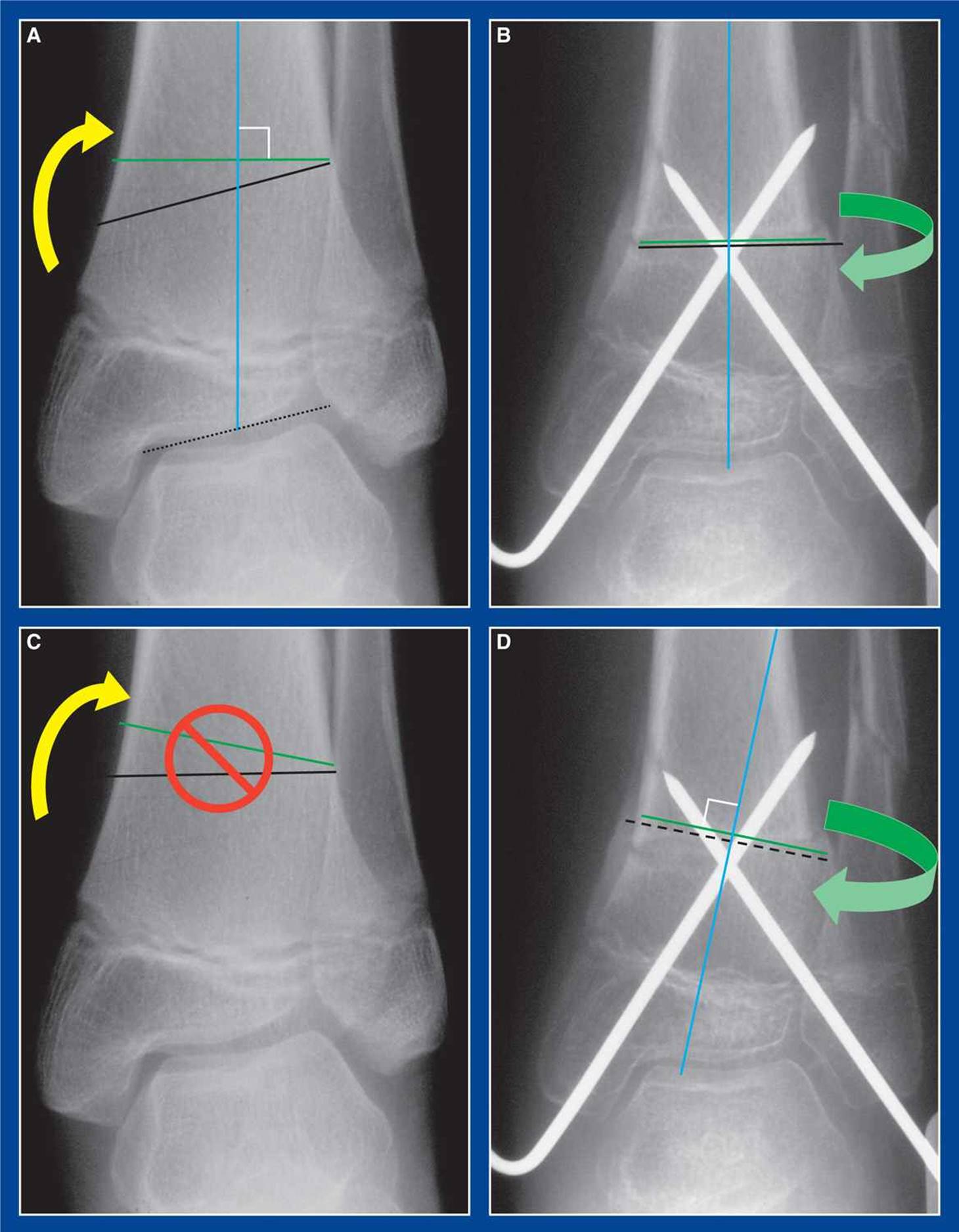
Figure 4-16. A and B. For a closing wedge and rotational osteotomy, the first cut (black line) must be the distal one and it must be parallel with the ankle joint (black dotted line). The second cut (green line) must be on the shaft fragment and it must be perpendicular to the axis of the shaft (blue line) or else rotation will result in undesired flexion, extension, varus, valgus, or combinations of these deformities. The crossed wires were inserted retrograde up to, but not across, the anticipated site of the distal osteotomy before the osteotomy was performed. This provided control of the distal fragment (see Management Principle #20-3, Figure 4-14, this chapter). The blue line is the axis of the tibia and the axis of rotation. C and D. If the shaft cut (green line) is anything other than perpendicular to the shaft, the axis of rotation is changed to a line perpendicular to that cut (blue line) and rotation of the distal fragment will create an undesired deformity: extension/varus with internal rotation, flexion/valgus with external rotation. The dashed black line represents the first (distal) cut in apposition with the second cut (green line).
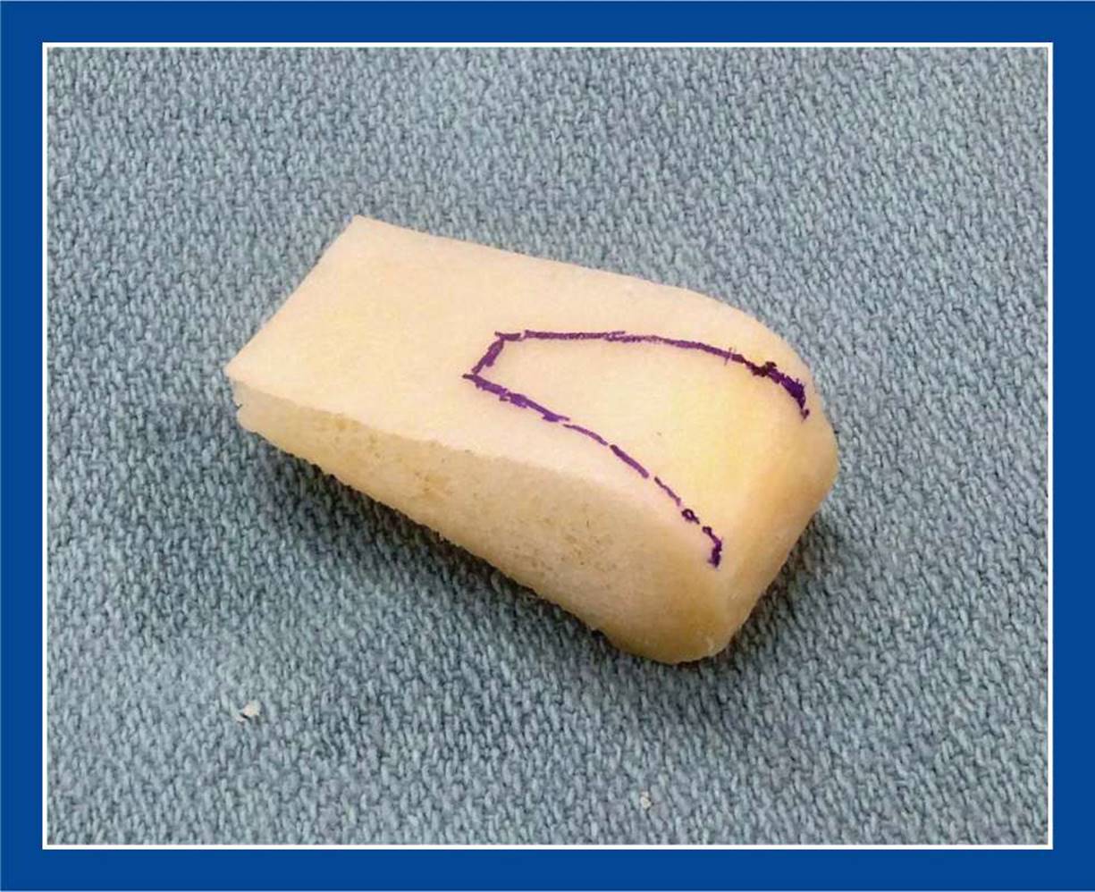
Figure 4-17. Tricortical iliac crest allograft is the ideal graft material for structural deformity correction surgery of the foot in children and adolescents.
The right location for a tendon transfer is based on several factors, including the axis of motion of the joint to be crossed (which in most cases means the subtalar joint), the presence and strength of all other agonist and antagonist muscles that cross the joint, the desired anchor structure (which could be a bone or the tendon of a weak or nonfunctioning muscle), and the ease with which the tendon can directly reach the desired location without curving around structures and losing additional strength (straight vector if possible).
The right tension is less about science and more about art. The tension is set statically with the assumption that the desired function will follow the new form, not unlike the way a puppeteer sets tension on the strings. The foot and ankle are held in a slightly overcorrected position with firm tension set on the tendon when anchored.
2. Tendon transfers will not correct structural deformities.
Muscle-balancing tendon surgery will correct dynamic deformities and will likely prevent or delay the development of structural deformities (see Management Principle #6, this chapter). Balanced muscles will also maintain deformity correction, though perhaps for only a limited time in progressive neuromuscular disorders (see Management Principle #7, this chapter). Importantly, balancing muscle forces by means of tendon transfers without concurrently correcting structural deformities creates structural deformities with good muscle balance. That is not the goal (see Management Principle #15-2, this chapter).
3. Tendon transfers are based on existing and anticipated patterns of muscle imbalance.
Knowledge of the underlying condition is important. Differentiation of static vs. progressive neurologic conditions will help determine some of the subtleties of tendon transfers and releases. Nevertheless, the rate of progression of a deformity that is due to muscle imbalance is rarely predictable. Correct the existing muscle imbalance and do your best to plan for the future.
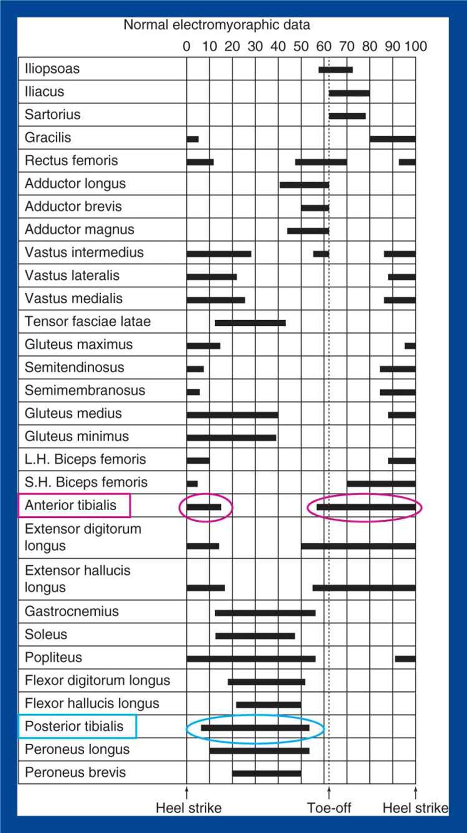
Figure 4-18. Chart of the timing of muscle activity during the gait cycle. The anterior tibialis contracts during the swing phase and at heel strike. The posterior tibialis contracts during the stance phase. It has not been shown conclusively that the phase of activity of a muscle can change if its tendon attachment site is changed.
4. Tendon transfers are much more challenging with joint preserving reconstructions.
But the challenge must be met. Following subtalar and triple arthrodeses, tendon transfers across the subtalar joint are of no value, because inversion and eversion motions are eliminated. The shock-absorbing function of the foot is likewise eliminated by those procedures, which is why they should be avoided (see Management Principle #13, this chapter).
MANAGEMENT PRINCIPLE #23
It is important to correct individual deformities in a complex multisegmental foot/ankle deformity in the proper order.
In some cases, the deformities are corrected sequentially in the same operative session, and sometimes sequential operations are required, either in close or remote proximity (Figure 4-19).
1. Cavovarus
Correct the forefoot deformity before the hindfoot. The forefoot becomes rigidly pronated (with plantar flexion of the 1st ray) before the hindfoot becomes rigidly inverted. That is the justification for performing the Coleman block test. If the forefoot is corrected before the hindfoot becomes rigidly deformed, no hindfoot deformity correction procedures are required. If the hindfoot is already rigidly deformed, it is still important to correct the forefoot first because the severity of forefoot deformity is most often greater than that of the hindfoot. Incomplete forefoot deformity correction results in the need for compensatory, rather than primary, hindfoot deformity correction procedures.
2. Equinocavovarus
Correct the cavus deformity at the first of two fairly closely staged operations. Correct the equinus at the second operation. This principle applies primarily to acquired deformities, though it should be considered in some congenital deformities as well. The justification for this recommendation has to do with the number of contracted tissues at the respective sites. The contracted soft tissues in a cavus or cavovarus deformity include plantar skin, plantar fascia, short toe flexor muscles, lowest muscle belly of the abductor hallucis muscle, posterior tibial neurovascular structures, long plantar ligament, posterior tibial tendon, and the midfoot plantar joint capsules. In acquired equinus, the only significantly contracted structure is the tendo-Achilles. The only structures that can be easily released in the plantar midfoot of a cavus deformity are the plantar fascia, short toe flexors, abductor hallucis, posterior tibial tendon, and plantar capsule of the talonavicular joint. By delaying tendo-Achilles lengthening for 2 to 3 weeks, the other contracted plantar soft tissues can be stretched into dorsiflexion against the unyielding calcaneus, which is being held in position by the tendo-Achilles. Once the plantar structures are stretched, the tendo-Achilles can be lengthened with less risk of converting an equinocavus deformity into a calcaneocavus deformity.
The exception to the rule is in congenital equinocavovarus, i.e., congenital clubfoot. Cavus and equinus can be released concurrently because there are multiple posterior as well as plantar soft tissue contractures.
3. Planovalgus
Correct the hindfoot deformity before the forefoot. In contrast to the cavovarus foot, the hindfoot in a flatfoot becomes structurally deformed before the forefoot. Following hindfoot deformity correction with the calcaneal lengthening osteotomy, forefoot rotation is assessed intraoperatively. In most cases, particularly in younger children and adolescents, the forefoot supination deformity corrects spontaneously following hindfoot deformity correction. The plane of the metatarsal heads aligns perpendicular to the axis of the hindfoot and tibia. However, if the plane of the metatarsal heads is supinated in relation to the axis of the hindfoot and tibia following hindfoot deformity correction, a plantar flexion osteotomy of the medial cuneiform is required to correct that second structural deformity. The degree of plantar flexion is determined after the hindfoot deformity has been fully corrected.
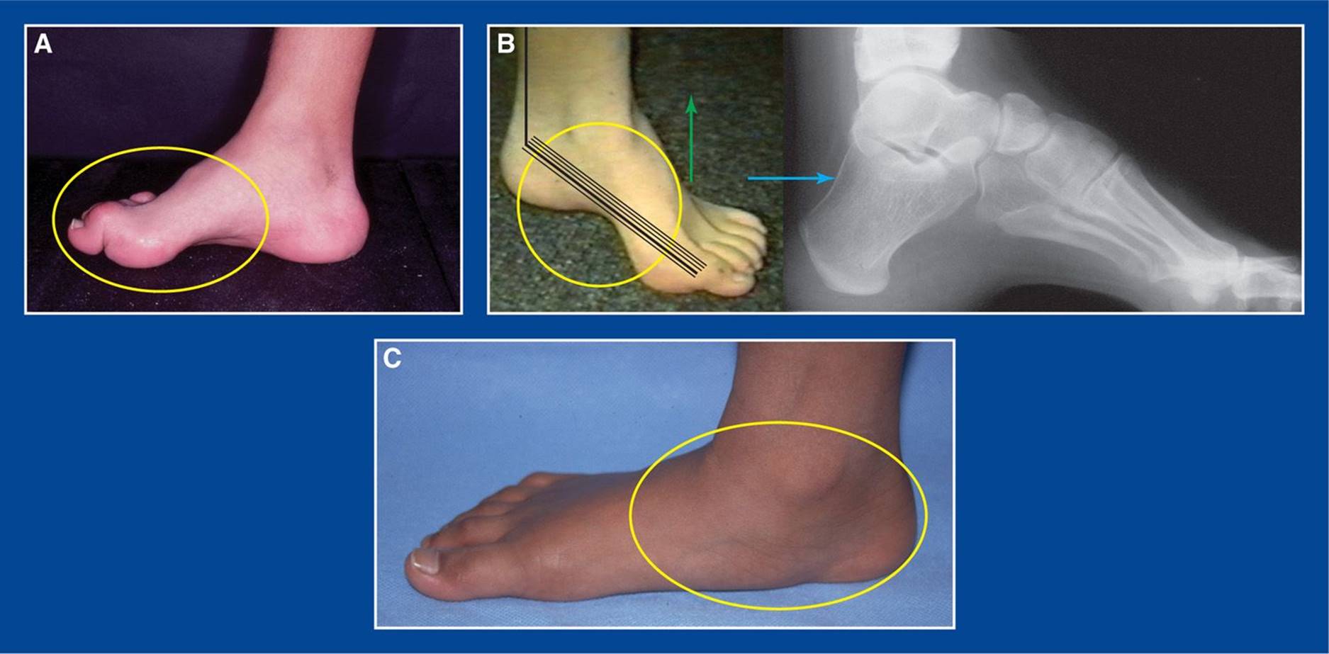
Figure 4-19. A. Correct the forefoot pronation (yellow oval) before the hindfoot varus in a cavovarus foot deformity. B. Correct the cavus (yellow circle) before the equinus in an acquired equinocavovarus foot deformity. Wait at least 2 weeks before correcting the equinus. There are multiple layers of plantar soft tissue contractures (thin black lines), only some of which can be released. The rest must be stretched against the calcaneus that is being held back firmly by the Achilles contracture. Concurrent release of the plantar fascia (thick black plantar line) and lengthening of the tendo-Achilles (thick black posterior ankle line) could convert an equinocavus deformity to a calcaneocavus deformity (follow the blue arrow). The lateral x-ray to the right of the foot image shows hyperdorsiflexion of the calcaneus with severe cavus, i.e., calcaneocavus. C. Correct the hindfoot valgus before the forefoot supination in a flatfoot deformity.
4. Equinoplanovalgus
It is extremely uncommon for surgery to be required for a planovalgus deformity without contracture of the gastrocnemius or the entire triceps surae (tendo-Achilles). It is the heel cord contracture that usually creates the pain which is the indication for surgery. In contrast to the equinocavovarus foot, lengthening of the gastrocnemius or tendo-Achilles must be performed at the time of correction of the foot deformities.
5. Planovalgus or cavovarus deformity with real or apparent ipsilateral tibial torsion
External rotation of the calcaneus/acetabulum pedis is a major component of eversion, the hindfoot deformity in a planovalgus/flatfoot deformity. Internal rotation of the calcaneus/acetabulum pedis is a major component of inversion, the hindfoot deformity in a cavovarus deformity (see Basic Principles #6 and 7, Chapter 2). There is only one easy to document normal rotational alignment of the subtalar joint/acetabulum pedis, i.e., essentially straight alignment of the axis of the talus and the axis of the 1st metatarsal on a weight-bearing AP radiograph (average 4° abducted, range of normal 12° abducted to 10° adducted [see Assessment Principle #18, Chapter 3]). Assessment of tibial torsion is less precise, both clinically and radiographically. Therefore, the inversion (internal rotation) or eversion (external rotation) deformity of the subtalar joint should be corrected to anatomic alignment first. Then any identified residual excessive rotation of the foot in relation to the leg (positive or negative thigh–foot angle) is due to tibial torsion. Significant tibial torsion can be corrected subsequently, if necessary, during the same anesthetic or at a later date. If a tibial rotational osteotomy is inappropriately performed in an attempt to avoid correction of the hindfoot rotational deformity, the axis of flexion and extension of the ankle will become mal-oriented. This could result in abnormal stresses in the ankle that could eventually lead to premature degenerative arthritis of that joint.
If significant external tibial torsion is identified/uncovered after a flatfoot deformity has been corrected by a CLO (a rare occurrence), distal tibia and fibula internal rotation osteotomies can be performed under the same anesthetic.
If significant external tibial torsion is identified/uncovered after complex reconstruction of a cavovarus foot deformity (a very common occurrence) (see Assessment Principle #7, Chapter 3), distal tibia and fibula rotational osteotomies should not be performed under the same anesthetic. The tendon transfers could potentially bind down in the scar tissue and fracture callus around the osteotomies, thereby causing tethering of the tendons. If the external tibial torsion proximal to a structurally well-corrected and muscularly well-balanced foot is later noted to be a problem, isolated derotational osteotomies of the tibia and fibula can be carried out safely.
6. Coincident subtalar joint and ankle joint valgus
Valgus deformity can exist in the ankle joint and in the subtalar joint. The frontal plane axis of the normal ankle joint is roughly perpendicular to the tibia and parallel to the floor in weight-bearing after the age of 3 to 4 years, except in children with myelomeningocele, lipomeningocele, early onset poliomyelitis, other early onset flaccid paralytic conditions, and approximately 66% of limbs with a clubfoot (see Assessment Principle#11, Figure 3-12, and Assessment Principle #21, Figure 3-27, Chapter 3). This is easy to assess radiographically. There is a wide range of normal values for subtalar joint alignment from neutral to valgus. If valgus deformity exists at both levels in a symptomatic hindfoot, the ankle valgus should be corrected first. Correction is technically easy (guided growth or supramalleolar osteotomy), and there is only one easy-to-assess anatomically normal correct alignment. Once an orthogonal ankle platform is established, correction of subtalar valgus can be undertaken at the time of hardware removal, if it is still deemed necessary.
7. Coincident subtalar joint varus and ankle joint valgus
This combination of deformities is often seen in a recurrent/residual clubfoot and in a cavovarus foot deformity in a child with myelomeningocele. In contrast to the situation in which valgus deformity exists at both levels (see preceding point), the subtalar joint deformity should be corrected first. This will expose the ankle valgus deformity that can subsequently be corrected either acutely or by guided growth. The time between procedures can be considered an opportunity for valgus weight-bearing to help maintain subtalar joint deformity correction, which is sometimes a challenge for a corrected varus hindfoot.
MANAGEMENT PRINCIPLE #24
Surgical efficiency and clinical outcomes can be improved by adhering to a specific order of events during complex foot reconstruction surgery:
1. Expose and prepare everything before completing anything.
Many exposures are gentle and nontraumatic, but some are vigorous and forceful. Osteotomies, for example, can be forceful and could potentially disrupt an already stabilized osteotomy at another site or a tensioned tendon transfer. Release all contracted soft tissues, perform all osteotomies, and move tendons to their intended sites of attachment before inserting bone grafts, internally fixing osteotomies, plicating soft tissues, or tensioning tendon transfers.
2. Perform and stabilize deformity corrections.
The foot needs to look like a foot before tendons are tensioned. The proper tensions will be different after the deformities are corrected. Therefore, the next step is to insert bone grafts or align osteotomy surfaces and stabilize the sites with internal fixation, if needed.
3. As you proceed, close incisions that no longer need to be accessed.
This is particularly true for incisions in which there has been minimal dissection and/or minimal expectation of the need for complete hemostasis. By so doing, there will be more rapid progress to cast application after the final tendon transfer incision is closed.
4. Set proper tension on tendon lengthenings/plications/transfers.
This is the last step, as it requires complete deformity correction for accuracy. Tendon transfers should then be performed in the order of most stable and secure to least stable and secure. An example is performing a peroneus longus to peroneus brevis transfer (using a Pulvertaft weave) before a Jones transfer.
MANAGEMENT PRINCIPLE #25
It is safe, reliable, and cosmetic to use absorbable subcuticular sutures for wound closures and no drains. Corollary: It is safe and reliable to use absorbable sutures for tendon lengthenings and transfers.
Operate carefully, but not slowly, achieving hemostasis along the way. Even complex foot reconstructions with one or more osteotomies and tendon transfers should take less than 2 hours of tourniquet time. Obtain final hemostasis after release of the tourniquet and before wound closure. It is rarely, if ever, necessary to use a drain. Use interrupted 3-0 absorbable sutures in the subcutaneous tissues and a running 4-0 absorbable subcuticular suture. Healing will be reliable and cosmetic. There is no need for cross-hatched scars. And avoiding ever having to remove sutures from children should be a professional goal and aspiration (Figure 4-20).
There are two exceptions to this principle. Nonabsorbable sutures should be used when serial casting will be required in the weeks after surgery to avoid initial excessive tension on a wound closure, as in a Cincinnati incision after a clubfoot operation in a severely deformed foot. Such a foot will have achieved full deformity correction before wound closure, but cannot assume that position after wound closure without blanching the wound edges or pulling the wound apart. The incision should be approximated with nonabsorbable vertical mattress sutures and the foot casted in mild equinovarus. The cast can be changed weekly under anesthesia and the skin stretched slowly (to avoid necrosis) until the foot assumes the fully corrected position. The final result should then be a thin cosmetic scar.
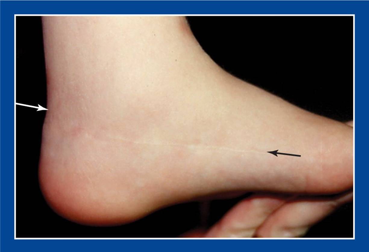
Figure 4-20. Cosmetic appearance of a healed Cincinnati incision that was used for a clubfoot operation years earlier (between the white and black arrows). A running 4-0 absorbable subcuticular suture was utilized.
The other role for a nonabsorbable skin closure is in the first of a two-stage reconstruction, in which the second stage involves utilization of an incision created in the first stage. An example is the medial foot incision used for a plantar-medial release in a cavovarus foot deformity. It is used again 2 weeks later in the second stage for a medial cuneiform opening wedge osteotomy. A running 3-0 monofilament subcuticular suture will create less reaction and scar tissue than an absorbable suture, making it easier to close the wound in the routine fashion the second time around.
The final point regarding suture material pertains to tendon lengthenings and transfers. Absorbable sutures work quite well in both situations in children, healing reliably as long as the foot and ankle are immobilized for at least 6 weeks; 8 weeks for adolescents. Tendon weaves (Pulvertaft) heal faster and more securely than side-to-side transfers.
MANAGEMENT PRINCIPLE #26
It is safe to apply a well-padded, bivalved fiberglass cast at the end of an even complex foot reconstruction that involves multiple bone and soft tissue procedures (Figure 4-21).
Fiberglass casts should be bivalved, rather than univalved, for the best circumferential relief of pressure and accommodation of swelling. The cuts should be medial and lateral at the opposite tangents of the cylinder. The bivalved cast is overwrapped with a loosely applied elastic bandage. Excessive swelling is rarely a problem. If it occurs, it is usually within the first 24 hours postoperatively and can often be managed by slight further spreading of the anterior and posterior shells of the cast. The bivalved cast is overwrapped with fiberglass before the child is discharged from the hospital. In most cases, there should be no reason to remove the cast and examine the foot for as long as 6 weeks.
A less desirable alternative immobilization device is a bulky overpadded splint. In most cases, a splint will not hold the foot in the ideal corrected position. Therefore, it will be necessary to change the splint into a cast in the first few weeks postoperatively. It may be unnecessarily painful to change the splint into a cast in the clinic in those first few weeks and it will be unnecessarily costly to make the change in the OR.
As a general rule, bivalve the cast if an osteotomy was performed, but not if only soft tissue procedures were performed.
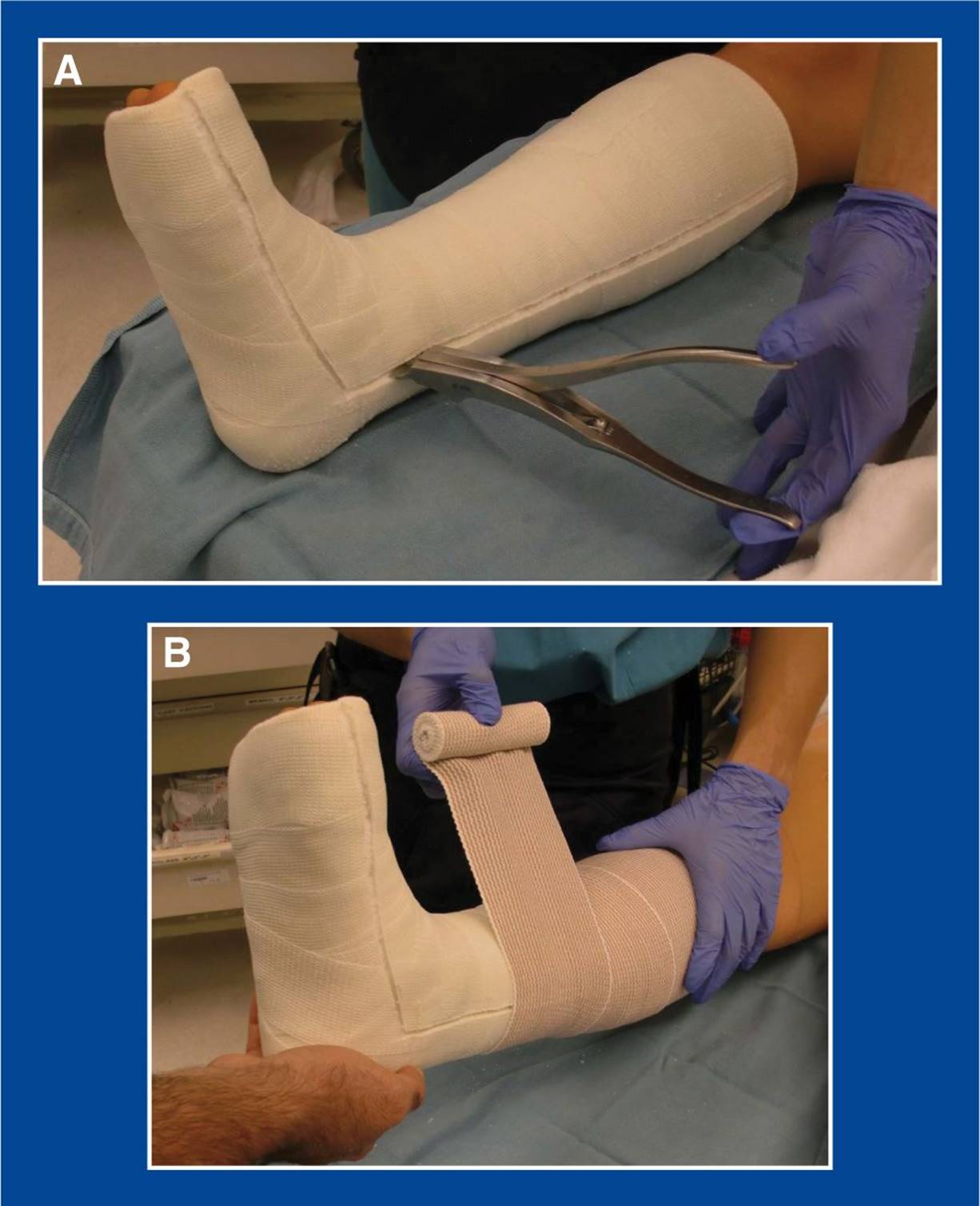
Figure 4-21. A. This short-leg cast was applied and immediately bivalved at the completion of a complex foot reconstruction operation that involved osteotomies and tendon transfers. B. The cast was loosely overwrapped with an elastic bandage. The following day, the elastic bandage was removed, the cast was overwrapped with fiberglass, and the patient was discharged from the hospital.
MANAGEMENT PRINCIPLE #27
Long-leg casts should be applied in two sections to ensure appropriate molding of the foot and protection of the soft tissues at the knee following both nonoperative and operative treatments (Figure 4-22).
The short-leg cast component is applied first, with attention focused on each of the segmental deformities of the foot. The cast is then extended above the knee after the short-leg component has hardened. It is too distracting to simultaneously focus on the position of the foot/ankle and the knee. With one-stage long-leg cast application, there is great risk that the foot molding will be inferior or that the cast padding and/or casting material will wrinkle in the popliteal fossa, creating skin ulceration. This principle applies to all long-leg casts, not just long-leg clubfoot casts.
MANAGEMENT PRINCIPLE #28
Formal physical therapy is appropriate for the successful rehabilitation of some, but not all, foot reconstructions in children and adolescents.
Children play for a living and are, therefore, their own very effective therapists. A few therapy sessions for instructions on gait retraining and strengthening are beneficial and worthwhile for some children. A good home program that is supplied by a therapist and monitored by parents is ideal.
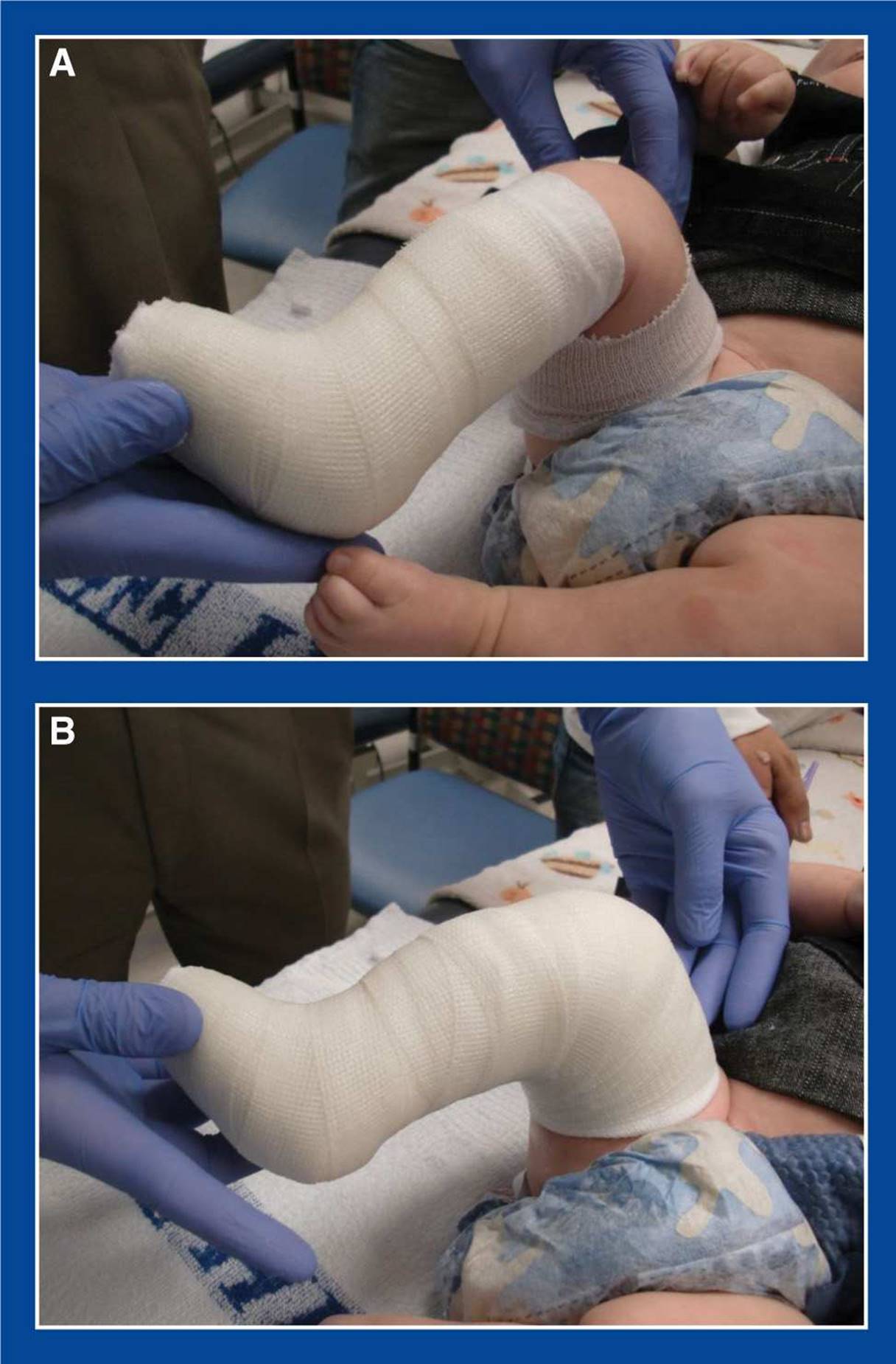
Figure 4-22. A. A short-leg (below the knee) cast is applied first with attention focused on the position of each of the segmental foot deformities. B. The cast is then extended above the 90° flexed knee. Care can be taken to ensure no bunching of the cast padding or cast material in the popliteal fossa. The appropriate thigh–foot angle can also be set.
MANAGEMENT PRINCIPLE #29
When it is not possible to make a malformed or deformed foot as comfortable and functional as a prosthesis, consider an amputation.
The technology of prosthetic design and function has advanced dramatically in the last two decades, particularly in very recent years. This has been influenced, in large part, by government-sponsored research stimulated by injuries sustained in wars abroad. Amputation, and Syme amputation in particular, is an almost routinely successful reconstructive procedure that can enable a high level of function, especially when performed early in life. Competitive sprinting, marathon running, triathlon participation, basketball, football, and other sports are all possible, even when the “disabled” athlete competes against able-bodied athletes. And the cosmetic appearance of a prosthetic can be, and usually is, better than a malformed or deformed limb, especially if the limb has undergone many operative reconstructive procedures (Figure 4-23).
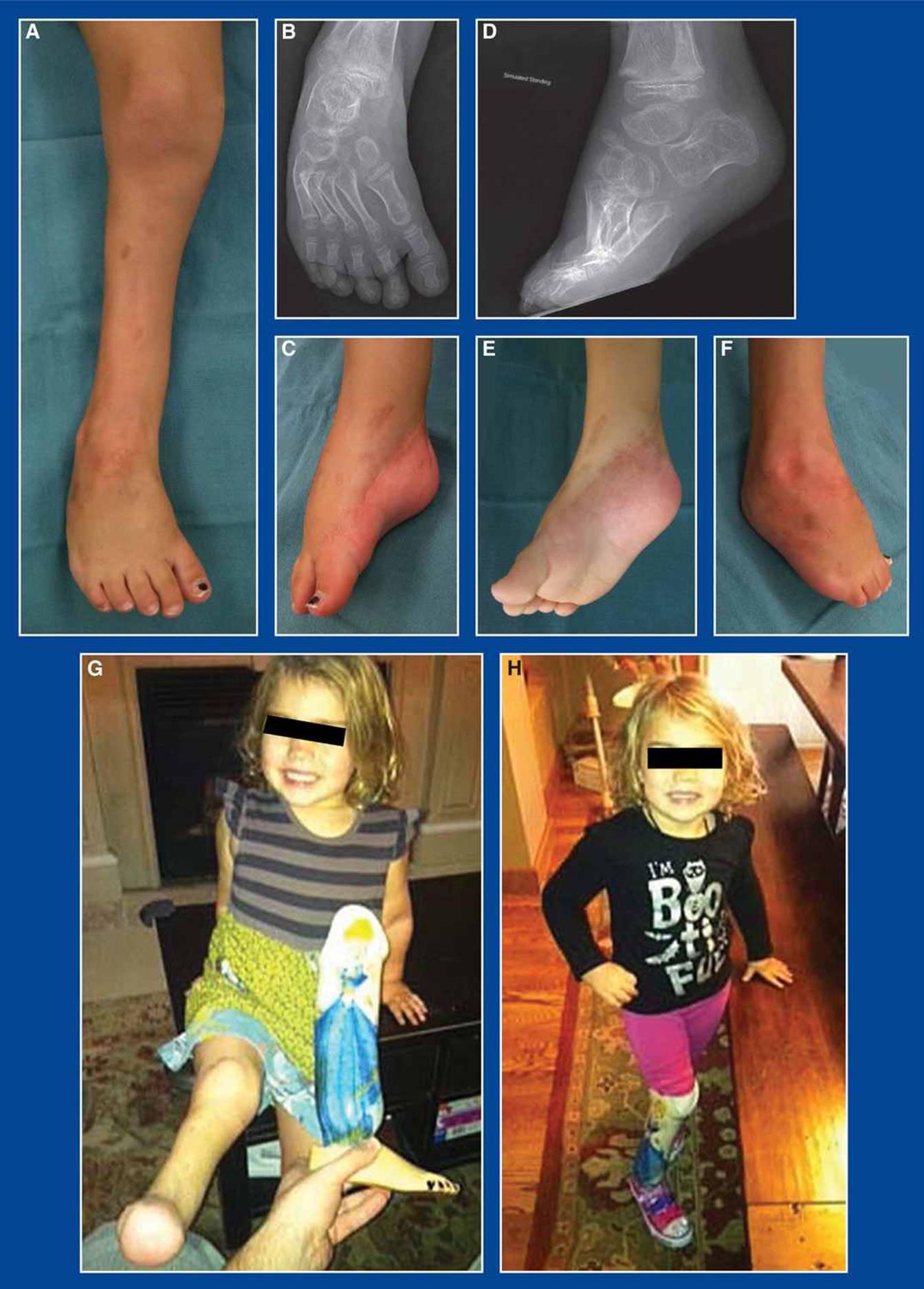
Figure 4-23. A–F. Completely rigid, severely deformed clubfoot in an otherwise normal, healthy 4-year-old girl. She had undergone five operative attempts by multiple surgeons to correct the deformity, including 6 months of gradual deformity correction in a three-dimensional external fixator. The deformity was overcorrected and held statically in the external fixator for several months. Following removal of the frame, the foot was casted in the overcorrected position for several weeks and then held in an AFO that she wore 23 hours per day. The deformity recurred within months after removal of the final cast, despite the use of the brace. G and H. Following a Syme amputation, she is now comfortable, happy, and participating in soccer, gymnastics, skiing, and other sports.