
Cavovarus Foot (Excluding Those Due to Cerebral Palsy—See Below)
1. Definition—Deformity
a. Acquired and usually progressive pronation deformity of the forefoot on the hindfoot that creates cavus deformity of the medial midfoot. There is secondary acquired and usually progressive varus/inversion deformity of the hindfoot. The ankle can be in dorsiflexion, plantar flexion, or neutral. It is the manifestation of a neuromuscular disorder, rather than a primary deformity, unless proven otherwise (Figure 5-5).
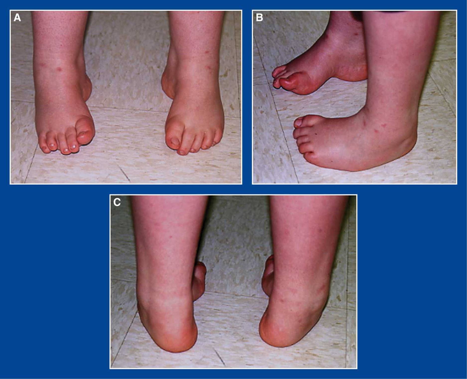
Figure 5-5. Cavovarus foot deformities in a young boy with CMT disease. A. Top/front view shows cavus with varus heels, visible medially. B. Side views show cavus of right foot and adductus of left foot. C. Posterior view shows varus heels and forefoot adductus.
2. Elucidation of the segmental deformities
a. Forefoot—pronated
b. Midfoot—adducted or neutral
c. Hindfoot—varus/inverted
d. Ankle—plantar flexed, neutral, or dorsiflexed
i. NOTE: It is uncommon for there to be contracture of the tendo-Achilles or the gastrocnemius in a cavovarus foot in a child with Charcot–Marie–Tooth (CMT) disease. The apparent ankle equinus (plantar flexion of the foot at the ankle) is, in fact, usually forefoot equinus, i.e., cavus (plantar flexion of the forefoot on the hindfoot) (see Assessment Principle #12, Figure 3-14, Chapter 3). The ankle is often hyperdorsiflexed with an exaggerated calcaneal pitch.
e. Tibia—external torsion
i. In most children with cavovarus foot deformities, regardless of the etiologic underlying neuromuscular disorder, there is coincident external tibial torsion (see Assessment Principle #7,Chapter 3).
f. Muscle imbalances (opposite those seen in dorsal bunion deformities) (Figure 5-6)
i. Weak anterior tibialis
ii. Strong peroneus longus
iii. Recruited and, therefore, stronger extensor hallucis longus (EHL) than flexor hallucis longus (FHL)
g. “Flexibility” classification for the forefoot and hindfoot (unpublished)
i. Flexible =
• dynamic deformity of the forefoot or hindfoot that corrects with tendon transfers
• dynamic and flexible deformity of the hindfoot that corrects following correction of the forefoot deformity and with tendon transfers
ii. Stiff = structural deformity of the forefoot or hindfoot that corrects with soft tissue releases
iii. Rigid = structural deformity of the forefoot or hindfoot that requires osteotomies and/or arthrodeses
h. Cavovarus Flexibility Classification System (Hindfoot–Forefoot) (unpublished)
i. Flexible–Flexible
ii. Stiff–Flexible
iii. Rigid–Flexible
iv. Rigid–Stiff
v. Rigid–Rigid
vi. Late Rigid–Rigid
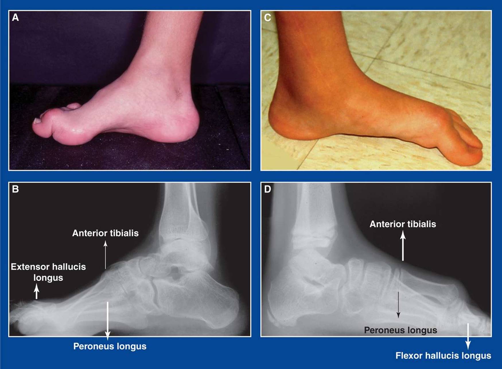
Figure 5-6. A. Cavovarus foot. B. Cavovarus foot muscle imbalances: weak anterior tibialis, relatively stronger peroneus longus, recruited extensor hallucis longus to compensate for weak anterior tibialis. C. Dorsal bunion deformity. D. Dorsal bunion muscle imbalances: strong anterior tibialis, weak peroneus longus, recruited FHL to compensate for weak peroneus longus.
3. Imaging
a. Standing AP and lateral of foot (see Assessment Principle #18, Figures 3-20 and 3-22, Chapter 3)
b. Standing AP block x-ray with 2.5-cm block under lateral forefoot (4th and 5th MT heads) (see Assessment Principle #19, Figure 3-24, Chapter 3)
c. Standing AP, lateral, and mortis of ankle
d. Standing AP and lateral thoracolumbar spine
e. AP pelvis (in patients with CMT or suspected CMT)
4. Natural history
a. Progressive increase in the severity and rigidity of the segmental deformities with pain, gait instability, and skin pressure injuries (inflammation, callus formation, blistering, ulceration) under the 1st and 5th MT heads and at the base of the 5th MT (see Assessment Principle #9, Figure 3-5, Chapter 3).
5. Nonoperative treatment
a. Accommodative shoe wear with over-the-counter soft arch supports pending results of neuromuscular workup—then operate.
6. Operative indications
a. Pain, gait instability, skin pressure injuries, and/or progressive deformity
i. following completion of a neuromuscular workup, with treatment of the underlying condition if treatment exists
7. Operative treatment, based on the Cavovarus Flexibility Classification System, with reference to the surgical techniques section of the book for each individual procedure (see Management Principles #13, 15, 16, 22, 23-1, and 24, Chapter 4). NOTE: If a gastrocnemius recession or a tendo-Achilles lengthening is needed, it should be performed in the second stage of a 2-stage procedure (see Management Principle #23-2, Chapter 4).
a. Flexible–Flexible
i. Peroneus longus to peroneus brevis transfer (see Chapter 7)
ii. Posterior tibialis tendon lengthening—Z-lengthening or intramuscular recession (see Chapter 7)
b. Stiff–Flexible
i. Superficial plantar-medial release (see Chapter 7)
ii. Posterior tibialis tendon lengthening—Z-lengthening or intramuscular recession (see Chapter 7)
iii. Peroneus longus to peroneus brevis transfer (see Chapter 7)
iv. Percutaneous tenotomy of FHL and FDL to toes 2 to 5 (see Chapter 7)
c. Rigid–Flexible
i. Stage 1
• Superficial plantar-medial release (see Chapter 7)
• Posterior tibialis tendon lengthening—Z-lengthening or intramuscular recession (see Chapter 7)
• Percutaneous tenotomy of FHL and FDL to toes 2 to 5 (see Chapter 7)
ii. Stage 2—2 weeks later
• Medial cuneiform (dorsiflexion) plantar-based opening wedge osteotomy (see Chapter 8)
• Peroneus longus to peroneus brevis transfer (see Chapter 7)
• Possible posterior calcaneus lateral displacement osteotomy (see Chapter 8)
• Possible split anterior tibialis tendon transfer (see Chapter 7)
• Possible Jones transfer of extensor hallux longus to 1st MT neck with hallux interphalangeal (IP) joint tenodesis (see Chapter 7) or arthrodesis (see Chapter 8)
• Possible Hibbs transfer of extensor digitorum communis to peroneus tertius or cuboid (see Chapter 7)
d. Rigid–Stiff
i. Stage 1
• Deep plantar-medial release (see Chapter 7)
• Percutaneous tenotomy of FHL and FDL to toes 2 to 5 (see Chapter 7)
ii. Stage 2—2 weeks later
• Medial cuneiform (dorsiflexion) plantar-based opening wedge osteotomy (see Chapter 8)
• Peroneus longus to peroneus brevis transfer (see Chapter 7)
• Possible posterior calcaneus lateral displacement osteotomy (see Chapter 8)
• Possible split anterior tibialis tendon transfer (see Chapter 7)
• Possible Jones transfer of extensor hallux longus to 1st MT neck with hallux IP joint tenodesis (see Chapter 7) or arthrodesis (see Chapter 8)
• Possible Hibbs transfer of extensor digitorum communis to peroneus tertius or cuboid (see Chapter 7)
e. Rigid–Rigid
i. Stage 1
• Deep plantar-medial release (see Chapter 7)
• Percutaneous tenotomy of FHL and FDL to toes 2 to 5 (see Chapter 7)
ii. Stage 2—2 weeks later
• Medial cuneiform (dorsiflexion) plantar-based opening wedge osteotomy (see Chapter 8)
• Peroneus longus to peroneus brevis transfer (see Chapter 7)
• Posterior calcaneus lateral displacement osteotomy (see Chapter 8)
• Possible split anterior tibialis tendon transfer (see Chapter 7)
• Possible Jones transfer of extensor hallux longus to 1st MT neck with hallux IP joint tenodesis (see Chapter 7) or arthrodesis (see Chapter 8)
• Possible Hibbs transfer of extensor digitorum communis to peroneus tertius or cuboid (see Chapter 7)
f. Late Rigid–Rigid
i. Stage 1
• Deep plantar-medial release (see Chapter 7)
• Percutaneous tenotomy of FHL and FDL to toes 2 to 5 (see Chapter 7)
ii. Stage 2—2 weeks later or concurrent
• Midfoot wedge resection/arthrodesis (see Chapter 8)
• or, Triple arthrodesis (see Chapter 8 and Management Principle #13, Chapter 4)
• Possible split anterior tibialis tendon transfer (see Chapter 7)
• Possible Jones transfer of extensor hallux longus to 1st MT neck with hallux IP joint tenodesis (see Chapter 7) or arthrodesis (see Chapter 8)
• Possible Hibbs transfer of extensor digitorum communis to peroneus tertius or cuboid (see Chapter 7)
• Possible posterior tibialis tendon transfer through the interosseous membrane to the dorsum of the foot. Best indication is a strong posterior tibialis and no other functional muscle power (see Chapter 7)
Cavovarus Foot (Due to Cerebral Palsy)
1. Definition – Deformity
a. Acquired and progressive varus deformity of the hindfoot with secondary pronation of the forefoot on the hindfoot creating a cavus midfoot deformity. The ankle is plantar flexed, because there is always associated contracture of the gastrocnemius or the entire triceps surae. The deformities are the result of muscle imbalances due to the cerebral injury rather than being primary deformities. Cavovarus is most commonly seen in children with spastic hemiplegia (Figure 5-7).
2. Elucidation of the segmental deformities
a. Forefoot—pronated
b. Midfoot—adducted or neutral
c. Hindfoot—varus/inverted
d. Ankle—plantar flexed (equinus)
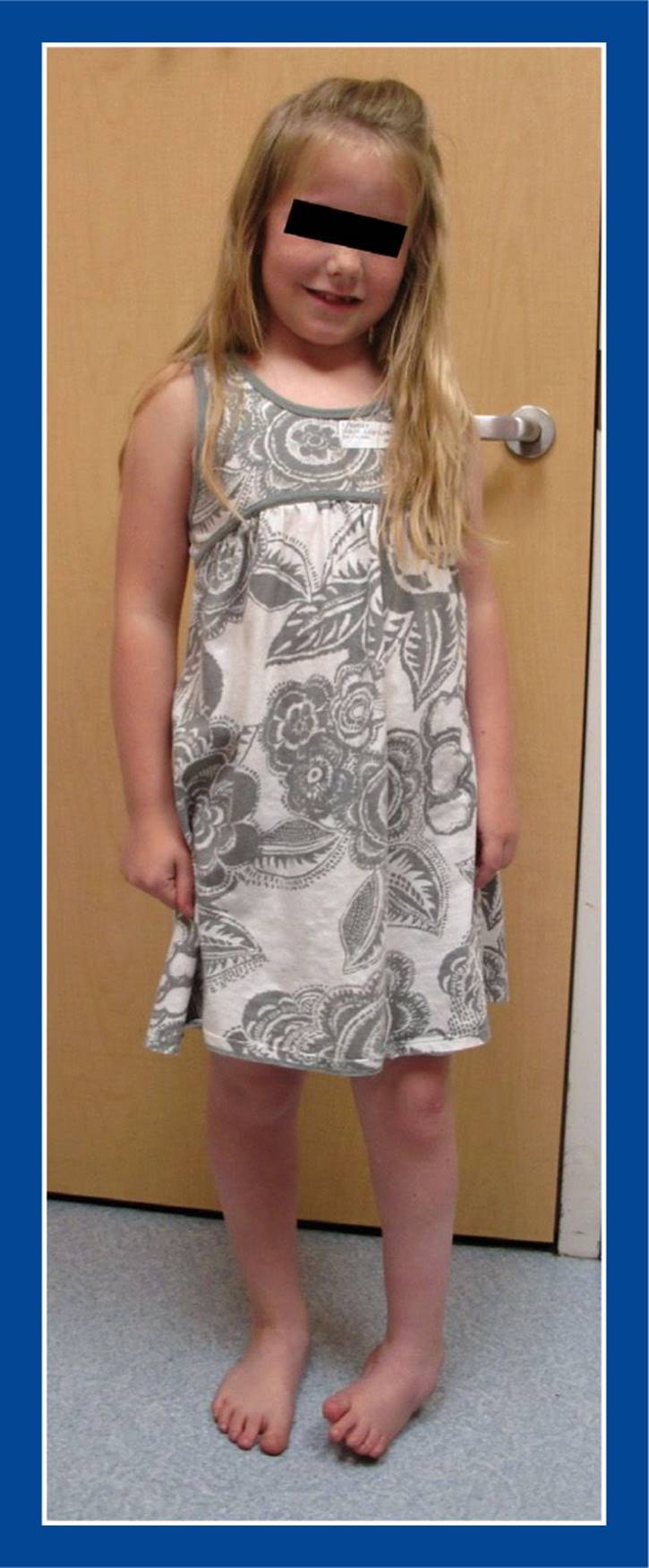
Figure 5-7. An 8-year-old girl with left hemiplegic cerebral palsy and with an equinocavovarus foot deformity.
e. Tibia—external torsion.
i. In most children with cavovarus foot deformities, including those with cerebral palsy, there is coincident external tibial torsion (see Assessment Principle #7, Chapter 3).
f. Muscle imbalances
i. Greater spasticity in the anterior tibialis and posterior tibialis than in the peroneal muscles
ii. Occasionally, the peroneus longus is overpowering the anterior tibialis
3. Imaging
a. Standing AP and lateral of foot (see Assessment Principle #18, Figures 3-20 and 3-22)
b. Standing AP block x-ray is not reliable in children with cerebral palsy, because the spastic inverters often do not relax sufficiently to allow the subtalar joint to evert (to reveal the true flexibility of the subtalar joint)
c. Standing AP, lateral, and mortis of ankle
4. Natural history
a. Progressive increase in the severity and rigidity of the segmental deformities with pain, gait instability, and skin pressure injuries (inflammation, callus formation, blistering, ulceration) at the base of the 5th MT, over the dorsolateral aspect of the talar head in the sinus tarsi region (related to rubbing in the AFO), and occasionally under the 1st MT head
5. Nonoperative treatment
a. Physical therapy—stretching
b. Bracing—AFO
c. Injection of botulinum toxin (BOTOX) into the most spastic muscles
d. Serial below-the-knee (short-leg) stretching casts
e. Tone-reducing medications, such as baclofen
6. Operative indications
a. Pain, gait instability, skin pressure injuries, and/or progressive deformity that are not controlled with nonoperative modalities
i. Ideally, in children over the age of 6 to 7 years
7. Operative treatment with reference to the surgical techniques section of the book for each individual procedure
a. Rancho procedure
i. Split anterior tibialis tendon transfer (see Chapter 7)
ii. Posterior tibialis tendon lengthening
iii. Strayer gastrocnemius recession (see Chapter 7)
• Rarely, if ever, a tendo-Achilles lengthening. The soleus is rarely contracted in children with cerebral palsy, and so a TAL should not be necessary. Overlengthening the tendo-Achilles results in a decrease/weakening of the ground reaction force and leads to lever arm dysfunction with an increased crouched gait (see Basic Principle #7, Figure 2-10, Chapter 2).
b. If rigid, severe forefoot pronation and hindfoot varus exist, those deformities must be corrected (see Management Principles #15, 16, and 22-2, Chapter 4) concurrent with muscle balancing procedures, as the latter will not correct the former (see Management Principles #15 and 22-2, Chapter 4).
i. If the hindfoot varus is flexible: superficial plantar-medial release (S-PMR) (see Chapter 7) plus posterior tibialis tendon lengthening—Z-lengthening or intramuscular recession (see Chapter 7)
ii. If the hindfoot varus is not flexible: Deep plantar-medial release (D-PMR) (see Chapter 7)
iii. If rigid forefoot pronation persists after S-PMR or D-PMR:
• Medial cuneiform (dorsiflexion) plantar-based opening wedge osteotomy (see Chapter 8)
• Peroneus longus to peroneus brevis transfer (see Chapter 7) rather than a split anterior tibialis tendon transfer, as the latter will potentiate the forefoot pronation in the face of a strong peroneus longus
Calcaneocavus (Transtarsal Cavus) Foot
1. Definition—Deformity
a. Plantar flexion of the entire forefoot on the hindfoot with hyperdorsiflexion of the hindfoot
i. due to muscle imbalance with weakness of the triceps surae, but preservation of strength in the posterior tibialis and peroneal muscles
ii. seen in some children with myelomeningocele, postpoliomyelitis, and other paralytic conditions (Figure 5-8)
2. Elucidation of the segmental deformities
a. Forefoot—plantar flexed
i. plantar flexion of the entire forefoot on the hindfoot, creating a transtarsal cavus
ii. MTs are parallel with each other in the sagittal plane.
b. Midfoot—neutral
c. Hindfoot—usually neutral with exaggerated calcaneal pitch
d. Ankle
i. Dorsiflexed
ii. often, valgus orientation
3. Imaging
a. Standing AP and lateral of foot
b. Standing AP, lateral, and mortis of ankle
c. Standing AP and lateral thoracolumbar spine
4. Natural history
a. Progressive increase in the severity and rigidity of the cavus deformity with increasing crouched gait along with pain and skin pressure injuries (inflammation, callus formation, blistering, ulceration) under the calcaneus and the MT heads as the weight-bearing pressures are concentrated under a progressively smaller plantar surface area
5. Nonoperative treatment
a. Tall arch support
6. Operative indications
a. Pain under the heel and/or the MT heads with weight-bearing (if the skin is sensate)
b. Ulceration, or skin at risk of ulceration, under the heel and/or the MT heads (if the skin is insensate)
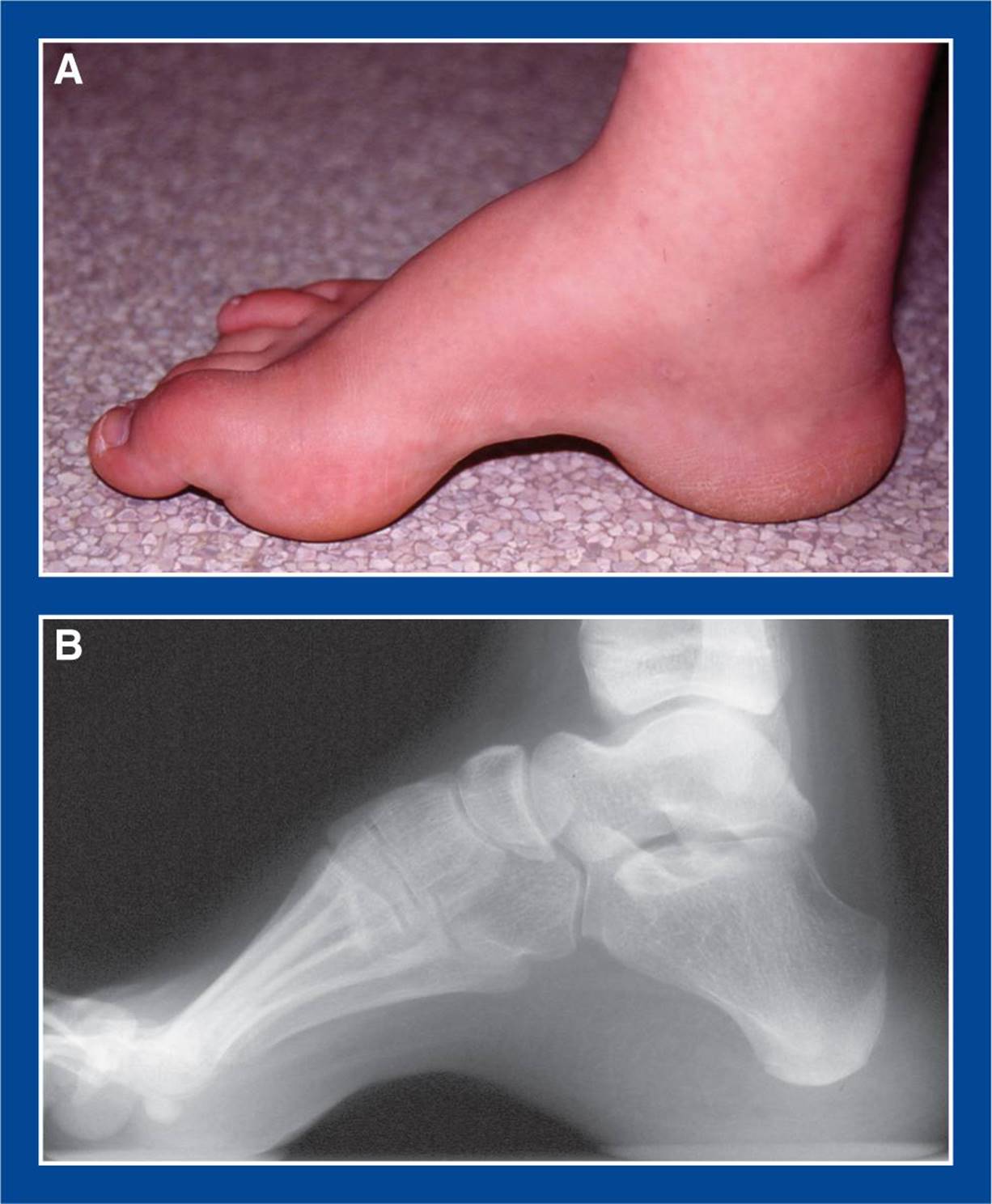
Figure 5-8. Calcaneocavus foot deformity in a teenager with S1 level myelomeningocele. A. Medial photo of foot shows exaggerated arch height across the entire midfoot. Though not visible in this photo, the hindfoot/subtalar joint is in neutral alignment. The soft tissues under the MT heads and the calcaneus are thick and callused. B. Standing lateral radiograph shows transtarsal cavus with relative parallelism of all MTs. In a cavovarus foot by contrast, the 1st MT would be hyperplantar flexed in relation to the 5th MT (Figure 3-25). The calcaneus, in this foot, is hyperdorsiflexed.
7. Operative treatment with reference to the surgical techniques section of the book for each individual procedure
a. Posterior calcaneus dorsal and posterior displacement osteotomy (see Chapter 8)
i. with plantar fasciotomy (see Chapter 7)
ii. with possible anterior tibialis tendon lengthening
b. Midfoot wedge resection/arthrodesis—perform this for the most severe and rigid cases (see Chapter 8)