
Congenital and Acquired Short Heel Cord
1. Definition—Deformity
a. Congenital or acquired contracture of the gastrocnemius or triceps surae (gastrocnemius and soleus) in an otherwise normal child with normal nerves, muscles, and bones (Figure 5-1)
b. Acquired contracture of the gastrocnemius or triceps surae (gastrocnemius and soleus) in a child with a neuromuscular disorder
c. Often associated with other idiopathic and acquired deformities
2. Elucidation of the segmental deformities
a. Ankle—plantar flexed (equinus)
i. Inability to dorsiflex the ankle to at least 10° above neutral with the subtalar joint held in neutral alignment (see Assessment Principle #12, Figure 3-13, Chapter 3)
• If it is possible to achieve 10° of dorsiflexion with the knee flexed but not with it extended, the gastrocnemius alone is contracted.
• If it is not possible to achieve 10° of dorsiflexion regardless of whether the knee is flexed or extended, the triceps surae (gastrocnemius and soleus) is contracted.
3. Imaging
a. None absolutely necessary
b. Standing anteroposterior (AP) and lateral of foot (optional)
c. Standing AP, lateral, and mortis of ankle (optional)
4. Natural history
a. Although never formally studied, congenital contracture of the gastrocnemius muscle or the triceps surae probably persists
b. Acquired contractures generally increase in severity or persist at an unacceptable degree
5. Nonoperative treatment
a. Accept it
b. Wear high heels
c. Twice (or more) daily heel cord stretching exercises along with nighttime dorsiflexion maintenance bracing
d. Serial short-leg stretching casts—for children up to around 5 years of age—followed by nighttime dorsiflexion maintenance bracing
6. Operative indications
a. Failure of nonoperative treatment to achieve and maintain at least 10° of ankle dorsiflexion above neutral with the subtalar joint in neutral alignment and the knee extended, if this lack of flexibility causes:
i. pain under the metatarsal (MT) heads,
ii. pain along the Achilles musculotendinous continuum, and/or
iii. functional disability with gait disturbance.
7. Operative treatment with reference to the surgical techniques section of the book for each individual procedure
a. Gastrocnemius recession (see Chapter 7)—perform this for an isolated contracture of the gastrocnemius, based on the Silfverskiold test (see Assessment Principle #12, Figure 3-13,Chapter 3)
b. Tendo-Achilles Lengthening (TAL) (see four techniques in Chapter 7)—perform this for contracture of the triceps surae (gastrocnemius and soleus), based on the Silfverskiold test (see Assessment Principle #12, Figure 3-13, Chapter 3). The considerations for which technique to use are elucidated in Chapter 7.
i. Percutaneous triple cut
ii. Mini-open double cut slide
iii. Open double cut slide
iv. Open Z-lengthening
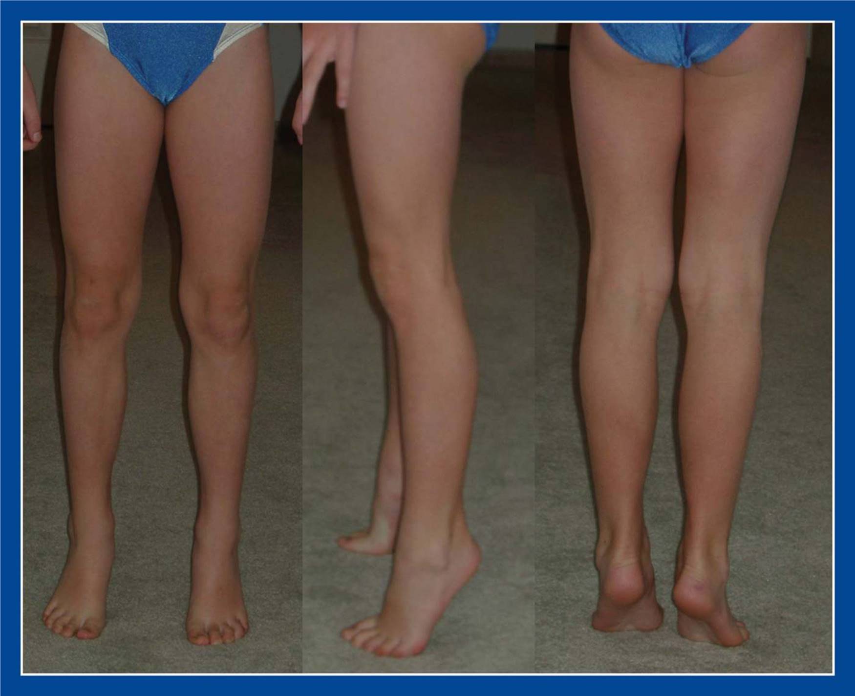
Figure 5-1. Toe-standing/walking due to congenital contracture of the gastrocnemius muscles in an otherwise normal child.
Positional Calcaneovalgus Deformit
1. Definition—Deformity
a. Congenital positional hyperdorsiflexion and valgus deformity of the hindfoot (Figure 5-2A)
b. Differential diagnosis is (Figure 5-2):
i. Congenital vertical/oblique talus
ii. Posteromedial tibial bowing
iii. Paralytic calcaneus deformity
2. Elucidation of the segmental deformities
a. Forefoot—neutral
b. Midfoot—neutral
c. Hindfoot—valgus/everted or neutral
d. Ankle—dorsiflexed (calcaneus)
3. Imaging
a. None, unless physical examination findings are equivocal
4. Natural history
a. 100% of these correct completely without intervention
5. Nonoperative treatment
a. None
b. Parents can be instructed to perform daily plantar flexion stretching exercises. It might not make any difference in the rate of correction of the deformity, but it does no harm. Formal physical therapy is notindicated!
6. Operative indications
a. None
7. Operative treatment with reference to the surgical techniques section of the book for each individual procedure
a. Not applicable
Acquired Calcaneus Deformity
1. Definition—Deformity
a. Calcaneus (hyperdorsiflexion) deformity of the ankle due to a weak triceps surae and a strong anterior tibialis (Figure 5-3)
b. Due to:
i. static muscle imbalance
• myelomeningocele, lipomeningocele, postpoliomyelitis
ii. acquired muscle imbalance
• tethered cord in myelomeningocele, lipomeningocele
iii. surgical overlengthening and/or weakening of the triceps surae, as in cerebral palsy and multiply operated clubfoot

Figure 5-2. Differential diagnosis for positional calcaneovalgus deformity. A. Positional calcaneovalgus. The longitudinal arch is present and the forefoot can be further plantar flexed on the hindfoot with gentle manipulation. Full passive ankle plantar flexion is not possible at birth. B. CVT. The longitudinal arch cannot be created by passive plantar flexion of the forefoot on the hindfoot. C. Posteromedial tibial bowing photo and x-ray. The deformity is actually in the tibia. The foot is well-shaped and flexible. D. Paralytic calcaneovalgus in a child with myelomeningocele. Weak/absent plantar flexors are noted, and there is the obvious lesion at the base of the spine.

Figure 5-3. A. Medial view of a calcaneus foot deformity in a child with myelomeningocele. The heel pad is large, thick, and callused from excessive load-bearing. B. Matching lateral x-ray. C. Plantar view of the foot showing the large, thick, and callused heel pad, but less than normal callus formation under the MT heads.
2. Elucidation of the segmental deformities
a. Ankle—dorsiflexion (calcaneus)
3. Imaging
a. Standing AP and lateral of foot (Figure 5-3)
b. AP, lateral, and mortis of ankle
4. Natural history
a. Persistence of deformity, with
i. progressive increase in callus formation on the plantar aspect of the heel
ii. eventual fissuring of the hypertrophic and callused skin of the heel pad
iii. ultimately, plantar heel ulceration
iv. Pain is rarely a clinical problem because this deformity occurs most commonly in children with myelomeningocele and lipomeningocele, who have insensate skin.
b. Progressive increase in crouched gait with gradual decrease in walking endurance. The underlying triceps surae muscle weakness, compounded by increased body weight with advancing age, leads to lever arm dysfunction (see Basic Principle #7, Figure 2-10, Chapter 2).
c. Poor brace integrity with rapid failure of the plastic at the ankle of the ankle-foot-orthotic (AFO)
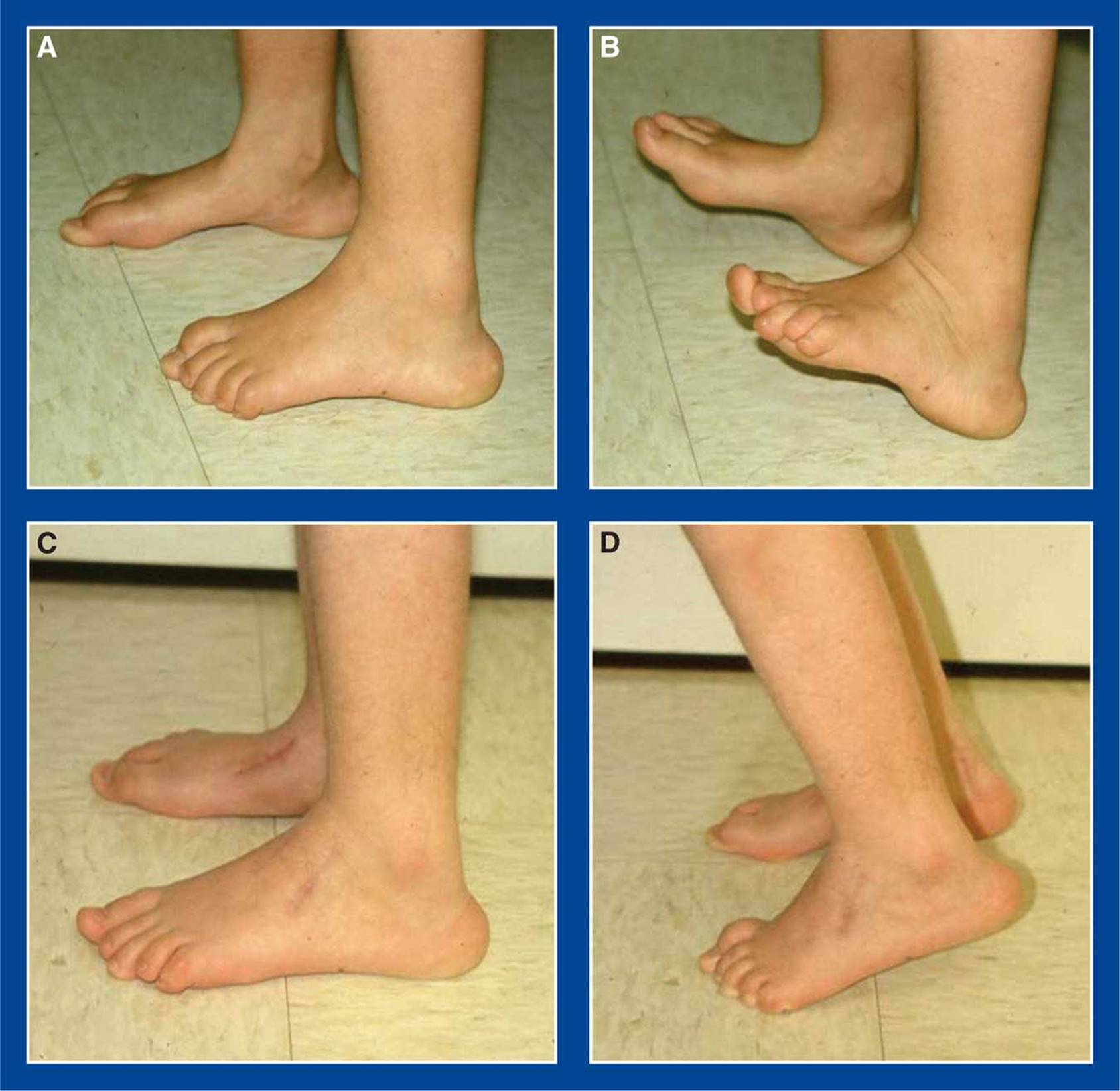
Figure 5-4. Child with L5 level myelomeningocele and calcaneus foot deformities. A. Preop lateral photos. Note the large, callused heel pads. B. She can easily heel stand, but cannot toe stand. C. One year after transfer of her anterior tibialis tendons to her tendo-Achilles, her heel pads are smaller and less callused. The peroneus tertius was released through the dorsolateral incision (not usually required). D. She was able to toe stand, though in only slightly greater than 5° of active plantar flexion. This transfer usually functions as a tenodesis that does not eliminate the need for ankle-foot orthoses, but it improves or eliminates the crouched gait and increases the useful life of the AFOs.
5. Nonoperative treatment
a. Body weight reduction
b. Increase rigidity of the AFO
6. Operative indications
a. Failure of nonoperative attempts to maintain walking endurance and heel skin integrity
7. Operative treatment with reference to the surgical techniques section of the book for each individual procedure
a. Anterior tibialis tendon transfer to the tendo-Achilles (see Chapter 7, Figure 7-27; Figure 5-4)
Valgus Deformity of the Ankle Joint
1. Definition—Deformity
a. Persistence of neonatal valgus orientation of the ankle joint after age 4 to 5 years (see Assessment Principle #11, Figure 3-12, and Assessment Principle #21, Figure 3-27, Chapter 3)
2. Elucidation of the segmental deformities
a. Ankle—valgus
i. Greater than 4° of valgus orientation of the articular surface of the distal tibia compared with the axis of the tibial shaft after the age of 4 to 5 years
3. Imaging
a. AP, lateral, and mortis of the ankle (see Assessment Principle #21, Figure 3-27, Chapter 3)
4. Natural history
a. Valgus orientation of the ankle joint is normal from birth (actually, from the time of in utero joint formation at 7 to 9 weeks’ gestation) until approximately age 4 to 5 years. The valgus alignment gradually corrects to neutral by that age in most normal children.
b. Valgus orientation of the ankle joint is reclassified as a deformity if it persists after approximately age 4 to 5 years
i. The average lateral distal tibia angle (LDTA) after age 4 to 5 years is 89° (1° of valgus), with the normal range of 86° to 92° (4° of valgus to 2° of varus); therefore, >4° of valgus is abnormal.
c. Congenital valgus orientation of the ankle joint persists as a deformity in:
i. up to 66% of limbs with clubfoot deformity
ii. fibula hemimelia, often as a ball-and-socket joint
iii. essentially all limbs affected by myelomeningocele, lipomeningocele, poliomyelitis, spinal cord tumor or injury, and other lower extremity paralyzing conditions (not cerebral palsy) that affect young children
d. Valgus deformity of the ankle can develop following:
i. injury to the lateral distal tibial physis and/or distal fibula physis
ii. fibula pseudarthrosis in congenital anterolateral bowing of the tibia and fibula, with or without tibial pseudarthrosis and with or without neurofibromatosis
e. Persistent and developmental valgus deformities of the ankle joint can cause:
i. lateral ankle/hindfoot pain from impingement of the lateral malleolus, peroneal tendons, and calcaneus
ii. medial ankle/hindfoot pain from stretch of the medial ankle joint and subtalar joint ligaments
iii. plantar–medial heel pain due to excessive loading on that area of the heel pad
iv. skin pressure irritation and/or pain under the medial malleolus due to weight-bearing on the firm shoe counter or on the hard plastic of an AFO (in children with paralytic conditions)
5. Nonoperative treatment
a. None indicated for asymptomatic cases
b. Over-the-counter, cushioned, semirigid arch supports (Figure 5-31) to invert the neutral subtalar joint into varus to compensate for the valgus deformity of the ankle joint. These are contraindicated if the gastrocnemius or entire triceps surae is contracted.
c. Adjust or modify the padding in an AFO in a child with an underlying paralytic condition
6. Operative indications
a. Failure of nonoperative treatment to relieve the:
i. lateral ankle/hindfoot pain from impingement of the lateral malleolus, peroneal tendons, and calcaneus
ii. medial ankle/hindfoot pain from stretch of the medial ankle joint and subtalar joint ligaments
iii. skin pressure irritation and/or pain under the medial malleolus due to weight-bearing on the shoe counter or the hard plastic of an AFO (in a child with an underlying paralytic condition)
b. Progressive valgus deformity due to injury to the lateral distal tibial physis and/or distal fibula physis
7. Operative treatment with reference to the surgical techniques section of the book for each individual procedure
a. Medial distal tibia guided growth with retrograde medial malleolus screw (see Chapter 8)—perform this in a skeletally immature child
b. Distal tibia and fibula valgus-correction osteotomies (see Management Principle #20, Figures 4-10 and 4-12, Chapter 4), (see Chapter 8)—perform this in a skeletally mature adolescent
c. Resection and fat grafting of the physeal bar (if appropriate) with or without concurrent distal tibia and fibula valgus-correction osteotomies (see Chapter 8)—perform this in a skeletally immature child with a small physeal bar
d. Completion of the distal tibial and fibula growth arrests (epiphysiodeses) with concurrent distal tibia and fibula valgus-correction osteotomies (see Chapter 8)—perform this in a skeletally immature child with a large, irresectable physeal bar
Valgus Deformity of the Ankle Joint and the Hindfoot
1. Definition—Deformities
a. Valgus orientation of the ankle joint after age 4 to 5 years (see Assessment Principle #11, Figure 3-12, and Assessment Principle #21, Figure 3-27, Chapter 3) and
b. Valgus deformity of the hindfoot, with or without eversion of the subtalar joint, as seen in:
i. Idiopathic flatfoot
ii. Congenital vertical talus (CVT)
iii. Congenital oblique talus (COT)
iv. Skewfoot
v. Tarsal coalition
vi. Congenital talocalcaneal synostosis associated with
• fibula hemimelia
• tibial hemimelia
• lower extremity hemiatrophy
• other syndromes and chromosome abnormalities
vii. Overcorrected clubfoot
• translational
• rotational
2. Elucidation of the segmental deformities
a. Hindfoot—valgus or valgus/eversion
b. Ankle—valgus
i. Greater than 4° of valgus orientation of the articular surface of the distal tibia compared with the axis of the tibial shaft after the age of 4 to 5 years
3. Imaging
a. Standing AP, lateral, and Harris axial views of the foot (see Assessment Principle #18, Figures 3-20, and 3-24, Chapter 3)
b. AP, lateral, and mortis of the ankle (see Assessment Principle #21, Figure 3-27, Chapter 3)
4. Natural history
a. Valgus orientation of the ankle joint is normal from birth (actually, from the time of in utero joint formation at 7 to 9 weeks’ gestation) until approximately age 4 to 5 years. The valgus alignment gradually corrects to neutral by that age in most normal children.
i. Valgus orientation of the ankle joint is reclassified as a deformity if it persists after approximately age 4 to 5 years
• The average LDTA after age 4 to 5 years is 89° (1° of valgus), with the normal range of 86° to 92° (4° of valgus to 2° of varus); therefore, >4° of valgus is abnormal.
ii. Congenital valgus orientation of the ankle joint persists as a deformity in:
• up to 66% of limbs with clubfoot deformity
• fibula hemimelia, often as a ball-and-socket joint
• essentially all limbs affected by myelomeningocele, lipomeningocele, poliomyelitis, spinal cord tumor or injury, and other lower extremity paralyzing conditions (not cerebral palsy) that affect young children
iii. Valgus deformity of the ankle can develop following:
• injury to the lateral distal tibial physis and/or distal fibula physis
• fibula pseudarthrosis in congenital anterolateral bowing of the tibia and fibula, with or without tibial pseudarthrosis and with or without neurofibromatosis
iv. Persistent and developmental valgus deformities of the ankle joint can cause:
• lateral ankle/hindfoot pain from impingement of the lateral malleolus, peroneal tendons, and calcaneus
• medial ankle/hindfoot pain from stretch of the medial ankle joint and subtalar joint ligaments
• plantar–medial heel pain due to excessive loading on that area of the heel pad
• skin pressure irritation and/or pain under the medial malleolus due to weight-bearing on the firm shoe counter or on the hard plastic of an AFO (in children with paralytic conditions)
b. Valgus deformity of the hindfoot, with or without eversion of the subtalar joint, can cause axial loading pain under the head of the talus and/or impingement-type pain in the sinus tarsi area
5. Nonoperative treatment
a. None indicated for asymptomatic cases
b. Over-the-counter, cushioned, semirigid arch supports (Figure 5-31) to invert the subtalar joint into varus to correct the subtalar valgus and to attempt to compensate for the valgus deformity of the ankle joint. These are contraindicated if the gastrocnemius or entire triceps surae is contracted.
c. Adjust or modify the padding in an AFO in a child with an underlying paralytic condition
6. Operative indications
a. Failure of nonoperative treatment to relieve the:
i. lateral ankle/hindfoot pain from impingement of the lateral malleolus, peroneal tendons, and calcaneus
ii. medial ankle/hindfoot pain from stretch of the medial ankle joint and subtalar joint ligaments
iii. skin pressure irritation and/or pain under the medial malleolus due to weight-bearing on the shoe counter or the hard plastic of an AFO (in a child with an underlying paralytic condition)
iv. axial loading pain under the head of the talus and/or impingement-type pain in the sinus tarsi area
b. Progressive valgus deformity due to injury to the lateral distal tibial physis and/or distal fibula physis
7. Operative treatment with reference to the surgical techniques section of the book for each individual procedure
a. Correct the ankle valgus first. There is only one, easy-to-assess, stable anatomic alignment of the ankle joint (see Management Principle #23-6, Chapter 4).
i. Medial distal tibia guided growth with retrograde medial malleolus screw (see Chapter 8)—perform this in a skeletally immature child
ii. Distal tibia and fibula valgus-correction osteotomies (see Chapter 8)—perform this in a skeletally mature adolescent
iii. Resection and fat grafting of the physeal bar (if appropriate) with or without concurrent distal tibia and fibula valgus-correction osteotomies (see Chapter 8)—perform this in a skeletally immature child with a small physeal bar
iv. Completion of the distal tibial and fibula growth arrests (epiphysiodeses) with concurrent distal tibia and fibula valgus-correction osteotomies (see Chapter 8)—perform this in a skeletally immature child with a large, irresectable physeal bar
b. Once the ankle joint is anatomically aligned, correct the subtalar joint valgus according to the type of valgus present
i. Idiopathic flatfoot—calcaneal lengthening osteotomy (see Chapter 8)
ii. CVT (in the older child)—naviculectomy (see Chapter 8)
iii. COT (in the older child)—calcaneal lengthening osteotomy (see Chapter 8)
iv. Skewfoot—calcaneal lengthening osteotomy (see Chapter 8)
v. Tarsal coalition—calcaneal lengthening osteotomy (see Chapter 8)
vi. Congenital talocalcaneal valgus synostosis associated with fibula hemimelia, tibial hemimelia, lower extremity hemiatrophy, other syndromes and chromosome abnormalities—posterior calcaneus displacement osteotomy (see Chapter 8)
vii. Overcorrected clubfoot
• Translational—posterior calcaneus displacement osteotomy (see Chapter 8)
• Rotational—calcaneal lengthening osteotomy (see Chapter 8)