
Karen C. Hayani
Arthur L. Frank
HIGH-YIELD FACTS
• Influenza usually causes yearly winter epidemics in temperate climates, but new strains that have the potential to cause infrequent but severe worldwide pandemics can arise.
• Influenza is transmitted by inhalation of infected droplets and aerosols (from persons with coughing or sneezing) or by direct contact with contaminated animals or objects.
• Viral shedding of influenza begins 24 hours before the onset of clinical illness and can last for 1 to 2 weeks.
• The most common symptoms in teenagers and adults include fever, cough, headache, sore throat, and malaise. Children may present with atypical symptoms that include lower respiratory tract symptoms. Infants may present with fever or apnea.
• In patients with lower respiratory symptoms, it may be difficult to distinguish primary influenza pneumonia from secondary bacterial pneumonia (both clinically and radiographically).
• Most influenza infections are self-limited and require only supportive care; however, antiviral medications can be considered for children at risk for severe or complicated infections (e.g., immunocompromised or with underlying cardiopulmonary disease), healthy children with severe symptoms, or children with special environmental circumstances (e.g., immunocompromised family members).
• If antiviral medications are started, they should be started within the first 24 to 48 hours of symptoms and given for 5 days. Evidence suggests they shorten the duration of symptoms but it is less clear whether they prevent serious complications, such as viral or secondary bacterial pneumonia.
• The website of the Centers for Disease Control (CDC), www.cdc.gov, is regularly updated, and provides information for parents and patients, and recommendations for physicians (e.g., immunization and antiviral medication information).
The name influenza originated in the fifteenth century from the Italian word influenza, meaning influence. It was thought that the disease was due to adverse astrological influences. Symptomatic influenza infection is commonly called “flu.” Flu can affect individuals of any age.
The virus is a member of the Orthomyxoviridae family, typically spheroid or ovoid in shape, and approximately 80 to 120 nm in diameter (Fig. 63-11 and 63-22). It has multiple segments of single-stranded, negative-sense RNA. Influenza A and B have eight gene segments, whereas influenza C has seven gene segments. The genome is surrounded by an envelope, made of a lipid bilayer, within which are embedded matrix proteins M1 (in both influenza A and B) and M2 (in only influenza A). The M1 proteins are located on the interior surface of the envelope and provide stability to the virus, whereas the M2 proteins serve as ion channels that facilitate uncoating of the virus in the host lysosome. The envelope is studded with glycoproteins, approximately 80% of which are hemagglutinin (HA) and approximately 20% are neuraminidase (NA). HA is the viral molecule that binds to the host sialic acid sugar, enabling attachment of the virus to the host epithelial cell. Once attachment has taken place, the process of endocytosis allows for entry of the virus into the cell. HA is the major stimulus for the host immune response. NA is involved in the release of newly formed virions from the host cell and it prevents reduced infectivity from aggregation of virions. The HA and NA glycoproteins determine the strain of the virus. Minor changes in these antigens are called antigenic drift, which usually occurs on a yearly basis. A major change in these glycoproteins is called antigenic shift, and this can cause worldwide pandemics. Antigenic drift and shift are important in the spread of influenza because new strains are able to evade the host immune system, which may not recognize the virus even after previous exposure to different influenza strains (Table 63-1).
|
TABLE 63-1 |
Terms Associated with Influenza |


FIGURE 63-1. Electron microscopic photograph of influenza virus. (Reproduced with permission from Longo DA, Harrison TR: Harrison’s Principles of Internal Medicine. 18th Ed. New York, McGraw-Hill; 2012.)

FIGURE 63-2. Diagram of influenza virus with key features shown. (Reproduced with permission from Willey J, Sherwood L, Woolverton C: Prescott’s Microbiology, 7th ed. New York, McGraw-Hill; 2008.)
EPIDEMIOLOGY AND TRANSMISSION
Influenza is perhaps the most familiar respiratory infection besides the common cold. The influenza virus is found worldwide, but the origins of many human strains could be traced to Asia, until the 2009 pandemic H1N1 (pH1N1) strain originated in Mexico. Person-to-person transmission, close contact, air travel, and migratory birds all play a role in the global spread of the virus. Each year in the United States, millions of people become symptomatically infected with one of the influenza strains despite massive immunization campaigns. In temperate areas, increased incidence of influenza is seen during winter months secondary to increased crowding indoors and easier transmission from person to person. The virus usually spreads through inhalation of infected droplets or aerosols from a cough or a sneeze but can also spread by direct contact with infected animals or fomites. Once infected, a person usually sheds the virus for 7 days, starting 24 hours prior to the onset of symptoms. Shedding can be prolonged in young children and immunocompromised patients.3 Annual epidemics have often been due to two predominant strains that circulate simultaneously. Since 1977, influenza A strains with the H1N1 and H3N2 serotypes have been present, with either type being predominant during any 1 year. For example, during the 2009 to 2010 season, more than 99% of isolates were the 2009 pandemic influenza A (H1N1) virus, but during the subsequent season, (2010–2011), this strain accounted for only 34% of activity while influenza A (H3N2) was the predominant strain.3 In addition, both influenza A and B viruses can concurrently circulate and other viruses, such as RSV or parainfluenza, may circulate simultaneously with influenza during any single season.4 When there is coinfection with human and animal influenza viruses, the segmented nature of the genome allows for reassortment between the viruses and the emergence of new serotypes with new HA or NA glycoproteins. Since populations lack immunity, worldwide pandemics can result.
PATHOPHYSIOLOGY
When the influenza virus is inhaled, it enters the respiratory epithelium and causes loss of ciliary function, decreased mucus production, and desquamation. This damaged lining of the respiratory tract is a potential portal of entry for secondary bacterial infections. The infection can spread to lower airways by inhalation into the alveoli or by contiguous spread. Systemic infection can occur causing a variety of clinical complications. The host responds with a humoral IgG as well as a mucosal IgA antibody response to the viral HA and NA antigens. Cytotoxic T lymphocytes also play a role.5 The host defenses include production of interleukin-6 and interferon-alpha, which result in many of the systemic clinical symptoms of influenza.4
CLINICAL PRESENTATION
The incubation period of influenza is 2 to 4 days and varies with viral strain and host factors, such as age and underlying conditions. Classically, the onset of symptoms is acute and usually includes fever, chills, headache, sore throat, dry cough, and myalgias. Otitis media and conjunctivitis can also occur. Any part of the respiratory tract can be affected, and although upper respiratory tract symptoms are most common, croup, bronchitis, and pneumonia can also develop. Pneumonia can be either due to the virus itself or due to a secondary bacterial infection and the two are difficult to distinguish. The development of productive cough and lobar consolidation on chest radiograph are suggestive of the latter. In older children, acute calf pain and refusal to walk may indicate benign and self-limited myositis, more often following influenza B than influenza A. Young children may present with fever alone (“rule out sepsis”) or may have gastrointestinal symptoms such as vomiting, abdominal pain, and diarrhea. Infants may present with apnea. Rarely, influenza can cause myocarditis, seizures, encephalopathy, encephalitis, transverse myelitis, or Guillain–Barré syndrome. In Japan, a severe, acute necrotizing encephalopathy due to influenza has been reported in young children. It is associated with rapid progression to seizures and coma.6 Reye syndrome can develop when aspirin is used during influenza infection but the incidence of this syndrome has decreased greatly with the widespread use of acetaminophen or ibuprofen for fever in children. Deaths usually occur due to complications, including secondary bacterial infections. In December 2006 and January 2007, for example, the CDC reported an increased number of deaths in children coinfected with influenza and community-acquired methicillin-resistant Staphylococcus aureus(CA-MRSA) pneumonia and an advisory was issued alerting health care professionals to this trend.7 During the 2009 to 2010 influenza A (H1N1) pandemic, secondary complications of influenza A (pH1N1) that were risk factors for mortality were myocarditis, encephalitis, and clinical diagnosis of early presumed CA-MRSA coinfection of the lung.8
DIFFERENTIAL DIAGNOSIS AND CLINICAL MANAGEMENT
The clinical presentation of influenza overlaps with other respiratory viruses (Table 63-2), especially RSV and human metapneumovirus, which often circulate in the community during the same season. Figure 63-3 summarizes an approach to the management of infants and children who may present to the clinic or emergency department (ED) with influenza.
|
TABLE 63-2 |
Respiratory Viruses to Consider in the Differential Diagnosis of Influenza (With Accompanying Clinical Tips) |


FIGURE 63-3. Management of suspected influenza in children.
LABORATORY AND RADIOGRAPHIC FINDINGS
During winter months in temperate climates, many children seen in the clinic or ED are diagnosed with “viral syndrome” depending on history and physical examination. In many cases, symptoms are mild, supportive care alone is appropriate, and no diagnostic testing is necessary. Making a specific diagnosis with respect to influenza or other respiratory viruses may be helpful and appropriate when patients are infants, have underlying diseases, or have severe symptoms. Specific identification may also be helpful in determining the epidemiology of influenza and other viruses in a community during a certain season. Table 63-3 outlines the features of the laboratory tests most commonly available. In general, nasopharyngeal swabs, aspirates or washes are preferred to throat swabs, and should be obtained within the first 24 to 72 hours of illness if possible.3 These specimens can be submitted for viral culture or rapid influenza-diagnostic tests which detect the influenza virus nucleoprotein by immunologic methods. These rapid tests are commonly commercially available, but vary with respect to the viral strains they detect, and have variable sensitivity (40%–80%) in part depending on the concentration of viral antigens in the sample.9 Influenza A or B polymerase chain reaction (PCR) tests done on nasopharyngeal specimens are the most sensitive and specific and are often part of an extended panel but may not be as widely available. Routine laboratory tests, such as CBC, are usually not needed but may be helpful in patients with lower respiratory tract symptoms. WBC counts of <5000/mm3 are often seen with influenza and other viruses and elevated WBC counts with predominantly polymorphonuclear cells suggest secondary bacterial infection. Serology is not useful acutely but may be helpful if a diagnosis needs to be made in retrospect. Chest radiographs are not routinely indicated, but if done due to suspected lower respiratory tract disease may help distinguish viral from secondary bacterial pneumonia.
|
TABLE 63-3 |
Laboratory Testing in Influenza Virus Infection |

TREATMENT
Most influenza infections require only supportive therapy. This includes fluids, rest, and ibuprofen or acetaminophen for fever. Aspirin (salicylates) should never be given to children with respiratory viral infections, because of the risk of Reye syndrome. If children are already taking aspirin (e.g., for Kawasaki disease), it should be stopped immediately. Over-the-counter decongestants are not recommended, especially in children younger than 2 years because these might be harmful. Several antiviral medications are approved for use in children (Table 63-4).2,10 These antiviral medications should not be given routinely, but can be considered in children who are at risk for severe or complicated infections, such as immunocompromised children or children with underlying cardiopulmonary disease, healthy children with severe symptoms, or children with special environmental circumstances (e.g., close contact with immunocompromised family members).3 Adamantanes (amantadine and rimantadine) and NA inhibitors (oseltamivir and zanamivir) will shorten the duration of symptoms if started within 24 to 48 hours of the onset of symptoms, but prevention of complications of influenza is less well documented in children. If treatment is indicated, the level of antiviral resistance in a community is important in determining which medication to prescribe, and enhanced surveillance for influenza antiviral resistance is ongoing at the CDC in collaboration with local health departments. In January 2006, for example, the CDC recommended that neither amantadine nor rimantadine be used for treatment or prophylaxis, because of a global spread of adamantine resistance.3 Since then, oseltamivir has become the drug of choice, and during the 2010 to 2011 season, only 1.3% of all influenza isolates tested were resistant to oseltamivir. 3 However, yearly fluctuations can occur; for example in February 2008, 8% of US influenza A (H1N1) isolates were oseltamivir resistant.11 Thus, in cases of life-threatening influenza illness, use of two antiviral medications (one from each class of drug) should be considered. In addition, if secondary bacterial pneumonia is suspected, physicians should be aware of the prevalence of CA-MRSA in their communities when choosing empiric antibiotics.7,8
|
TABLE 63-4 |
Antiviral Medications used for Influenza Prophylaxis and Treatmenta |

PREVENTION: IMMUNIZATION AND PROPHYLAXIS
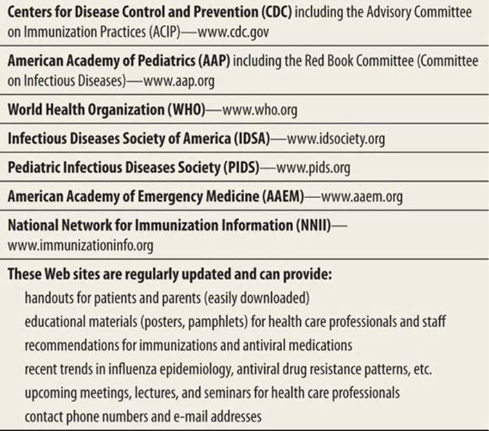
Influenza vaccines are newly formulated every year in the summer depending on predictions of the three most likely strains to circulate the following winter. Each year the trivalent inactivated (TIV) and the live attenuated (LAIV) influenza vaccines are based on the same strains and this usually includes two influenza A strains and one influenza B strain. Usually, some strains are new and some are repeated from vaccines in the previous years. For example, the 2012 to 2013-influenza vaccine formulation contains influenza A/California/7/2009 (H1N1)-like strain, which is repeated from previous vaccines, and two new strains: influenza A/Victoria/361/2011 (H3N2)-like and influenza B/Wisconsin/1/2010 strains.12 Prevention against influenza can also include prophylaxis with antiviral medications in certain circumstances.13The medications approved for prophylaxis in various age groups are outlined in Table 63-4. As the CDC recommendations for vaccination and prophylaxis change frequently depending in part on epidemiological conditions, the CDC website should be consulted. Additional helpful websites for up-to-date information on influenza are listed in Table 63-5.
|
TABLE 63-5 |
Helpful Websites |
REFERENCES
1. Fauci AH, Braunwald E, Kasper DL, et al. Harrison’s Principles of Internal Medicine. 17th ed. New York, NY: McGraw-Hill; 2008.
2. Brooks GF, Butel JS, Morse SA. Jawetz, Melnick & Adelberg’s Medical Microbiology. 24th ed. New York, NY: McGraw-Hill; 2007.
3. American Academy of Pediatrics. Influenza. In: Pickering LK, Long SS, eds. Red Book: 2012 Report of the Committee on Infectious Diseases. 29th ed. Elk Grove Village, IL: American Academy of Pediatrics; 2012:439–453.
4. Dawood FS, Subbarao K, Fiore AE. Influenza viruses. In: Long SS, Pickering LK, Prober CG, eds. Principles and Practice of Pediatric Infectious Diseases. 4th ed. Philadelphia, PA: Elsevier Churchill Livingstone; 2012:1149–1159.
5. Glezen WP. Orthomyxoviridae. In: Feigin RD, Cherry JD, Demmler-Harrison GJ, Kaplan SL, eds. Textbook of Pediatric Infectious Diseases. 6th ed. Philadelphia, PA: Saunders; 2009:2395–2413.
6. Grose C. The puzzling picture of acute necrotizing encephalopathy after influenza A and B virus infection in young children. Pediatr Infect Dis J. 2004;23:253–254.
7. Centers for Disease Control and Prevention Severe methicillin-resistant staphylococcus aureus community-acquired pneumonia associated with influenzaᾰLouisiana and Georgia, December 2006–January 2007. MMWR Morb Mortal Wkly Rep. 2007;56(14):325–329.
8. Randolph AG, Vaughn F, Sullivan R,, et al. Critically ill children during the 2009-2010 influenza pandemic in the United States. Pediatrics. 2011. 128(6):e1450–e1458.
9. Treanor JJ. Influenza viruses, including avian influenza and swine influenza. In: Mandell GL, Bennett JE, Dolin R, eds. Principles and Practice of Infectious Diseases. 7th ed. Philadelphia, PA: Elsevier Churchill Livingstone; 2010:2265–2288.
10. Taketomo CK, Hodding JH, Kraus DM, eds. Pediatric and Neonatal Dosage Handbook. 19th ed. Hudson, OH: Lexi-Comp. Inc; 2012.
11. Centers for Disease Control and Prevention. Update: influenza activity—United States, September 30, 2007–February 9, 2008. MMWR Morb Mortal Wkly Rep. 2008;57:179–183.
12. Centers for Disease Control and Prevention. Prevention and control of influenza with vaccines: recommendations of the Advisory Committee on Immunization Practices (ACIP) – United States 2012-2013 influenza season. MMWR Morb Mortal Wkly Rep. 2012;61(32):613–618.
13. Committee on Infectious Diseases, American Academy of Pediatrics. Recommendations for prevention and control of influenza in children, 2012-2013. Pediatrics. 2012;130:780–792.