
Approach to the Problem
Discoloration around the eye, the periorbital skin and eyelids, and of the eye may result from multiple causes. Children and adults with darker skin pigmentation—those of African, Asian, or Hispanic descent—may also have increased pigmentation of the bulbar conjunctivae. This discoloration is often bilateral, but may be asymmetric and patchy and most noticeable in the interpalpebral area. Conjunctival hyperemia, injection, and inflammation, inclusively referred to as conjunctivitis, can occur from numerous causes, including infection, trauma, allergies, foreign bodies in the eye, reaction to drugs or other toxins, and mechanical irritation from eyelashes and eyelids. Discoloration below the eyes can be seen with “allergic shiners,” which results from venous congestion due to sinus mucosal edema from systemic allergic inflammation. Chalazions are granulomas formed from blocked meibomian glands along the eyelash margin. They are not infectious and do not require antibiotic treatment. Cellulitis, orbital (postseptal) and periorbital (or preseptal), can cause discoloration and swelling of the periorbital skin as well as eyelids and can cause hyperemia of bulbar conjunctivae. In orbital cellulitis, the infection extends past the orbital septum, resulting in eye motility restriction, optic nerve compression and vision loss, and/or secondary extension into the central nervous system. Certain tumors can cause discoloration around and in the eyes. Neuroblastoma, a tumor of embryonic sympathetic neuroblasts typically originating in the abdomen, can cause unilateral or bilateral proptosis, periorbital swelling, and lid or infraorbital ecchymosis as part of the classic presentation for orbital metastasis. Rhabdomyosarcoma, the most common primary malignant orbital tumor in children, may present with rapidly progressive proptosis and eyelid redness not associated with localized warmth or fever, distinguishing it from cellulitis. Capillary hemangiomas are vascular orbital tumors of proliferating capillary endothelial cells. They can cause a bluish discoloration of the overlying skin or the classic strawberry mark. A nevus flammeus, or port-wine stain, which can be seen in Sturge–Weber syndrome, is a vascular malformation of dilated capillaries causing a reddish stain to the skin. Melanocytic lesions can affect conjunctiva as well as surrounding tissues of the eye and include conjunctival nevus, congenital nevocellular nevus of skin, and oculodermal melanocytosis or nevus of Ota. Conjunctival nevi may be flat or elevated and are typically brown and commonly found at the temporal and nasal limbi. Congenital nevocelluar nevus of skin, which occurs on eyelids, has potential for malignant transformation. Ocular and oculodermal melanocytosis, or nevus of Ota, causes slate-gray or bluish discoloration to sclera, and can involve eyelid and adjacent skin producing a brown, bluish, or black discoloration. Icterus is a yellow discoloration of the sclera that occurs with jaundice and results from bilirubin binding to the bulbar conjunctiva. In some connective tissue disorders such as Ehler–Danlos syndrome, Marfan syndrome, and osteogenesis imperfecta, the sclera may appear blue because of the underlying blue-brown uveal tissue (retina and ciliary body) showing through the thinner and more transparent sclera. In blunt trauma to the eye, there can be significant swelling and bruising that affect surrounding tissues.
Key Points in the History
• Itching and tearing are the hallmark of allergic eye disease. Patients will often report other allergy symptoms of rhinorrhea and nasal congestion, and a history of other atopic diseases such as asthma and eczema. A family history of atopic disease may also be reported.
• Viral conjunctivitis usually occurs in epidemic outbreaks in schools and child care settings. It may also be associated with concurrent or recent viral upper respiratory tract infections.
• Periorbital cellulitis may follow superficial eyelid trauma, conjunctivitis, skin infection, or upper respiratory tract or sinus infection.
• Orbital cellulitis in children is most commonly associated with sinus disease. A history of rapid onset of swelling, blurred vision, and double vision is common.
• Capillary hemangiomas are typically not evident at birth, but exhibit rapid growth over the first several months to about 6 to 9 months of age. After the first year of life, they show signs of regression at a variable rate.
• Vision loss, eye pain, or double vision may be reported with trauma to the eye and its surrounding tissues.
• Poor and/or double vision may occur with infiltrative disease of the orbit and extraocular muscles.
Key Points in the Physical Exam
• Visual acuity should always be checked and documented with age appropriate tools when possible.
• Preauricular lymphadenopathy may be seen with viral conjunctivitis.
• If bullous mucosal lesions are seen with conjunctivitis, consider Stevens–Johnson syndrome in the differential diagnosis.
• Proptosis, decreased ocular movement, decreased vision, and/or abnormal pupillary response to light suggest orbital involvement and may help to distinguish orbital from periorbital cellulitis. In periorbital cellulitis, the periorbital skin becomes taut and inflamed, but there is no orbital involvement.
• The ecchymotic discoloration associated with metastatic neuroblastoma can precede diagnosis of the primary tumor, which is typically found in the thorax or abdomen.
• Computed tomography (CT) or magnetic resonance imaging (MRI) may be needed to distinguish hemangioma from plexiform neurofibroma, lymphatic malformation, and rhabdomyosarcoma, which all can exhibit rapid growth and proptosis.
• In evaluating traumatic injuries, it is important to rule out any intraocular injuries that may be present. Presence of subconjunctival hemorrhage along with hyphema raises concern for ruptured globe.
|
PHOTOGRAPHS OF SELECTED DIAGNOSES |
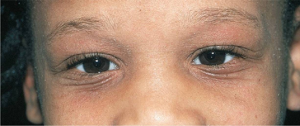
Figure 14-1 Allergic shiners. Note the darkening of the periorbital areas, the lichenification, and the characteristic double fold (Dennie–Morgan fold) that extends from the inner to the outer canthus of the lower eyelid. (Used with permission from Goodheart HP. Goodheart's Photoguide of Common Skin Disorders. 2nd ed. Philadelphia, PA: Lippincott Williams & Wilkins; 2003:48.)
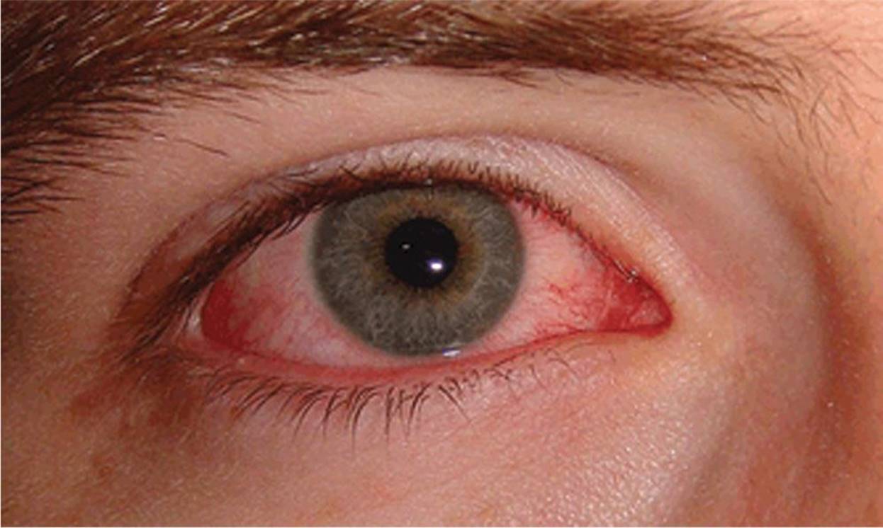
Figure 14-2 Conjunctivitis, viral. (Used with permission from McDonagh DO, Lereim I, Micheli LJ, et al. FIMS Sports Medicine Manual. Philadelphia, PA: Lippincott Williams & Wilkins; 2011.)
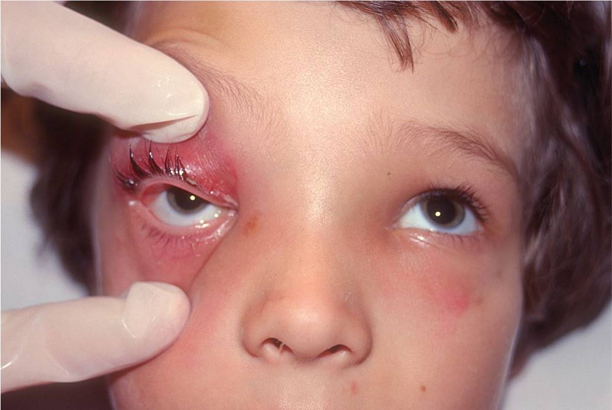
Figure 14-3 Periorbital cellulitis. Note the absence of proptosis and symmetric upward gaze. (Courtesy of James W. McManaway, III, MD, Hershey Pediatric Ophthalmology Associates.)

Figure 14-4 Orbital cellulitis. (Courtesy of the Penn State Hershey Eye Center.)
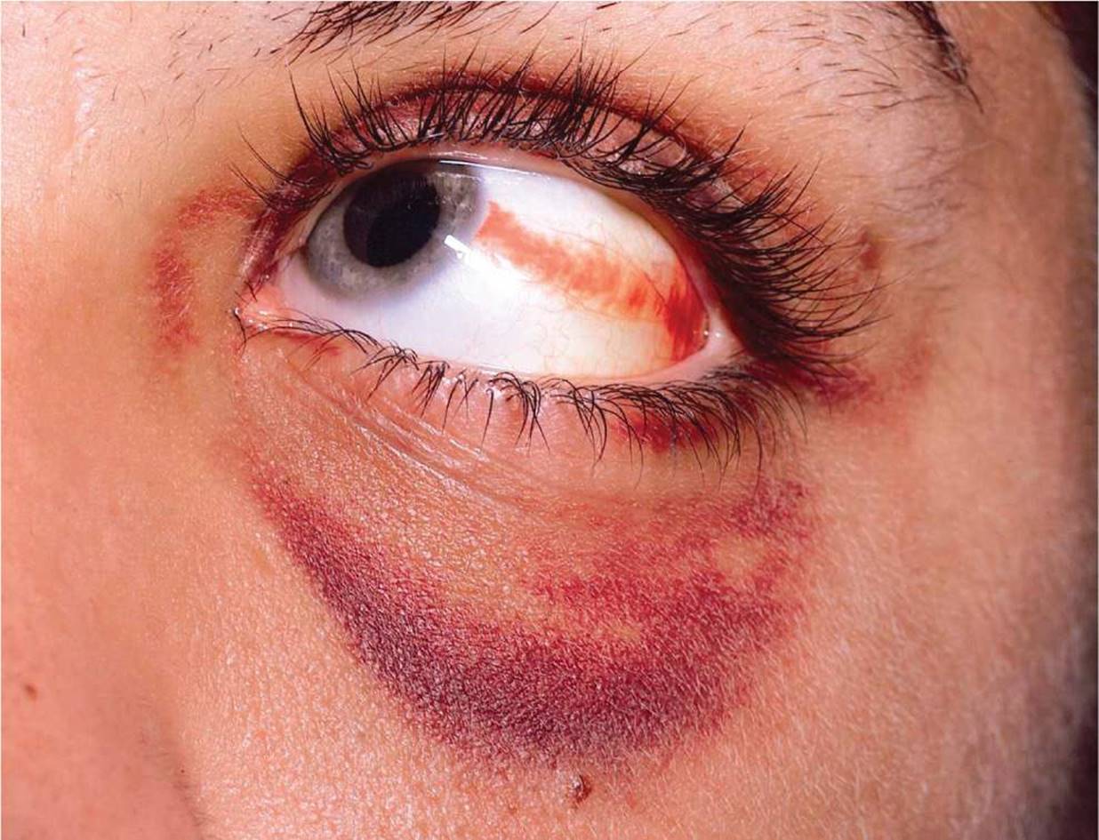
Figure 14-5 Subconjunctival hemorrhage and black eye from trauma. (Courtesy of Dean John Bonsall, MD, MS, FACS.)
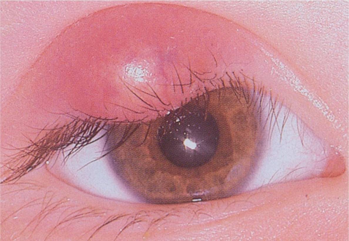
Figure 14-6 Chalazion. This child has a red, elevated, erythematous nodular mass located on lid margin. (Used with permission from Berg D, Worzala K. Atlas of Adult Physical Diagnosis. Philadelphia, PA: Lippincott Williams & Wilkins; 2006.)
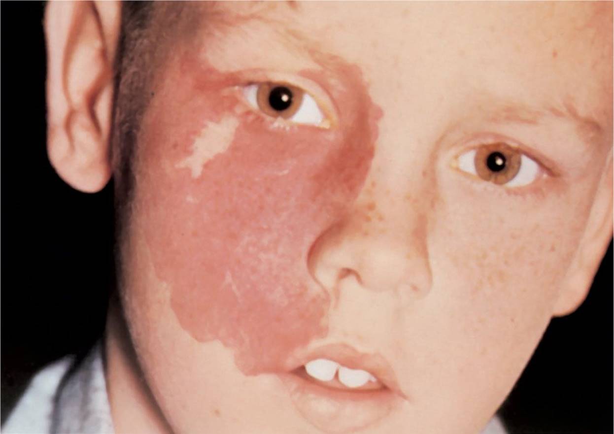
Figure 14-7 Nevus flammeus (port-wine stain). This child has a large, irregular, macular patch along the right side of his face that has the dark red appearance of port-wine stain. (Used with permission from Weber J, Kelley J. Health Assessment in Nursing. 2nd ed. Philadelphia, PA: Lippincott Williams & Wilkins; 2003.)

Figure 14-8 Nevus of Ota. This child has blue/gray macular patch involving most of their sclera and around the eyelid consistent with nevus of Ota. (Courtesy of Dean John Bonsall, MD, FACS.)
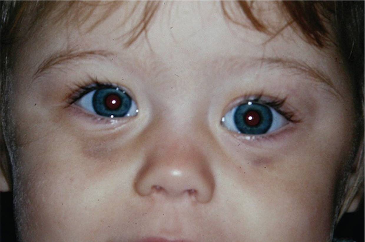
Figure 14-9 Metastatic neuroblastoma. (Courtesy of Julia L. Stevens, MD, University of Kentucky.)
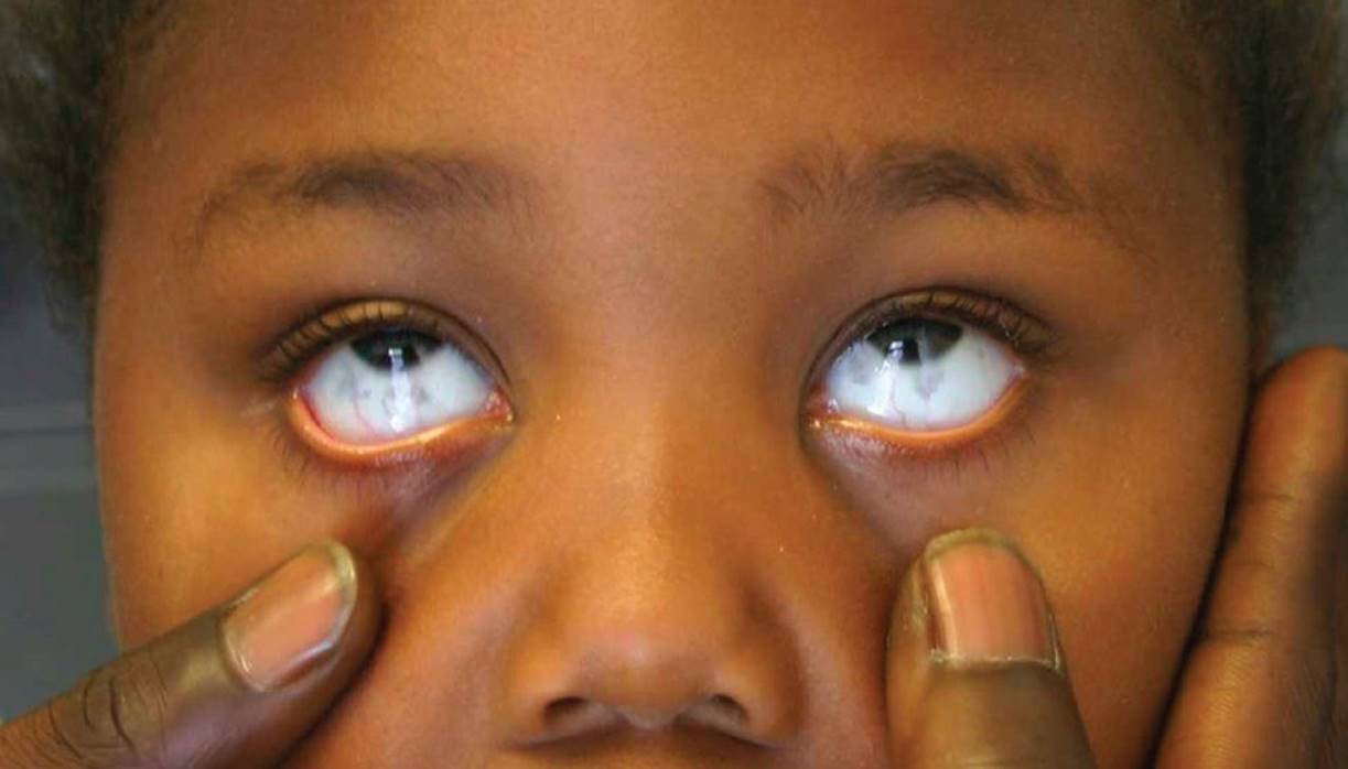
Figure 14-10 Scleral epithelial melanosis. (Courtesy of Julie A. Boom, MD.)
DIFFERENTIAL DIAGNOSIS


Other Diagnoses to Consider
• Uveitis
• Episcleritis/scleritis
• Other orbital tumors, including metastatic disease or extraocular extension of intraocular tumors (retinoblastoma)
• Dacryocystitis
• Dacryoadenitis
• Mucocele
• Encephalocele
When to Consider Further Evaluation or Treatment
• Eye discoloration associated with decreased vision should be evaluated by an ophthalmologist promptly.
• Scleral icterus should be evaluated with serum total and direct bilirubin levels, and underlying causes of jaundice should be considered.
• Children with periorbital cellulitis may be treated with oral systemic antibiotics and closely monitored. If there are no signs of improvement within 48 to 72 hours, CT imaging and changing to IV antibiotics should be considered.
• Children with suspected orbital cellulitis should have an orbital CT and started on systemic antibiotics. Otolaryngology and/or ophthalmology should be consulted for evaluation of possible surgical drainage.
• Children with suspected orbital disease should be evaluated with CT and/or MRI.
• Suspected connective tissue diseases should be referred to genetics for further evaluation.
SUGGESTED READINGS
Helveston EM, Ellis FD. Pediatric Ophthalmology Practice. 2nd ed. St Louis, MO: Mosby; 1984.
Nelson LB, Olitsky SE, Harley RD, eds. Harley's Pediatric Ophthalmology. 5th ed. Philadelphia, PA: Lippincott Williams & Wilkins; 2005:201–216, 367–419.
Shields CL, Fasiudden A, Mashayekhi A, et al. Conjunctival nevi: Clinical features and natural course in 410 consecutive patients. Arch Ophthalmol. 2004;122:167–175.
Traboulsi E. Pediatric ophthalmology. In: McMillan JA, ed. Oski's Pediatrics Principles and Practice. 4th ed. Philadelphia, PA: Lippincott Williams & Wilkins; 2006:801–827.
Wright KW, Spiegel PH, eds. Pediatric Ophthalmology and Strabismus. 2nd ed. New York, NY: Springer; 2003.