
Approach to the Problem
There are various abnormalities of the pupils, iris, and lens in children. In most cases, timely diagnosis and management are critical. The assessment of visual acuity is the most integral facet of the ophthalmologic examination. More than half of the visual abnormalities in children are first discerned by their primary care physician. Many diagnoses, such as leukocoria, require prompt referral to an ophthalmologist. When in doubt, referral is a prudent approach in managing many of these diagnoses.
Key Points in the History
• Congenital cataracts are associated with intrauterine infections, such as congenital rubella and congenital varicella syndrome, and metabolic disorders. Congenital cataracts may be associated with Down, Edward, and Turner syndromes.
• One third of cataracts are hereditary, and nearly one third of cataracts in children have no identifiable etiology.
• Brushfield spots occur in up to 85% to 90% of children with Down syndrome, but they may be seen in normal children as well.
• Colobomas may occur in normal children or as part of genetic syndromes, such as CHARGE syndrome.
• Iritis and uveitis raise suspicion for conditions associated with systemic inflammation as in juvenile idiopathic arthritis (JIA).
• Hyphemas are often the result of blunt trauma to the globe.
Key Points in the Physical Examination
• Small, centrally located cataracts are often clinically stable without an impact on vision.
• Leukocoria, or a white pupillary reflex, is an important clinical sign of intraocular tumors, such as retinoblastoma. Retinoblastoma is the leading malignant ocular tumor in children.
• Leukocoria is bilateral in 30% to 40% of cases.
• It is important to rule out scleral rupture or the presence of a foreign body when chemosis is present.
• Kaiser–Fleischer rings are rims of brown-green pigment in the cornea. Although occasionally visible to the naked eye, slit lamp examination is sometimes necessary to visualize these rings.
• Iritis is characterized by pain, tearing, photophobia, and decreased visual acuity. Symptoms may be acute and develop rapidly over 1 to 2 days. Iritis may be asymptomatic in children with rheumatologic disease such as JIA.
• Blunt traumatic injuries to the eye warrant an inspection of the anterior chamber, the space between cornea and iris, for hyphemas.
• Small hyphemas require slit lamp examination, whereas larger ones may be visible to the naked eye.
• When blood pools in the inferior portion of the eye from a hyphema, it often causes elevated intraocular pressure and decreased visual acuity.
|
PHOTOGRAPHS OF SELECTED DIAGNOSES |

Figure 15-1 Aniridia. This photograph shows a child with bilateral aniridia. (Courtesy of Brian Forbes, MD.)

Figure 15-2 Aniridia. (Courtesy of Sophia M. Chung, MD.)
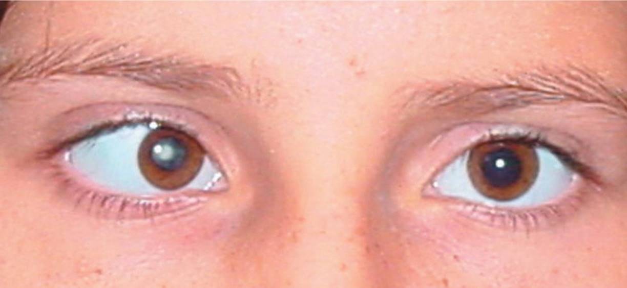
Figure 15-3 Cataract. Note the central haze in the right eye of this patient. (Courtesy of Brian Forbes, MD.)

Figure 15-4 Bilateral central cataracts. (Courtesy of Brian Forbes, MD.)

Figure 15-5 Leukocoria. (Used with permission from Rubin E, Farber JL. Pathology. 3rd ed. Philadelphia, PA: Lippincott Williams & Wilkins; 1999:765.)
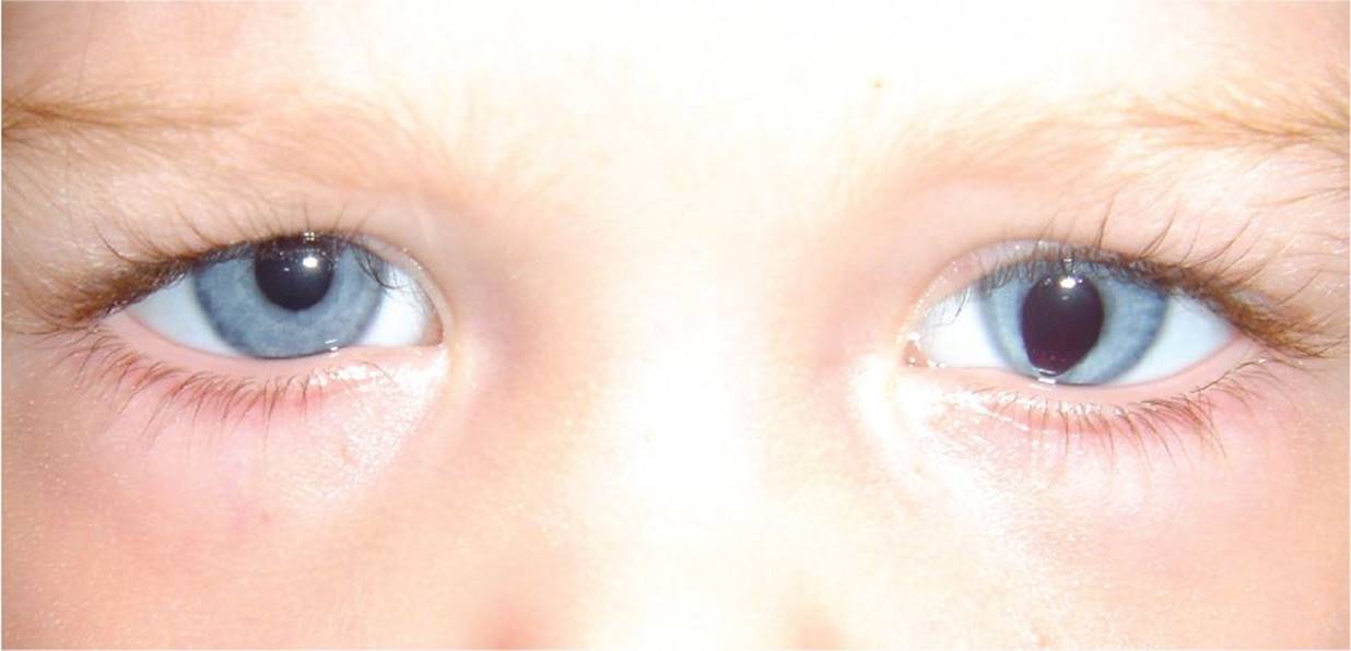
Figure 15-6 Coloboma in the left eye. (Courtesy of Brian Forbes, MD.)

Figure 15-7 Heterochromia iridium. (Courtesy of Brian Forbes, MD.)

Figure 15-8 Iritis. (Used with permission from Harwood-Nuss AL, Wolfson AB, Linder CH, et al. The Clinical Practice of Emergency Medicine. 3rd ed. Philadelphia, PA: Lippincott Williams & Wilkins; 2001:66.)
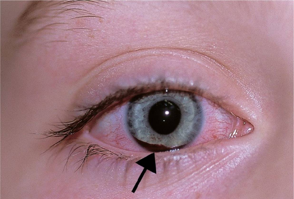
Figure 15-9 Hyphema. This 7-year-old girl was struck by a hard rubber ball and presented with blurred vision. The 1-mm hyphema was only visible when she was upright. (Used with permission from Fleisher GR, Ludwig S, Baskin MN. Atlas of Pediatric Emergency Medicine. Philadelphia, PA: Lippincott Williams & Wilkins; 2004:403.)
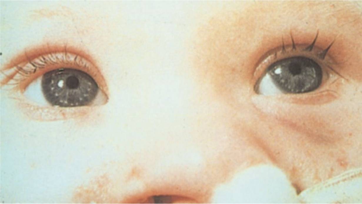
Figure 15-10 Brushfield spots. (Used with permission from Bickley LS, Szilagyi P. Bates’ Guide to Physical Examination and History Taking. 8th ed. Philadelphia, PA: Lippincott Williams & Wilkins; 2003:771.)

Figure 15-11 Kaiser–Fleischer ring. (Used with permission from Tasman W, Jaeger E. The Wills Eye Hospital Atlas of Clinical Ophthalmology. 2nd ed. Philadelphia, PA: Lippincott Williams & Wilkins; 2001:466.)
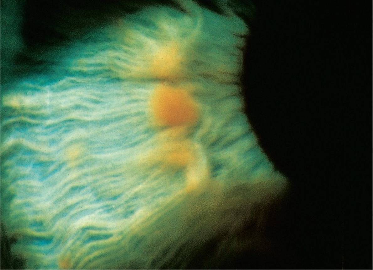
Figure 15-12 Lisch Nodule. Lisch nodule of iris characterized by its elevation, smooth contour, and soft translucency. (Used with permission from Gold DH, Weingeist TA. Color Atlas of the Eye in Systemic Disease. Baltimore, MD: Lippincott Williams & Wilkins; 2001).

Figure 15-13 Iris Melanoma. (Used with permission from Tasman W, Jaeger E. The Wills Eye Hospital Atlas of Clinical Ophthalmology. 2nd ed. Philadelphia, PA: Lippincott Williams & Wilkins, 2001.)
DIFFERENTIAL DIAGNOSIS


Other Diagnoses to Consider
• Pupil abnormalities:
• Horner syndrome
• Adie syndrome
• Third cranial nerve palsy
• Persistent pupillary membrane
• Lens abnormalities:
• Lens subluxation
• Dystrophy
• Cystinosis
• Lenticular myopia
• Iris abnormalities:
• Ocular albinism
• Iridodialysis
• Cyclodialysis
When to Consider Further Evaluation or Treatment
• Leukocoria is a diagnosis warranting immediate attention and referral as it may result in visual impairment and may be life threatening.
• Retinoblastoma, the most common intraocular tumor in childhood, typically presents with leukocoria. Mean age at diagnosis is 12 months for bilateral and 24 months for unilateral tumors.
• Iritis and uveitis require prompt attention and a slit lamp examination. In addition, patients should be evaluated for other systemic diseases to determine the cause of inflammation.
• An iris nevus that changes shape, size, or thickness requires prompt attention as iris nevi and melanomas are the most common primary tumors of the iris.
SUGGESTED READINGS
Halder S, Oureshia W, Ali A. Leukocoria in children. J Pediatr Ophthalmol Strabismus. 2008;45(3):179–180.
Krishnamurthy R, VanderVeen DK. Infantile cataracts. Inter Ophthalmol Clin. 2008;48(2):175–192.
Lai JC, Ferkat S, Barron Y, et al. Traumatic hyphema in children: Risk factors for complications. Arch Ophthalmol. 2001;119(1):64–70.
McLaughlin JP, Fung AT, Shields JA, Shields CL. Iris melanoma in children: Current approach to management. Oman J Ophthalmol. 2013;6:53–55.
Melamud A, Palekar R, Singh A. Retinoblastoma. Am Fam Physician. 2006;73(6):1039–1044.
Patel H. Pediatric uveitis. Pediatr Clin North Am. 2003;50:125–136.
Savar A, Cestari DM. Neurofibromatosis type I: Genetics and clinical manifestations. Semin Ophthalmol. 2008;23(1):45–51.
Tasman W, Jaeger E. The Wills Eye Hospital Atlas of Clinical Ophthalmology. 2nd ed. Philadelphia, PA: Lippincott Williams & Wilkins; 2001:466.