
Approach to the Problem
Acute otitis media (AOM) is one of the most common diagnoses and reasons for antibiotic prescriptions in children. With more than 5 million cases diagnosed annually, it is associated with individual discomfort, family disruption, financial costs, serious sequelae, and antimicrobial resistance. For these reasons, it is important to make the correct diagnosis when evaluating the tympanic membrane (TM).
Pneumatic otoscopy allows the visualization of TM characteristics: color, contour (normal, retracted, full, bulging), position, and mobility. A normal TM is described as translucent, pearly gray, and mobile. A light reflex and boney landmarks, such as the arm of the malleus, are generally easily viewed. The examination requires that the child be restrained or held still and have patent and clear ear canal. Also, a pneumatic otoscope with a good seal and light source must be available.
Key Points in the History
• Acute onset, hyperpyrexia, and otalgia are features of AOM and not otitis media with effusion (OME).
• Concomitant or recent upper respiratory tract infections or allergies are commonly seen with AOM and OME.
• Hearing loss is a nonspecific finding that may be caused by middle ear (ME) fluid (AOM, OME), as well as by structural damage of the TM or ossicles (severe tympanosclerosis, TM perforation, or cholesteatoma).
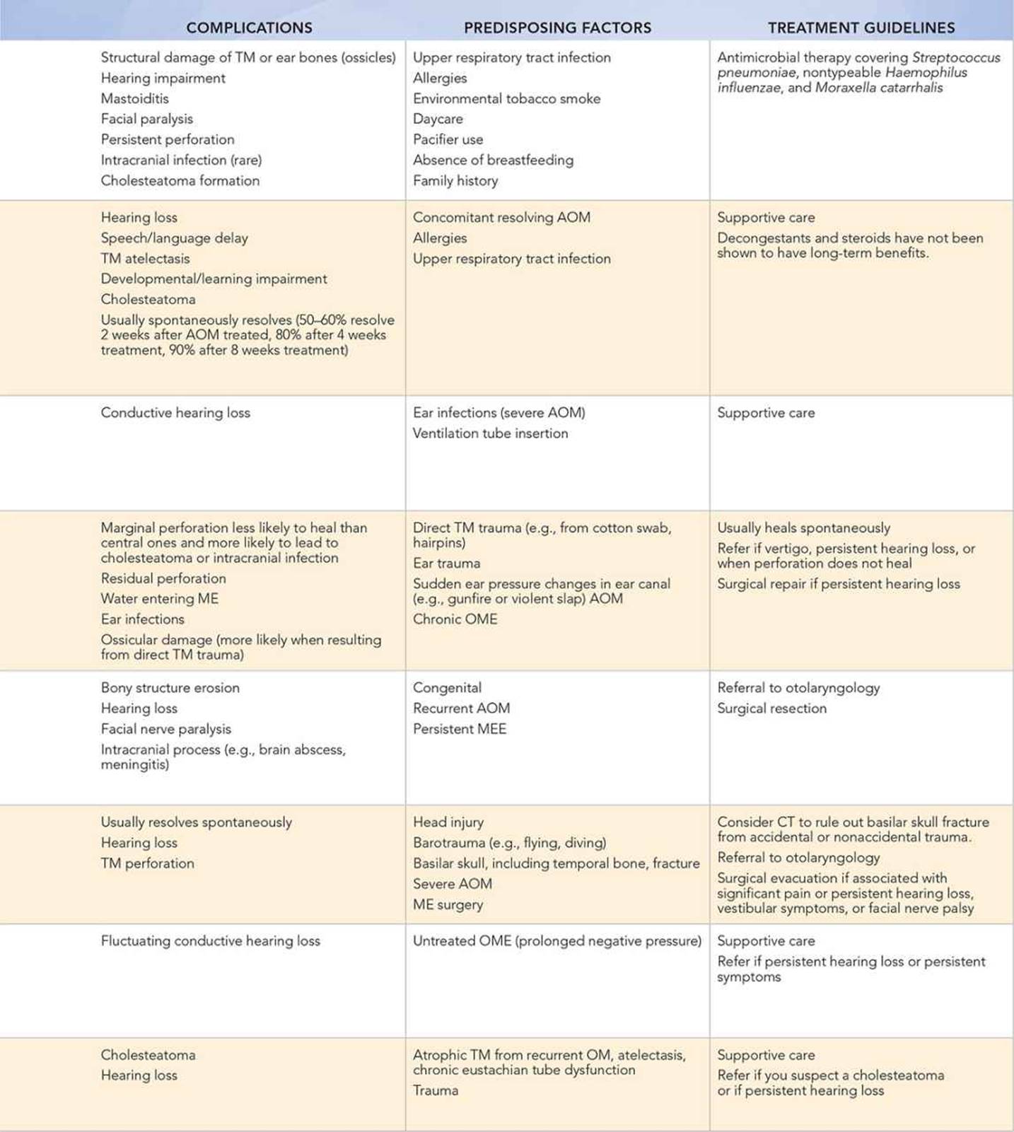
• Refer children to a pediatric otolaryngologist whenever TM perforation is accompanied by hearing loss or vertigo, or when ME fluid is chronic and associated with hearing loss and/or speech delay.
• Suspect cholesteatoma if persistent middle ear effusion (MEE) or hearing impairment, greasy and/or whitish mass, or no clinical response is present when treating another suspected TM problem.
• When a cholesteatoma is associated with ataxia or headaches, neuroimaging should be considered to evaluate for the presence of a brain abscess.
Key Points in the Physical Examination
• One must immobilize the head carefully and firmly when evaluating the TM and ear canal, while using a snug-fitting ear speculum. The small (2.5-mm diameter) ear speculum should be used in infants and preschool children, whereas the large (4-mm diameter) ear speculum should be used in school-aged children and adolescents.
• The light reflex may be absent in some normal children.
• Mobility, assessed by pneumatic otoscopy, should be measured, especially when the history and/or physical examination suggest a problem. Poor TM mobility is associated with AOM, MEE, TM perforation, or TM structural damage as with tympanosclerosis.
• Mild TM erythema can occur in association with fever, crying, upper respiratory tract infections, or irritation from cerumen or foreign objects.
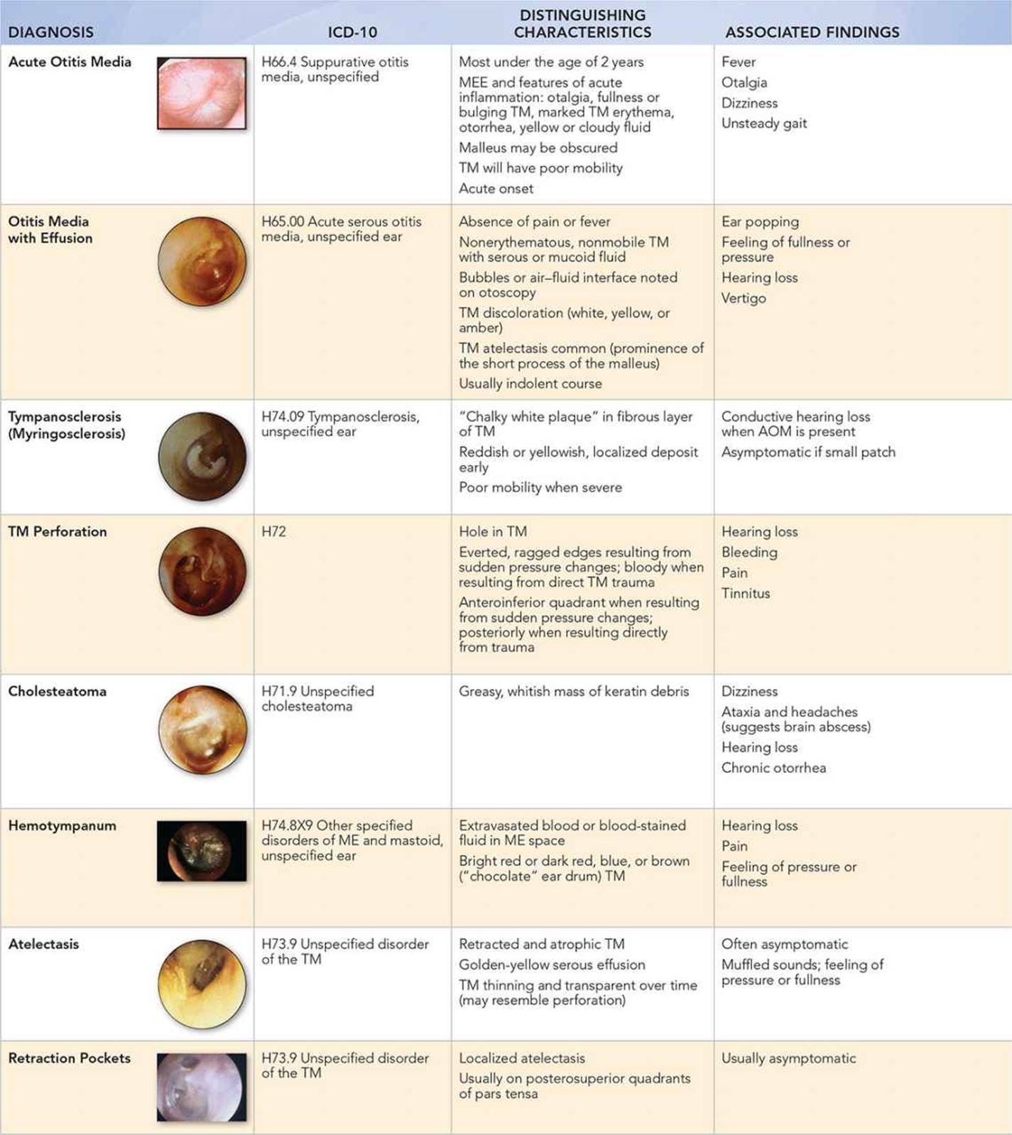
• AOM should have evidence of MEE and acute inflammation, including TM bulging or fullness, marked erythema, otorrhea, or yellow or cloudy fluid.
• Air bubbles and amber TM discoloration are associated with serous ME fluid or OME.
• Blood in the ME causes a bluish, deep red, or brown (“chocolate”) appearance of the TM.
• Chalky white plaques on the TM (tympanosclerosis) are seen with healed inflammation.
• TM mobility is absent or decreased with TM perforation.
• Manipulation of the ear pinna to ensure proper visualization of TM varies with age. As in adults, the pinna should be lifted posterosuperiorly in older children. The pinna should be pulled horizontally backward in infants and younger children.
• Localized TM atelectasis, especially in the posterosuperior quadrant of the pars tensa, is seen with retraction pockets.
• Excessive localized mobility reflects a healed perforation or TM thinning.
• OME is evidenced by fluid bubbles and air–fluid levels or by at least two of the following TM changes: abnormal color including white, yellow, amber, or blue; opacification; decreased mobility.
|
PHOTOGRAPHS OF SELECTED DIAGNOSES |
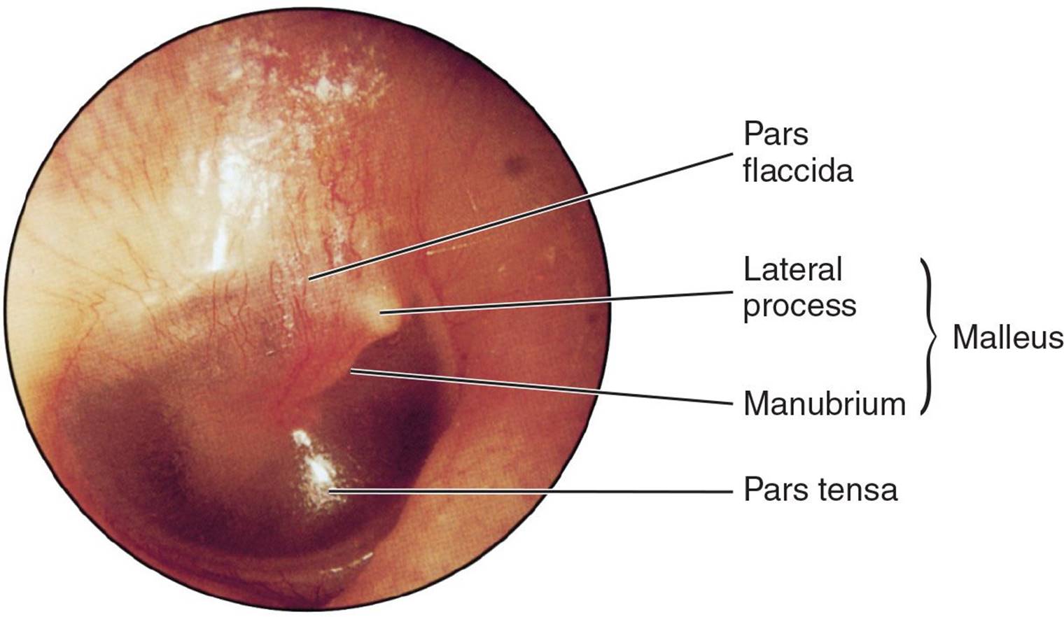
Figure 21-1 Normal tympanic membrane. (Photo used with permission from Handler SD, Myer CM. Atlas of Ear, Nose and Throat Disorders in Children. Ontario: BC Decker; 1998:28.)
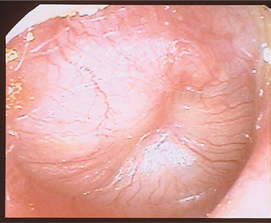
Figure 21-2 Acute otitis media. Typical acute otitis media with a red, distorted, bulging tympanic membrane in a highly symptomatic child. (Courtesy of Alejandro Hoberman, Children’s Hospital of Pittsburgh, University of Pittsburgh.)
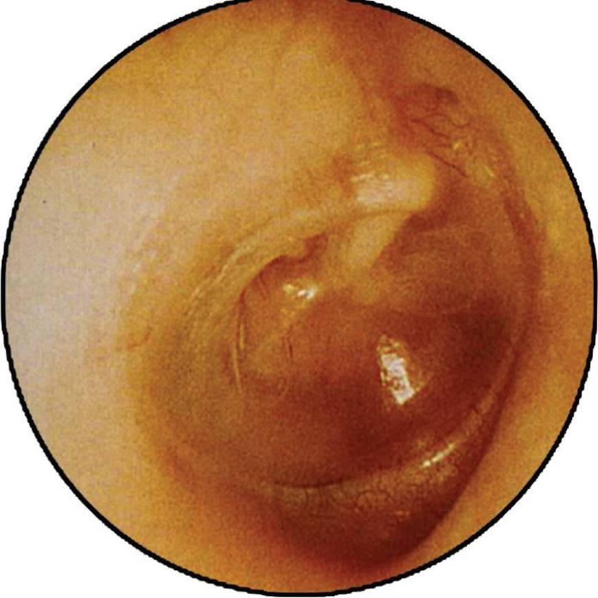
Figure 21-3 Otitis media with effusion. (Courtesy of Glenn Isaacson, MD.)
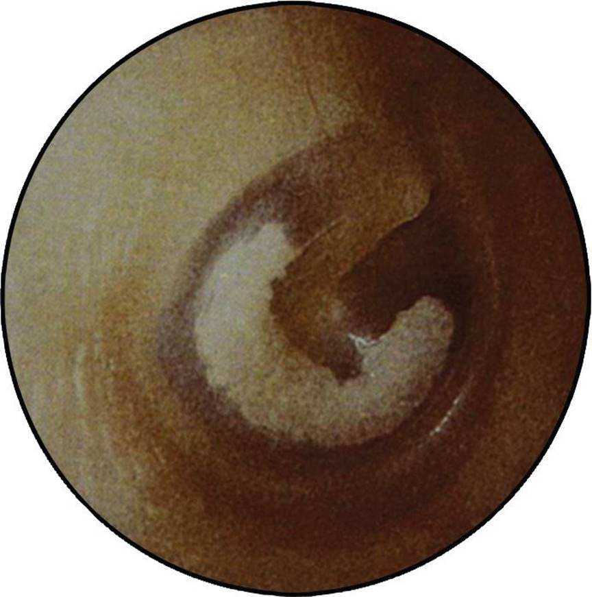
Figure 21-4 Tympanosclerosis. (Courtesy of Steven D. Handler, MD, MBE.)

Figure 21-5 Tympanic membrane perforation. (Courtesy of Steven D. Handler, MD, MBE.)
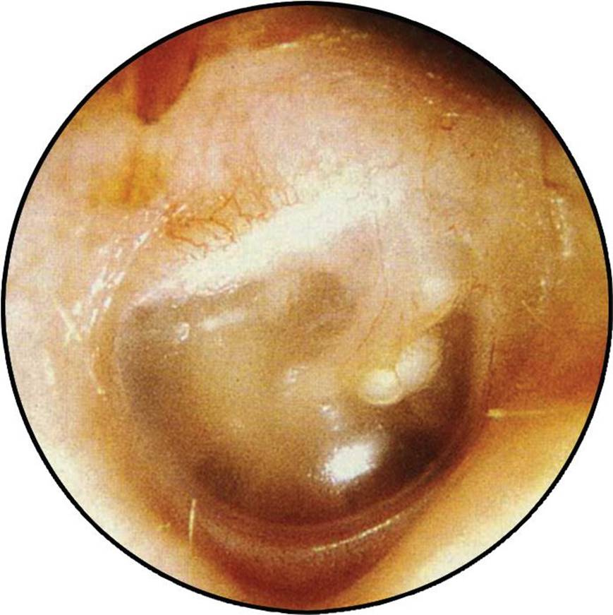
Figure 21-6 Cholesteatoma. (Used with permission from Handler SD, Myer CM. Atlas of Ear, Nose and Throat Disorders in Children. Ontario: BC Decker; 1998:30.)
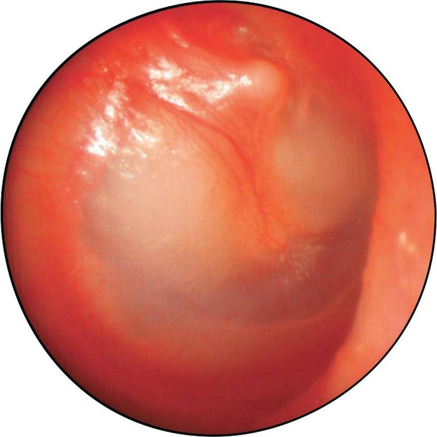
Figure 21-7 Cholesteatoma. Note the white, pearly lesion seen behind the TM in the anterior and posterior superior quadrants. (Courtesy of John A. Germiller, MD, PhD.)
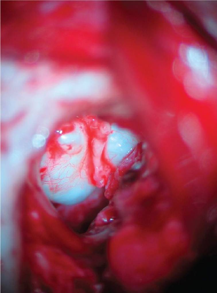
Figure 21-8 Cholesteatoma. Intraoperative view of the lesion that corresponds with the previous figure. (Courtesy of John A. Germiller, MD, PhD.)
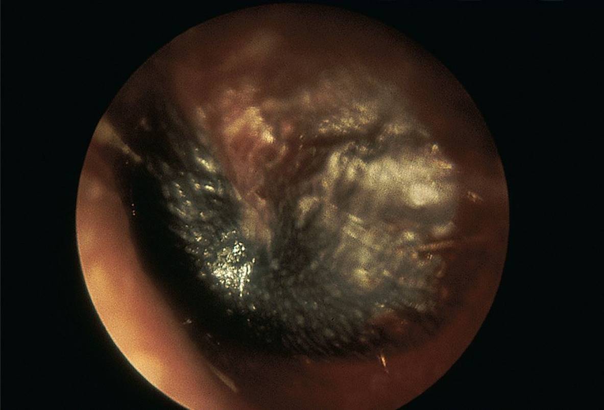
Figure 21-9 Hemotympanum. This hemotympanum was seen in association with a left temporal bone fracture. (Courtesy of Ellen Deutsch, MD.)

Figure 21-10 Atelectasis, severe. (Courtesy of Ellen Deutsch, MD.)
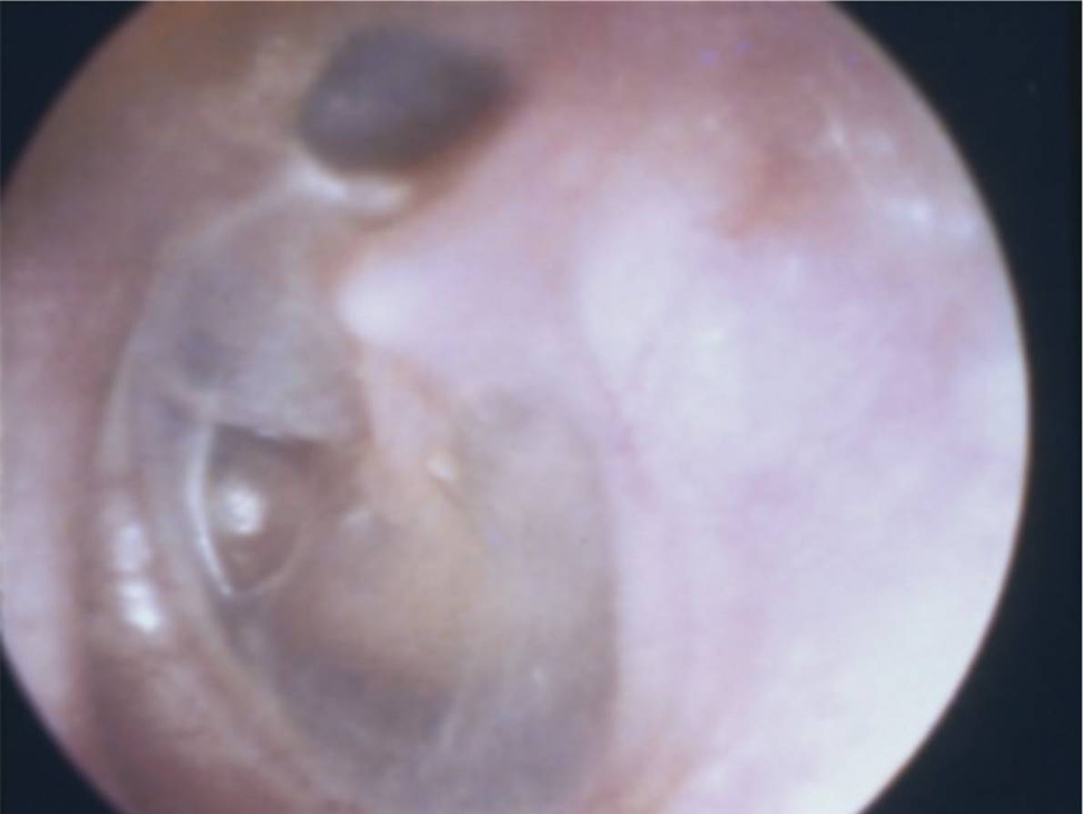
Figure 21-11 Retraction pocket. (Courtesy of Steven D. Handler, MD, MBE.)
DIFFERENTIAL DIAGNOSIS
Other Diagnoses to Consider
• Bullous myringitis
• Improper technique (e.g., inadequate light resource, poor speculum seal)
• Cerumen
• Mastoiditis
• Trauma to temporal bone
• Foreign body in ear canal
• Bleeding disorder
• Glomus tympanicum or glomus jugulare tumor
• Otosclerosis (Schwartze sign)
When to Consider Further Evaluation or Treatment
• Consider antibiotic treatment when MEE is associated with acute inflammation, as evidenced by TM bulging or fullness, marked erythema, otorrhea, or yellow or cloudy fluid.
• Chronic MEE associated with hearing loss and/or speech delay should be referred to a pediatric otolaryngologist.
• Investigate for a cholesteatoma if MEE persists or AOM does not respond to antibiotic therapy.
• Suspect a brain abscess when a cholesteatoma is associated with ataxia or headaches.
• Consider an atypical or resistant organism in a child who is immunocompromised or has been exposed to frequent or recent antimicrobial therapy.
• Persistent TM perforation or TM perforation associated with hearing loss or vestibular symptoms (i.e., nausea, vomiting, nystagmus, ataxia, vertigo) should be referred to an otolaryngologist.
SUGGESTED READINGS
American Academy of Family Physicians, American Academy of Otolaryngology—Head and Neck Surgery, American Academy of Pediatrics Subcommittee on Otitis Media with Effusion. Otitis media with effusion. Pediatrics. 2004;113:1412–1429.
Bluestone CD, Klein JO, Definitions, terminology, and classification. In: Bluestone CD, Klein JO, eds. Otitis Media in Infants and Children. 4th ed. Hamilton: BC Decker, 2007:1–19.
Coker TR, Chan LS, Newberry SJ, et al. Diagnosis, microbial epidemiology, and antibiotic treatment of acute otitis media in children: A systematic review. JAMA. 2010;304:2161–2169.
Hoberman A, Paradise JL, Rockette HE, et al. Treatment of acute otitis media in children under 2 years of age. N Engl J Med. 2011;364:105–115.
Lieberthal AS, Carroll AE, Chonmaitree T, et al. The diagnosis and management of acute otitis media. Pediatrics. 2013;131:e964–e999.
Shaikh N, Hoberman A, Kaleida PH, et al. Otoscopic signs of otitis media. Pediatr Infect Dis J. 2011;30:822–826.