
Approach to the Problem
Swelling of the nasal bridge is an uncommon problem that can present a diagnostic dilemma to the practitioner. Lesions resulting in swelling can be divided into congenital and acquired etiologies. Congenital lesions of the nose occur in 1 in 20,000 to 40,000 live births. The most common congenital lesions to consider in the differential diagnosis include hemangiomas, dacryocystoceles, dermoid cysts, gliomas, and encephaloceles. Teratomas, lymphangiomas, lipomas, and angiofibromas are less frequently encountered. Acquired lesions occur secondary to trauma or infection.
Key Points in the History
• Rapid growth of the lesion in the first weeks to months of life is suggestive of a hemangioma.
• Multiple cutaneous hemangiomas are associated with diffuse neonatal hemangiomatosis.
• Increased size when crying or straining raises the possibility of a connection to the central nervous system consistent with an encephalocele.
• Intermittent discharge of sebaceous material can occur with nasal dermoid cysts.
• Fever can be evidence of an infected congenital cystic lesion or meningitis complicating lesions that extend to the central nervous system.
• Anterior encephaloceles are associated with hydrocephalus, agenesis of the corpus callosum, and other brain malformations.
• A history of trauma may result in a nasal fracture or contusion.
Key Points in the Physical Examination
• Reddish or bluish discoloration and telangiectasias are most consistent with hemangiomas, but may be seen in gliomas.
• Cystic lesions located on the lateral aspect of the nose just under the medial canthus indicate the presence of a dacryocystocele.
• Dermoid cysts are midline lesions.
• Hair protruding from a midline mass most likely represents a dermoid cyst.
• Compressible lesions suggest encephaloceles and hemangiomas.
• Transillumination may be evident in encephaloceles.
• A positive Furstenberg sign, represented by enlargement of the mass with compression of the jugular veins, makes the diagnosis of an encephalocele probable.
|
PHOTOGRAPHS OF SELECTED DIAGNOSES |
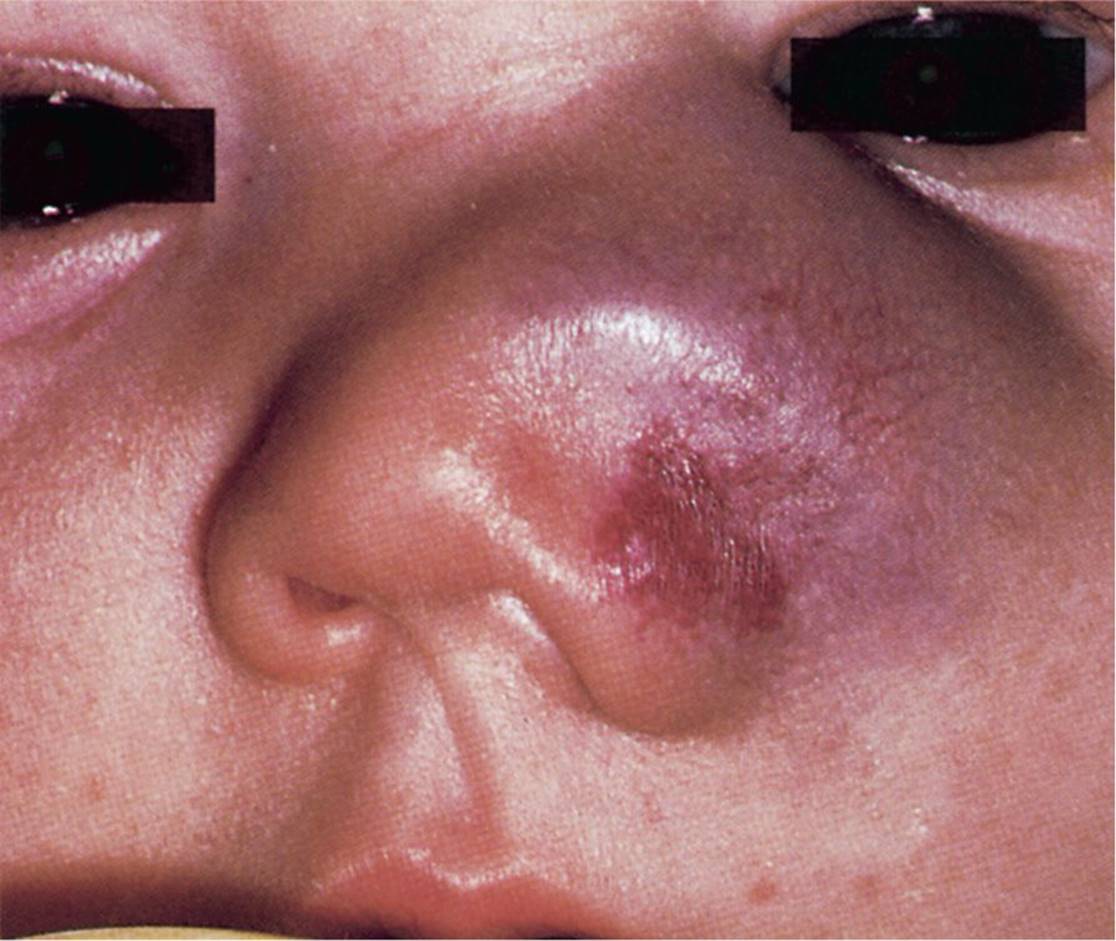
Figure 22-1 Nasal hemangioma in a child 6 months of age. Note the vascularity of this nasal mass. (Used with permission from Handler SD, Myer CM. Atlas of Ear, Nose, and Throat Disorders in Children. Ontario: BC Decker; 1998:55.)

Figure 22-2 Resolving hemangioma. A compressible mass without obvious vascularity on the nasal bridge of a child. (Courtesy of E. Douglas Thompson, Jr, MD.)

Figure 22-3 Dermoid cyst. A midline mass on the upper nasal bridge. (Courtesy of Kathleen Cronan, MD.)
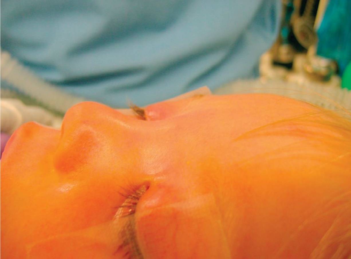
Figure 22-4 Dermoid cyst. Preoperative view. (Courtesy of John A. Germiller, MD, PhD.)
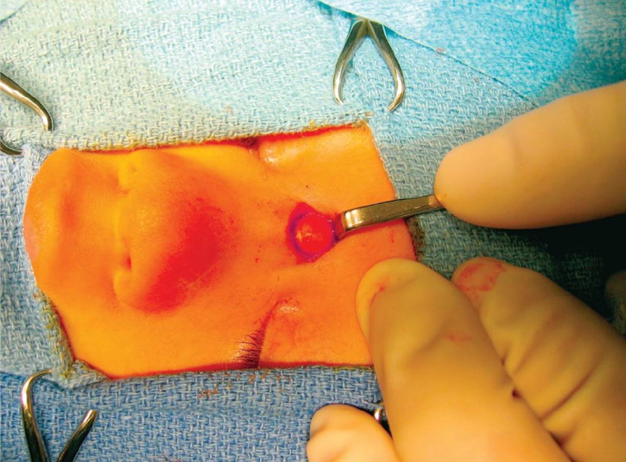
Figure 22-5 Dermoid cyst. Intraoperative view of a well-circumscribed dermoid cyst that corresponds with the previous figure. (Courtesy of John A. Germiller, MD, PhD.)
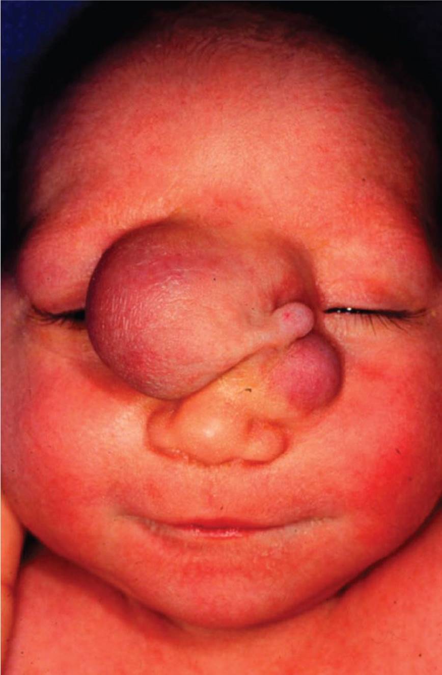
Figure 22-6 Nasal bridge encephalocele. Large nasal bridge mass in a neonate. (Courtesy of Joseph Piatt, MD.)
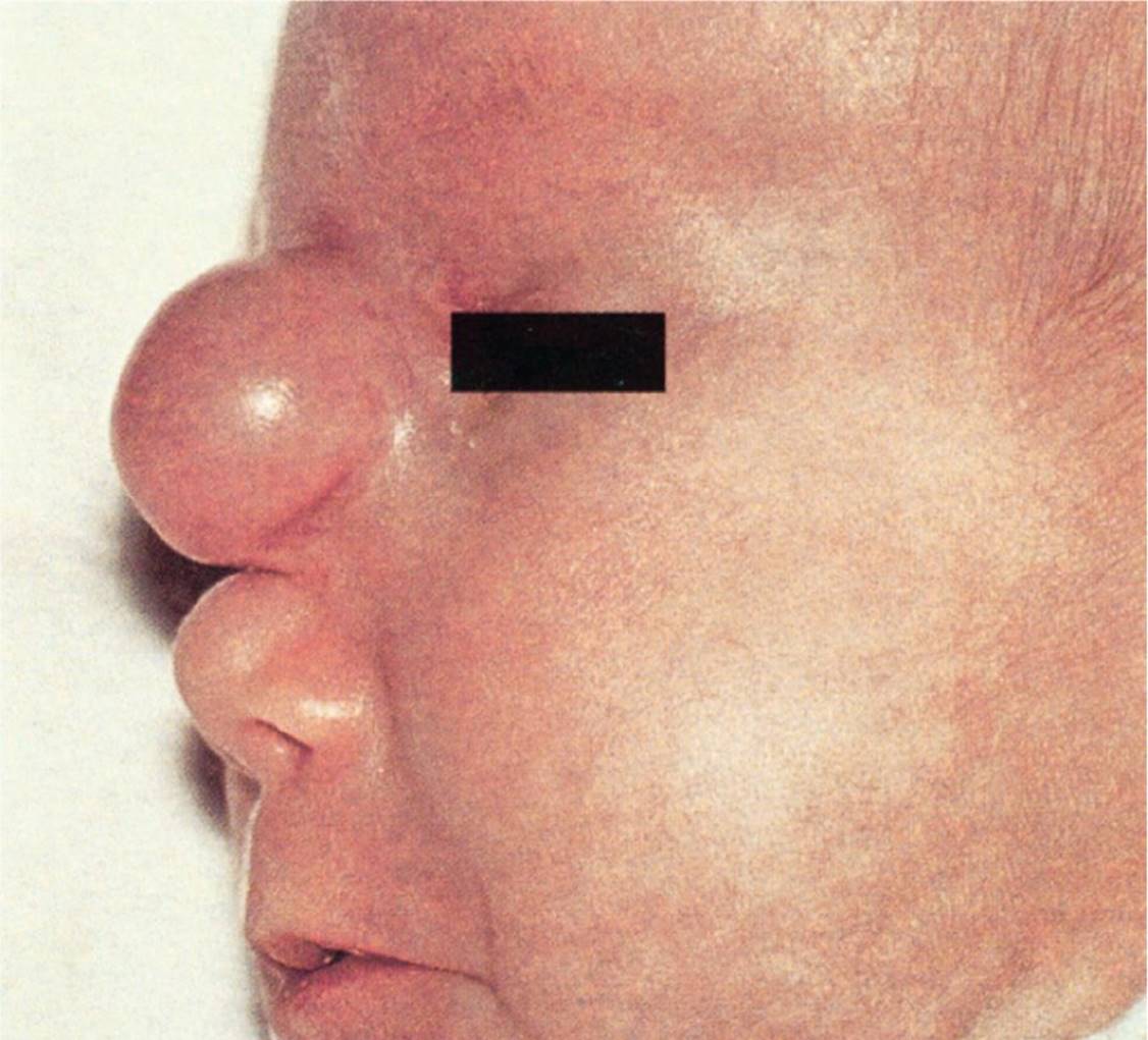
Figure 22-7 Large frontal encephalocele. A midline nasal protrusion in a newborn. (Used with permission from Handler SD, Myer CM. Atlas of Ear, Nose, and Throat Disorders in Children. Ontario: BC Decker; 1998:48.)
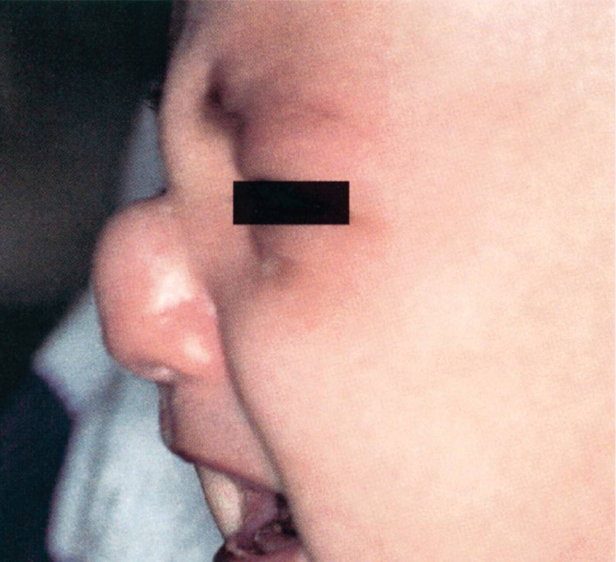
Figure 22-8 Large glioma under the nasal dorsum. Deformity of the nasal bridge in a newborn. (Used with permission from Handler SD, Myer CM. Atlas of Ear, Nose, and Throat Disorders in Children. Ontario: BC Decker; 1998:48.)
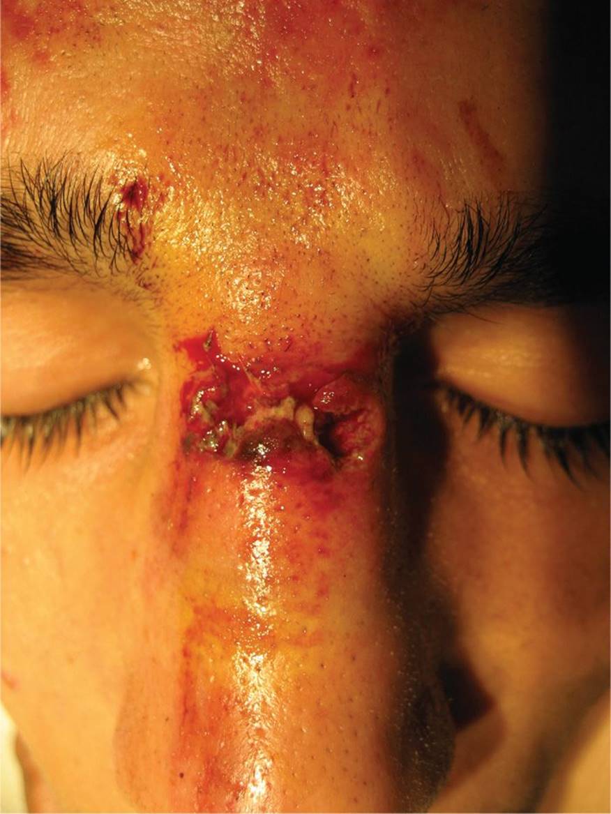
Figure 22-9 Nasal trauma from an assault. Note the swelling, ecchymosis, and laceration. (Courtesy of Brooke Burkey, MD.)

Figure 22-10 Hypertrophic scar. Nasal bridge scarring due to use of a continuous positive airway pressure machine. (Courtesy of E. Douglas Thompson, Jr, MD.)
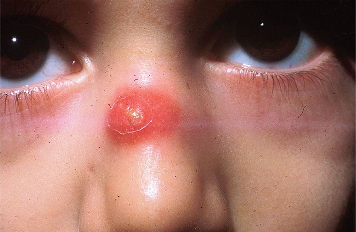
Figure 22-11 Nasal bridge abscess. Painful, erythematous lesion with purulent center on a child’s nasal bridge. (Courtesy of the late Peter Sol, MD.)
DIFFERENTIAL DIAGNOSIS

Other Diagnoses to Consider
• Diffuse neonatal hemangiomatosis
• PHACE syndrome (posterior fossa malformations, hemangiomas, arterial anomalies, cardiac defects, and eye abnormalities)
• Complex craniofacial anomalies
• Hypertrophic scar
When to Consider Further Evaluation or Treatment
• Nasal hemangiomas resulting in obstruction of the airway, impairment of vision, or significant cosmetic concerns should be referred to a pediatric dermatologist or plastic surgeon for treatment.
• Hemangiomas obscuring vision should be referred to a pediatric ophthalmologist for evaluation for amblyopia.
• Oral propranolol is the preferred treatment for hemangiomas. Propanolol is more effective with fewer adverse effects than traditional therapies such as surgical excision or systemic/intralesional corticosteroids.
• Laser therapy is a promising alternative therapy for some hemangiomas, but the evidence is more anecdotal.
• MRI of the head and neurosurgical referral are indicated if a dermoid cyst, glioma, or encephalocele is suspected.
• Nasal fractures with deformity, septal hematoma, septal deviation, or difficult to control epistaxis should be referred to an ENT specialist.
SUGGESTED READINGS
Chen TS, Eichenfield LF, Friedlander SF. Infantile hemangiomas: An update on pathogenesis and therapy. Pediatrics. 2013;131(1):99–108.
Dasgupta NR, Bentz ML. Nasal gliomas: Identification and differentiation from hemangiomas. J Craniofac Surg. 2003;14:736–738.
Lee WT, Koltai PJ. Nasal deformity in neonates and young children. Pediatr Clin North Am. 2003;50(2):459–467.
Mahapatra AK, Suri A. Anterior encephaloceles: A study of 92 cases. Pediatr Neurosurg. 2002;36(3):113–118.
Wong RK, VanderVeen DK. Presentation and management of congenital dacryocystocele. Pediatrics. 2008;122(5):e1108–e1112.
Wright RJ, Murakami CS, Ambro BT. Pediatric nasal injuries and management. Facial Plast Surg. 2011;27(5):483–490.
Zapata S, Kearns DB. Nasal dermoids. Curr Opin Otolaryngol Head Neck Surg. 2006;14(6):406–411.