
Approach to the Problem
Children of all ages may present with a variety of swellings within the mouth, ranging from benign lesions to very serious infections. Differentiating mouth swellings may be difficult owing to a lack of available diagnostic and laboratory testing. Common benign lesions can be diagnosed using characteristic locations and distinguishing physical features. Serious swellings of the mouth will typically present with associated systemic signs of illness. Peritonsillar abscess and Ludwig angina have potentially life-threatening complications.
Key Points in the History
• Bohn nodules and Epstein pearls are present in newborns (see Chapter 24: Mouth Sores and Patches and Chapter 25: Focal Gum Lesions).
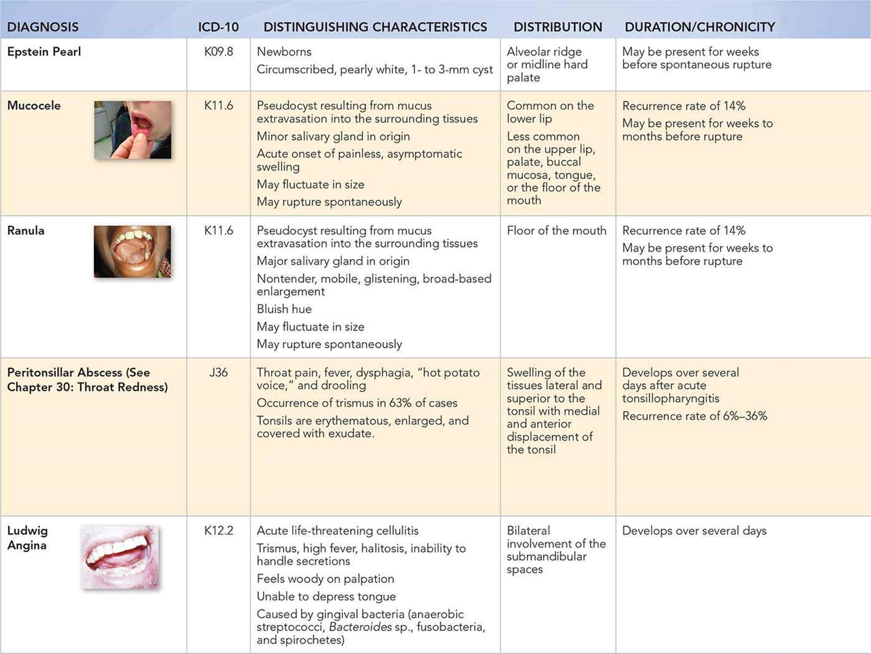
• Mucoceles and ranulas arise acutely, rupture spontaneously, and are painless and asymptomatic.
• Systemic signs of infection, such as fever and throat pain, help differentiate benign swellings of the mouth from more serious infections.
• Peritonsillar abscess is generally preceded by acute tonsillopharyngitis.
• Patients with Ludwig angina have a history of high fever and an inability to handle secretions.
Key Points in the Physical Examination
• Epstein pearls are smooth, nontender, translucent, pearly white, 1- to 3-mm cysts on the palate near the midline of the roof of the mouth. When such lesions occur on the gums, they are referred to as Bohn nodules.
• Mucoceles and ranulas are fluid-filled, nontender, mobile, and glisten and have a bluish hue.
• Mucoceles are most common on the lower lip.
• Ranulas are found on the floor of the mouth.
• Peritonsillar abscess is characterized by swelling of tissues lateral and superior to the affected tonsil, anterior and medial displacement of the affected tonsil, and displacement of the uvula toward the contralateral side.
• Patients with peritonsillar abscess can have a muffled, or “hot potato,” voice.
• Ludwig angina always involves the bilateral submandibular spaces.
• Ludwig angina is characterized by elevation and posterior displacement of the tongue.
• Trismus, or difficulty in opening the mouth, is a frequent finding in patients with peritonsillar abscess and Ludwig angina.
|
PHOTOGRAPHS OF SELECTED DIAGNOSES |
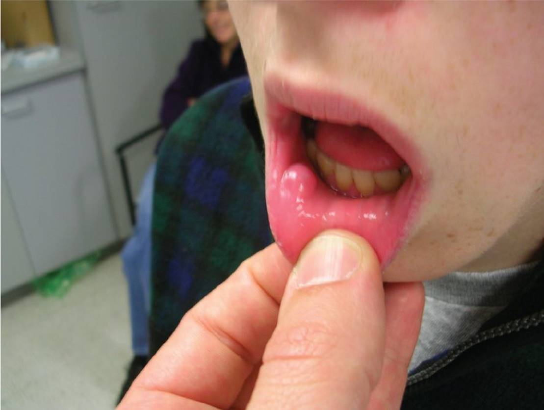
Figure 29-1 Mucocele. Fluid-filled pseudocyst protruding from lower lip. (Courtesy of Paul S. Matz, MD.)

Figure 29-2 Mucocele. Fluid-filled pseudocyst protruding from lower lip. (Courtesy of Michael Lemper, DDS.)

Figure 29-3 Mucocele of soft palate. Whitish pseudocyst on the right side of the soft palate. (Used with permission from Handler SD, Myer CM. Atlas of Ear, Nose and Throat Disorders in Children. Ontario: BC Decker; 1998:85.)

Figure 29-4 Ranula. Fluid-filled mass on the floor of the mouth. (Courtesy of Kathleen Cronan, MD.)

Figure 29-5 Ranula. Fluid-filled cyst on the base of the tongue. (Courtesy of George A. Datto, III, MD.)
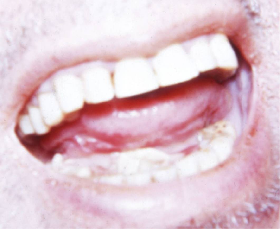
Figure 29-6 Ludwig angina. Note elevation of tongue secondary to swelling of the floor of the mouth. (Used with permission from Greenberg MI. Greenberg's Atlas of Emergency Medicine. Philadelphia, PA: Lippincott Williams & Wilkins; 2005:7.)
DIFFERENTIAL DIAGNOSIS

Other Diagnoses to Consider
• Hemangioma
• Lymphangioma
• Fibroma
• Parulis (gum abscess)
When to Consider Further Evaluation or Treatment
• Most mucoceles and ranulas rupture spontaneously and will not require further intervention.
• Large ranulas that interfere with speech, swallowing, or respiration should be referred for evaluation by otolaryngology.
• Peritonsillar abscesses require antibiotic administration and pain management in a hospital setting in addition to concurrent consultation with otolaryngology for possible drainage.
• Ludwig angina is a life-threatening condition that requires immediate hospitalization, evaluation for airway compromise, intravenous antibiotics, and consultation by otolaryngology.
SUGGESTED READINGS
Brierly DJ, Chee CKM, Speight PM. A review of paediatric oral and maxillofacial pathology. Int J of Paediatr Dent. 2013;23(5):319–329.
Cathcart RA. Inflammatory swellings of the head and neck. Surgery. 2012;30(11):597–603.
Delaney J, Keels MA. Pediatric oral pathology. Pediatr Clin North Am. 2000;47(5):1125–1147.
Krol DM, Keels MA. Oral conditions. Pediatr Rev. 2007;28:15–22.
Patel NJ, Sciubba J. Oral lesions in young children. Pediatr Clin North Am. 2003;50:469–486.