
Approach to the Problem
Throat redness is a familiar complaint to the general pediatrician or family practitioner. Erythema of the posterior oropharynx suggests an inflammatory or infectious process, but can also be caused by exposure to environmental allergens, airborne irritants, or acid from chronic laryngopharyngeal reflux. The majority of pediatric infections involving the posterior oropharynx are of viral origin. Careful attention to details provided in the patient’s history, and to findings observed on the physical examination, will help to identify patients for whom additional testing, such as group A streptococcus rapid antigen detection, should be performed. It is imperative to recognize the acutely ill patient with pharyngeal erythema, and direct the medical evaluation and treatment plan accordingly. For patients with a persistent or recurrent complaint of throat redness, one must broaden the differential diagnosis to include less common etiologies or pathogens.
Key Points in the History
• Pharyngitis may cause neck pain and stiffness.
• Throat redness associated with upper respiratory tract symptoms (rhinorrhea, cough, and conjunctivitis) and/or lower gastrointestinal tract manifestations (vomiting with diarrhea) are characteristic of viral infection and rarely represent a bacterial throat infection.
• Throat pain due to postnasal drip is often worse at night or in the early morning, but improves during the day.
• Infection with adenovirus is associated with conjunctivitis and/or otitis media.
• In group A streptococcal pharyngitis, symptom onset is typically acute. Sore throat, dysphagia, fever, headache, abdominal pain, and vomiting commonly occur.
• Younger children with streptococcal pharyngitis may present with fever, headache, vomiting, abdominal pain, and decreased oral intake. They may not identify sore throat as a primary complaint.
• Incidence of group A beta-hemolytic streptococcal pharyngitis peaks in the late winter and early spring.
• Epidemics of group A beta-hemolytic streptococcal pharyngitis occur in patients who live in close quarters such as military units, dormitories, mental health facilities, schools, homeless shelters, and group homes.
• Otalgia, with referred pain due to sensory innervation of the glossopharyngeal and vagus nerves supplying both the throat and the ear, can be a presenting symptom of streptococcal pharyngitis.
• Retropharyngeal abscesses are most common in children between 2 and 4 years of age.
• Consider infectious mononucleosis, due to Epstein–Barr virus (EBV) infection, in adolescents with fever, throat pain, enlarged posterior lymph nodes, and significant fatigue.
• Adolescents may not disclose sexual risk behaviors; therefore, test teens with persistent pharyngitis for sexually transmitted diseases, such as gonorrhea.
• Candida may be associated with inhaled steroid use for persistent asthma, or seen in patients who are immunosuppressed or immunocompromised.
• Mycoplasma infection is usually associated with cough.
• Increased throat pain after meals or when supine suggests pain related to gastroesophageal reflux.
• Chronic mouth breathing associated with obstructive sleep apnea leads to dry, irritated mucosae and a sore throat, which is worse in the morning and improves throughout the day as the patient drinks fluids. Asking about ambient room temperatures, especially in the winter months, can provide useful history to support this diagnosis.
Key Points in the Physical Examination
• Severe viral pharyngitis and streptococcal pharyngitis may present with fever, pharyngeal erythema with or without exudates, and cervical lymphadenopathy.
• Hoarseness suggests vocal cord inflammation due to a viral process, or laryngopharyngeal reflux.
• Children younger than 3 years of age with throat redness and exudate are more likely to have viral pharyngitis than group A beta-hemolytic streptococcal infection.
• Children with a peritonsillar abscess may have pharyngeal erythema accompanied by unilateral tonsillar hypertrophy, uvular deviation toward the unaffected side, fever, and trismus (the inability to open the mouth fully). In addition, patients typically have a “hot potato” voice, speaking as if they have a mouthful of hot potatoes, due to an inflamed throat.
• Ulcerations are typically seen with viral pharyngitis and stomatitis. Ulcerations on the tonsillar pillars suggest the diagnosis of herpangina caused by coxsackievirus or echovirus. Ulcerations from herpes simplex virus are typically more anterior but may occur posteriorly and in association with gingivitis.
• Infectious mononucleosis may present with pharyngeal and tonsillar erythema, fever, difficulty swallowing, and posterior cervical lymphadenopathy. Fatigue is a prominent symptom, and may persist in up to 22% of cases beyond 2 to 3 weeks of illness.
• Drooling results from the inability to swallow one’s secretions. Watch for drooling in patients with severe pharyngitis, pharyngeal ulcerations, or a retropharyngeal abscess.
• Sitting in a tripod position, inspiratory stridor, difficulty breathing, and dehydration are collective findings that require evaluation for epiglottitis.
• Diphtheria is a rare cause of pharyngitis in well-immunized populations. An asymmetric gray pharyngeal membrane, which extends beyond the borders of the anterior tonsillar pillars onto the soft palate and/or the uvula, suggests this diagnosis.
|
PHOTOGRAPHS OF SELECTED DIAGNOSES |
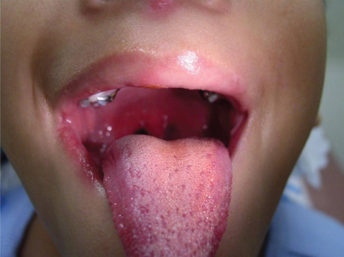
Figure 30-1 Group A streptococcal pharyngitis. Note the marked erythema posteriorly in this patient with scarlet fever. (Courtesy of Esther K. Chung, MD, MPH.)
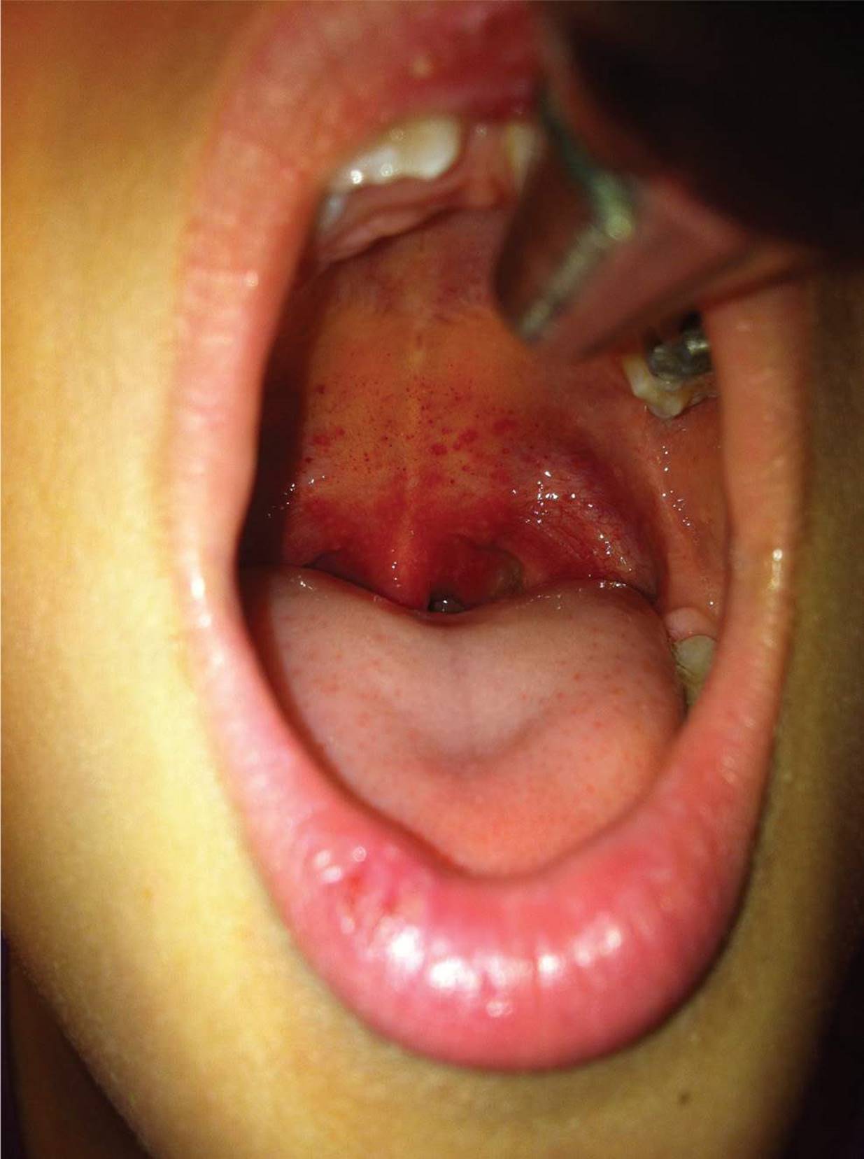
Figure 30-2 Group A streptococcal pharyngitis. Note the marked erythema of the uvula and tonsillar pillars and the palatal petechiae. (Courtesy of Naline Lai, MD.)
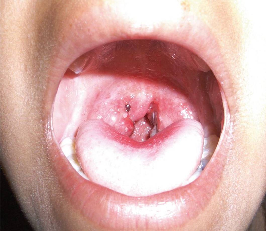
Figure 30-3 Viral pharyngitis. Mild erythema and erythematous papules on both tonsils. (Courtesy of Paul S. Matz, MD.)

Figure 30-4 Erythematous ulcers on the posterior pharynx. Ulcerations on the tonsillar pillars suggest the diagnosis of herpangina caused by coxsackievirus or echovirus. (Used with permission from Neville BW, Damm DD, White DK. Color Atlas of Clinical Oral Pathology. 2nd ed. Baltimore, MD: Williams & Wilkins; 1998.)
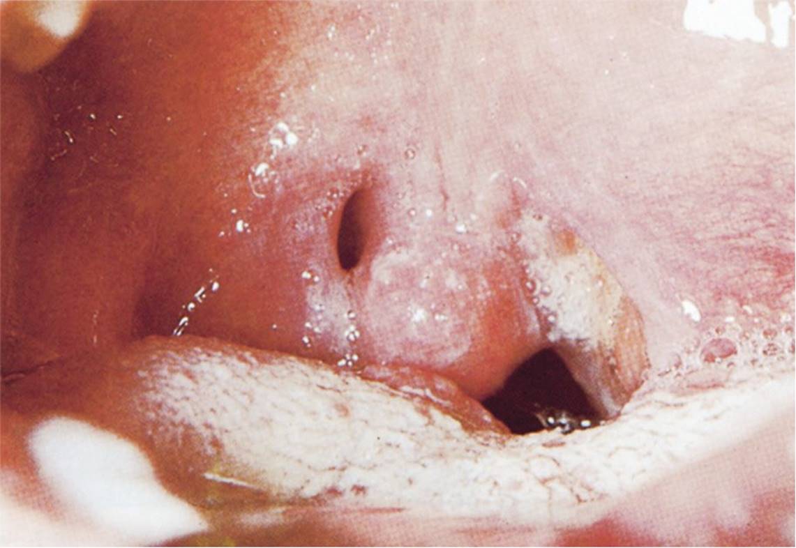
Figure 30-5 Peritonsillar abscess of the left tonsil. (Used with permission from Handler SD, Myer CM. Atlas of Ear, Nose and Throat Disorders in Children. Ontario: BC Decker; 1998:90.)

Figure 30-6 Peritonsillar abscess of the right tonsil. Note the swelling and distortion of the area around the right tonsil. (Courtesy of the late Peter Sol, MD.)
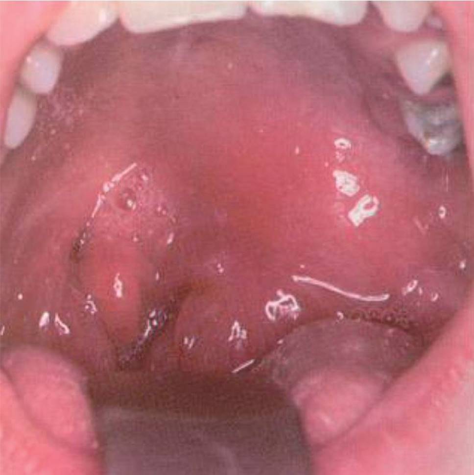
Figure 30-7 Peritonsillar abscess. Note the erythema and swelling on the left and the right deviation of the uvula. (Courtesy of Seth Zwillenberg, MD.)

Figure 30-8 Acute tonsillitis secondary to infectious mononucleosis. Note the marked tonsillar enlargement with erythema and the large white-gray patches. (Used with permission from Handler SD, Myer CM. Atlas of Ear, Nose and Throat Disorders in Children. Ontario: BC Decker; 1998:91.)
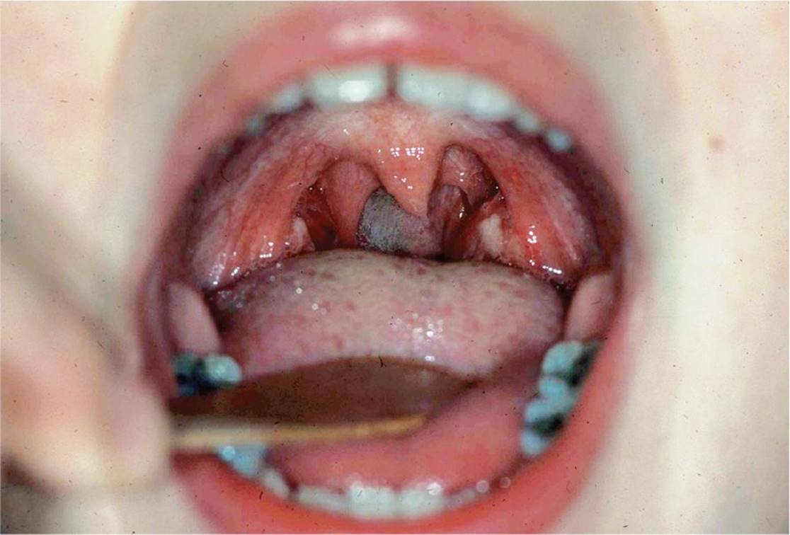
Figure 30-9 Acute tonsillar inflammation due to infectious mononucleosis. Note the tonsillar enlargement with erythema and white exudates. (Courtesy of the late Peter Sol, MD.)
DIFFERENTIAL DIAGNOSIS
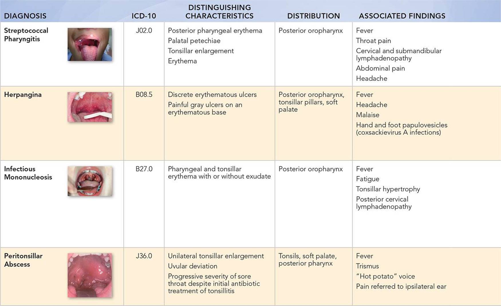

Other Diagnoses to Consider
• Sexually transmitted diseases (e.g., syphilis [primary or secondary], primary HIV infection, or those caused by Neisseria gonorrhoeae, and Chlamydia trachomatis)
• Candidal infection
• Mycoplasma pneumonia
• Irritation due to chemical exposure or inhalants
• Gastroesophageal or laryngopharyngeal reflux
• Postnasal drip from allergic rhinitis or viral upper respiratory tract infection
• Obstructive sleep apnea
• Diphtheria
When to Consider Further Evaluation or Treatment
• Patients with persistent streptococcal pharyngitis symptoms after 48 hours of appropriate antimicrobial therapy should be seen to rule out suppurative complications. Broad-spectrum antibiotics should be considered.
• A peritonsillar abscess or cellulitis requires emergent otolaryngology evaluation for possible incision and drainage and parenteral antibiotics.
• Viral pharyngitis and herpangina require repeat evaluation if the symptoms persist for more than 10 days.
• Ulcers may be a presenting manifestation of inflammatory bowel disease.
• Urgent evaluation of infectious mononucleosis and treatment with corticosteroids is warranted if signs of airway compromise are evident.
• The development of jaundice, irritability, mental status change, chest pain, or limp may indicate complications of mononucleosis.
SUGGESTED READINGS
Alcaide ML, Bisno AL. Infections of the head and neck: Pharyngitis and epiglottitis. Infect Dis Clin N Am. 2007;21(2):449–469.
Chan TV. Otolaryngology for the internist: The patient with sore throat. Med Clin N Am. 2010;94(5):923–943.
Choby B. Diagnosis and treatment of streptococcal pharyngitis. Am Fam Physician. 2009;79(5):383–390.
Del Mar C. Once-daily amoxicillin eradicates group A beta-hemolytic strep as well as penicillin twice a day. Pediatrics. 2008;153:725–725.
Handler SD, Myer CM. Atlas of Ear, Nose and Throat Disorders in Children. Ontario: BC Decker; 1998:90–91.
Schwartz B, Marcy SM, Phillips WR, et al. Pharyngitis—principles of judicious use of antimicrobial agents. Pediatrics. 1998;101(suppl 1):171–174.
Shaikh N, Swaminathan N, Hooper E. Accuracy and precision of the signs and symptoms of streptococcal pharyngitis in children: A systematic review. J Pediatr. 2012;160:487–493.
Shulman ST, Bisno AL, Clegg HW, et al. Clinical practice guideline for the diagnosis and management of group A streptococcal pharyngitis: 2012 update by the Infectious Diseases Society of America. Clin Infect Dis. 2012;55(10):e86–e102.