
Approach to the Problem
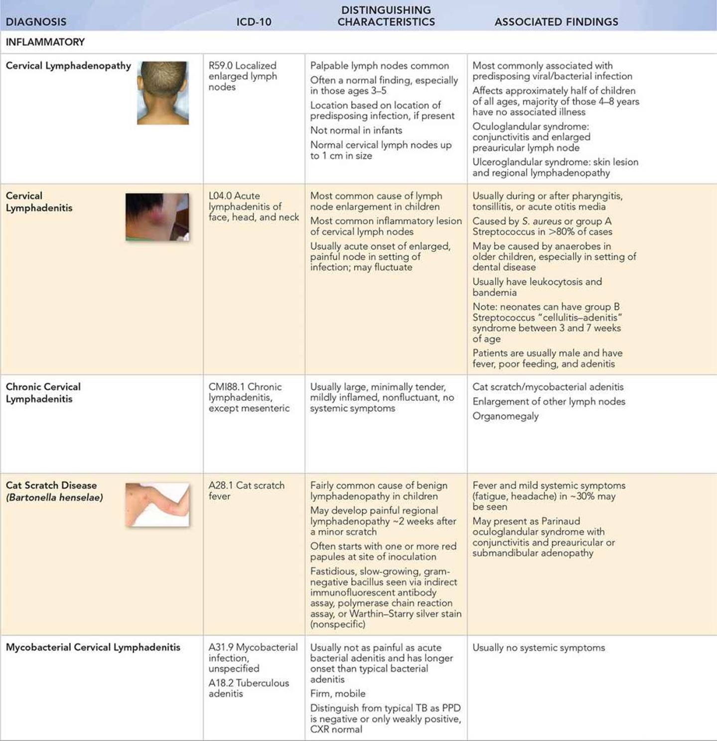
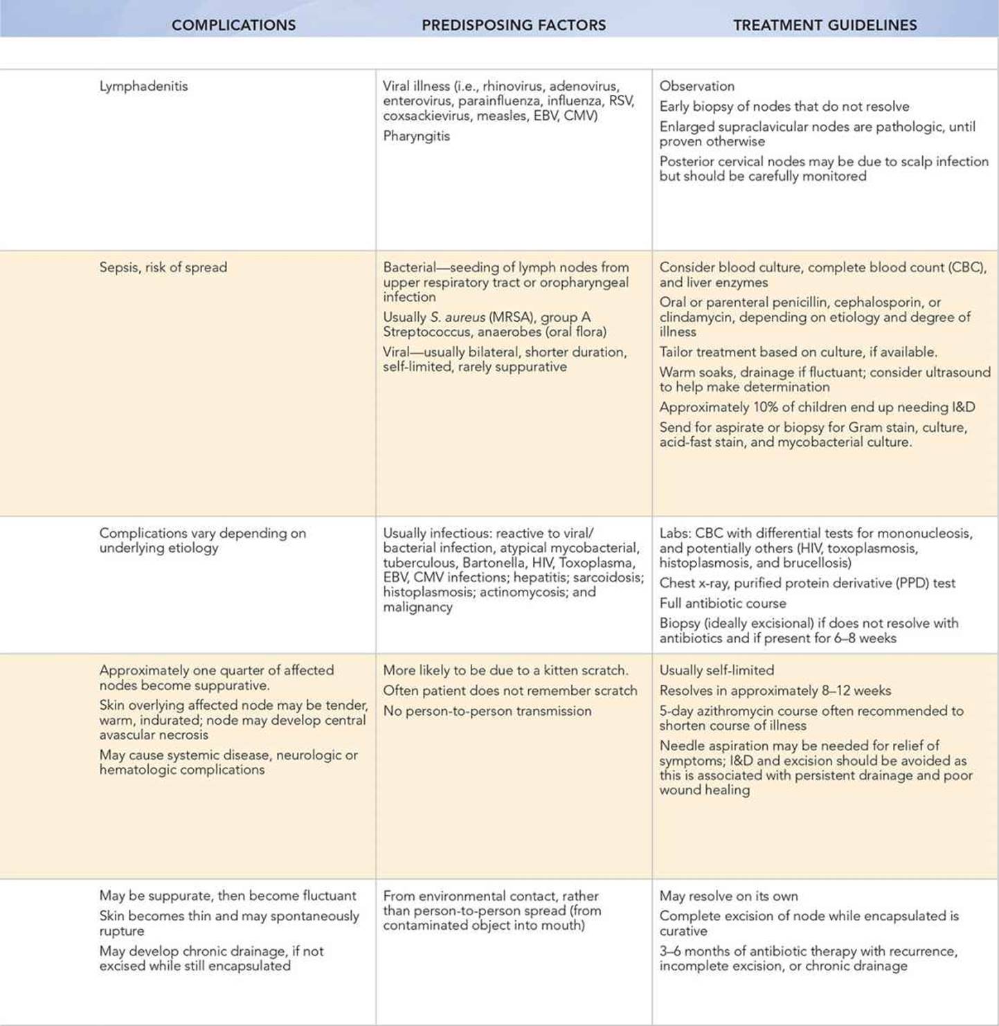
Causes of neck masses and swelling can be broken down into three categories: inflammatory, congenital, and neoplastic; though some may be in more than one category, such as teratoma/dermoid cyst, or infected congenital lesions such as an infected branchial cleft cyst. Clinicians make the majority of diagnoses by taking a careful history and performing a physical examination. Although most neck masses in children are due to inflammatory conditions, followed by congenital conditions, the clinician must be wary of more ominous causes, such as malignancy. Although 80% to 90% of head and neck masses in children are benign, 5% of all malignancies in children are in the head and neck area.
Key Points in the History
• Carefully assess factors such as age; onset, duration, and progression of symptoms; presence or absence of systemic symptoms including fever, fatigue, weight loss, night sweats, joint pain, or swelling; recent upper respiratory tract infection (URI) or sick contacts; animal or food contacts, especially animal bites or scratches or exposure to uncooked meats and unpasteurized milk; immunization status, immunocompromised; recent travel; and medications.
• Acute or subacute enlargement, pain, erythema, fluctuance, and/or recent URI suggest inflammatory conditions.
• Lesions present since birth or shortly thereafter are likely congenital. Think of underlying anatomic anomaly when there is recurrent infection in the same location.
Key Points in the Physical Examination
• Make note of size, location, including sidedness, consistency, mobility, pain, overlying skin changes, and whether the swelling is localized to the neck region or more generalized as in diffuse adenopathy.
• Examine all other nodes and complete a full HEENT (Head, Eyes, Ears, Nose, and Throat) examination, including an oropharyngeal, dental, face, scalp, ear, and eye exam. Perform a general examination, including cardiovascular, pulmonary, abdominal, and extremities examination.
• Consider serial assessments of the neck mass or swelling, ideally performed by the same physician each time.
• Note some diagnosis-specific findings on examination, including the following:
• Viral adenopathy may be seen in association with findings such as rash, pharyngeal erythema, oral mucosal vesicles, or conjunctival injection.
• Cervical adenitis is usually rapid in onset, unilateral, tender, warm, and red. The child may have fever, fatigue, and irritability.
• Patients with bartonella infection usually only have one enlarged node in the area that drains the site of inoculation. A papule or pustule may have been seen at the inoculation site approximately 2 weeks before lymphadenopathy.
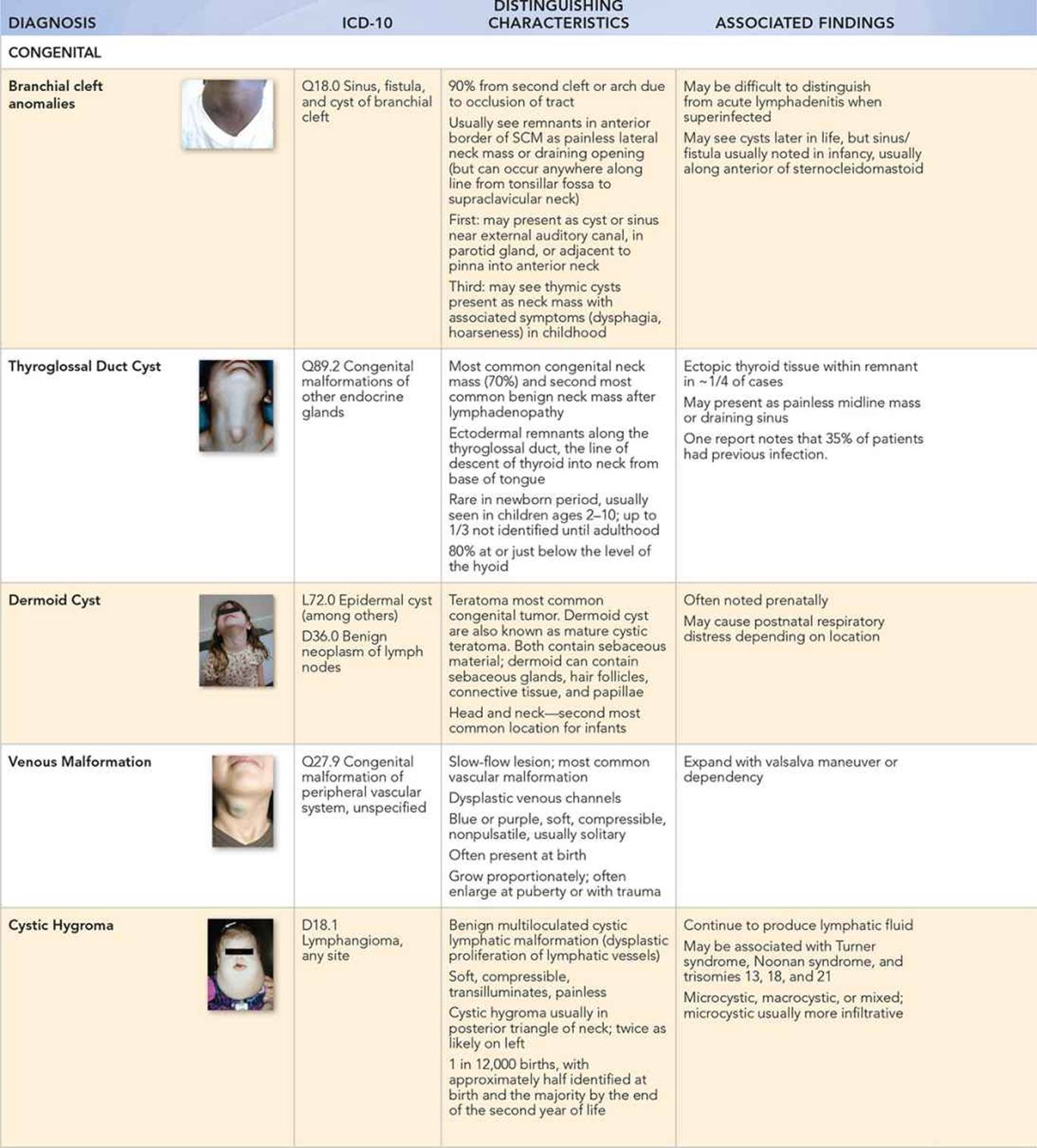
• Branchial cleft cysts are usually deep to the upper third anterior border of the sternocleidomastoid, painless, and with clear drainage from the opening along the sternocleidomastoid. The tract may be palpable.
• Thyroglossal duct cysts are soft, smooth, nontender, and in the midline, usually below the level of the hyoid. Only 20% are above and 15% are at the level of the hyoid. They typically do not have a primary external opening unless infected. Look for upward movement of the mass with swallowing or tongue protrusion, due to connection with the base of the tongue.
• Dermoid cysts are usually mobile, though they may adhere to underlying bone, and be nontender. They may be in the midline and confused with thyroglossal duct cysts. Dermoid cysts can be distinguished by depth in that they are generally superficial, by the presence of sebaceous material, and by anatomy as they typically have no connection to the hyoid or tongue.
• A soft, spongy, compressible swelling with a bluish hue may be a venous malformation.
• Cystic hygromas can be as small as a few millimeters but are often much larger. They are discrete, soft, nontender, and mobile.
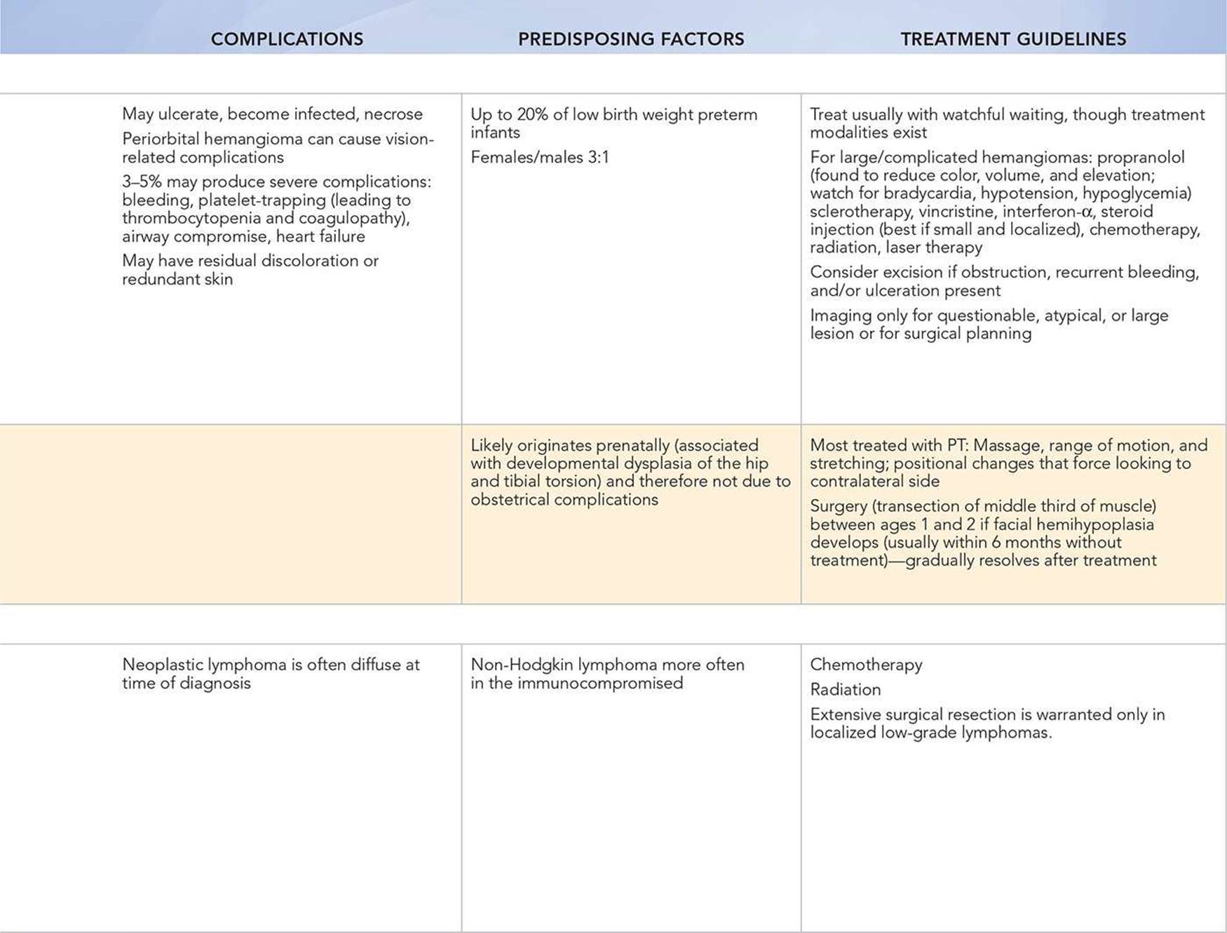
• Hemangiomas are initially blue or bright red and soft, mobile, and nontender. When regressing, they may have a grayish hue.
• In congenital torticollis, the face and the chin tilt away from the affected side, while the head tilts toward the ipsilateral shoulder. A mass may be felt.
• A firm, painless mass is concerning for malignancy. The risk of malignancy increases with increasing node size. Look for associated petechiae, which may indicate malignancy.
|
PHOTOGRAPHS OF SELECTED DIAGNOSES |
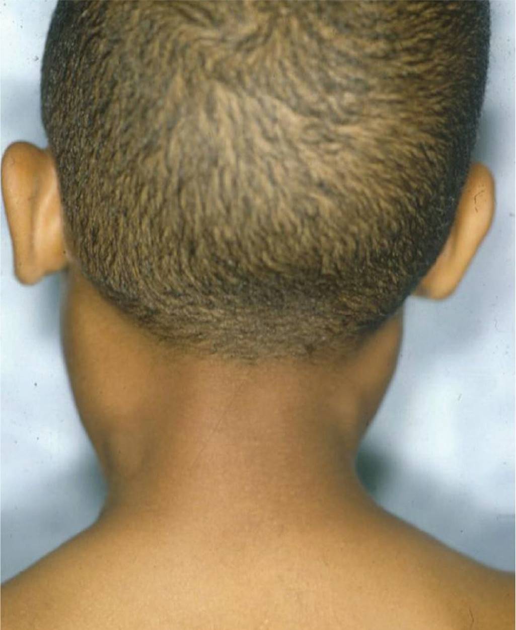
Figure 31-1 Cervical adenopathy. A posterior view of bilateral adenopathy in a 7-year-old male with a 1- to 2-week history of malaise, sore throat, and low-grade fevers. (Courtesy of Ellen Deutsch, MD.)
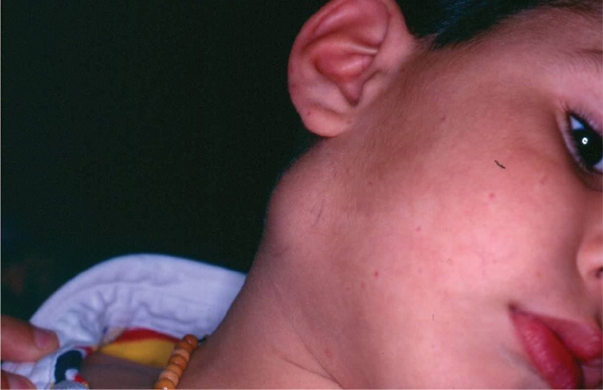
Figure 31-2 Cervical adenitis. This case of acute unilateral adenitis in this 3-year-old child is most likely caused by Staphylococcus aureus or group A streptococcus. (Courtesy of Jan Edwin Drutz, MD.)
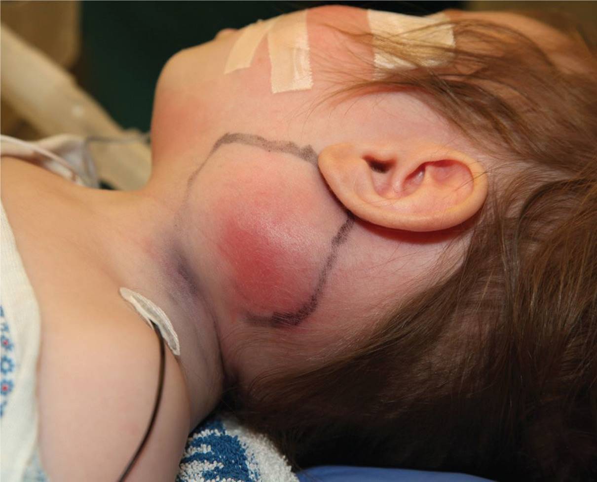
Figure 31-3 Staphylococcal cervical lymphadenitis. (Courtesy of Eden Palmer and Jonathan Perkins, DO.)
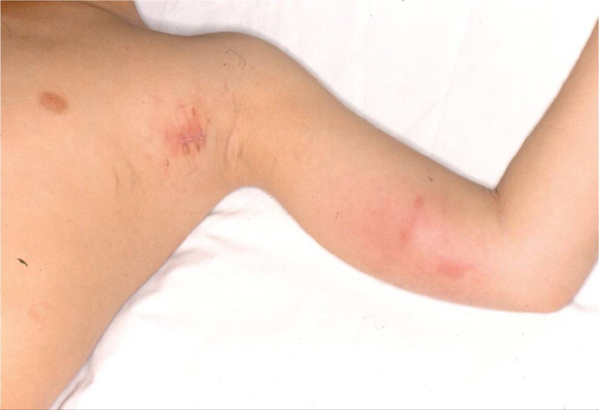
Figure 31-4 Cat scratch adenopathy. Epitrochlear and axillary adenopathy that developed proximal to the inoculation site on the finger shown in Figure 31-5. (Courtesy of Mark A. Ward, MD.)

Figure 31-5 Cat scratch inoculation site on the extremity of the 9-year-old child shown in Figure 31-4. (Courtesy of Mark A. Ward, MD.)
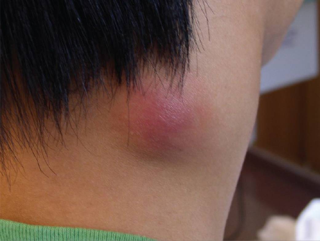
Figure 31-6 Tuberculous adenitis. Note the erythematous swelling in this 13-year-old recent immigrant from southeast Asia who presented with bilateral posterior cervical neck masses. (Courtesy of Esther K. Chung, MD, MPH.)
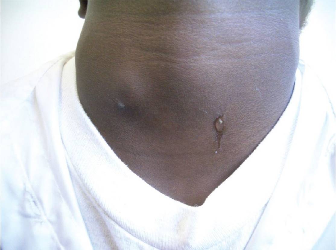
Figure 31-7 Branchial cleft cyst. Draining branchial cleft sinus. (Courtesy of Paul S. Matz, MD.)
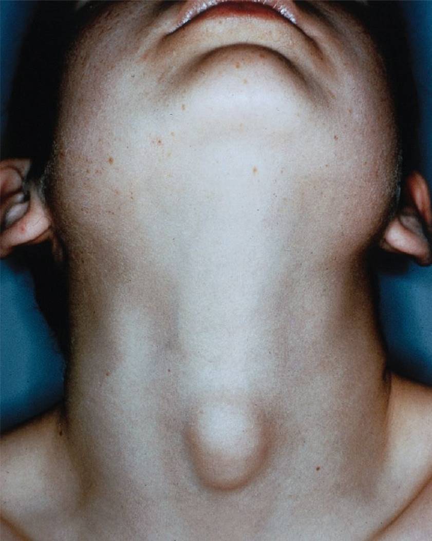
Figure 31-8 Thyroglossal duct cyst. A midline cervical mass presenting in a 6-year-old child. (Used with permission from Snell RS. Clinical Anatomy. 7th ed. Baltimore, MD: Lippincott Williams & Wilkins; 2005:CD418.)

Figure 31-9 Dermoid cyst. A mass found midline overlying the hyoid bone in a 4-year-old child. (Courtesy of Mary L. Brandt, MD.)
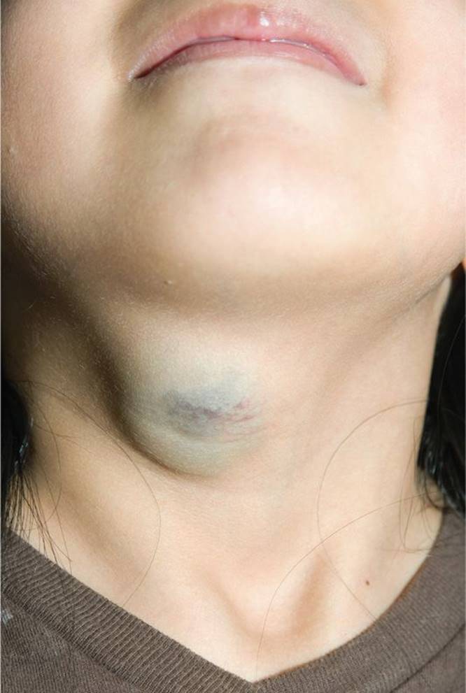
Figure 31-10 Venous malformation. (Courtesy of Eden Palmer and Jonathan Perkins, DO.)
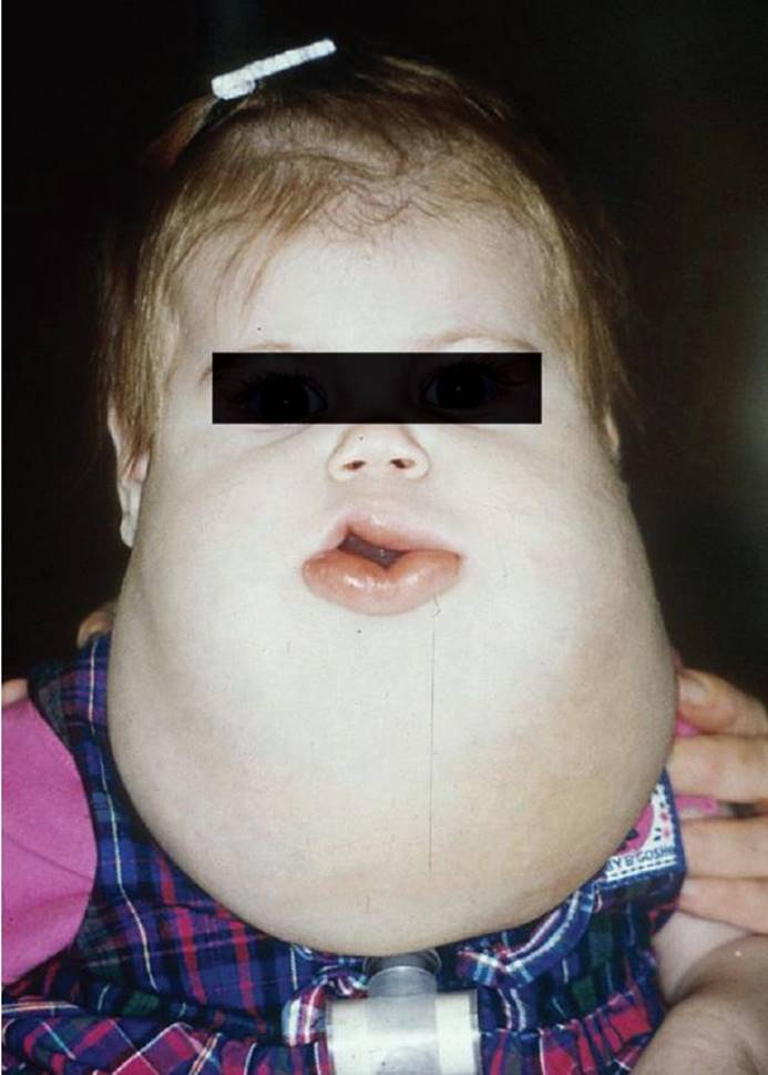
Figure 31-11 Cystic hygroma. A large, soft cervical mass in a 9-month-old infant. Tracheostomy was placed at birth. (Courtesy of Ellen Deutsch, MD.)
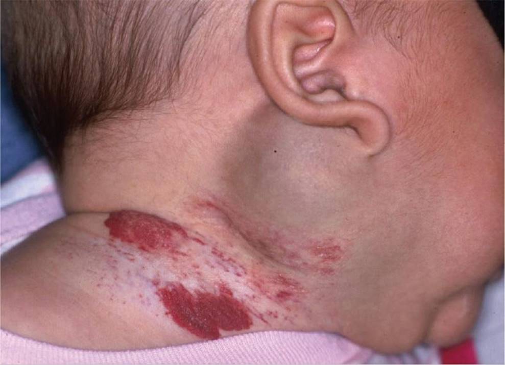
Figure 31-12 Hemangioma. A soft, nontender mass with bluish as well as bright-red aspects of color presents in this 1-month-old infant. (Courtesy of Ellen Deutsch, MD.)
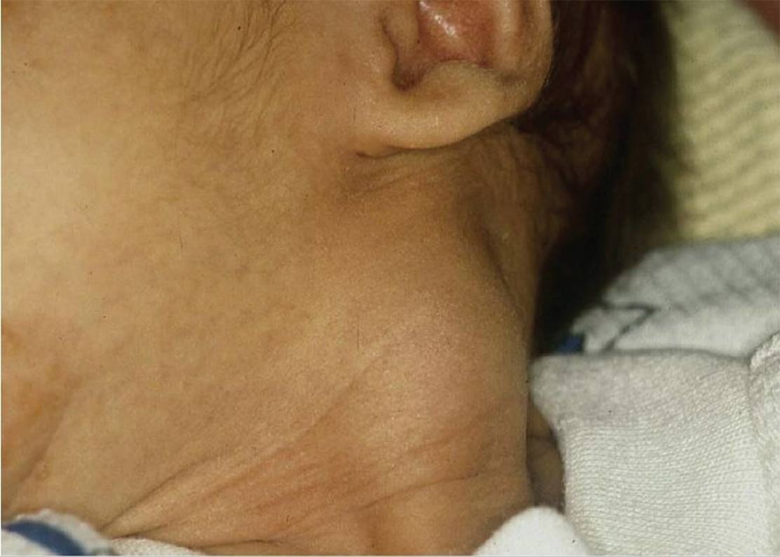
Figure 31-13 Congenital torticollis. A fibrotic mass located in the sternocleidomastoid muscle (SCM) of a 1-month-old infant whose mother was concerned about the infant’s head tilt to one side. (Courtesy of Ellen Deutsch, MD.)
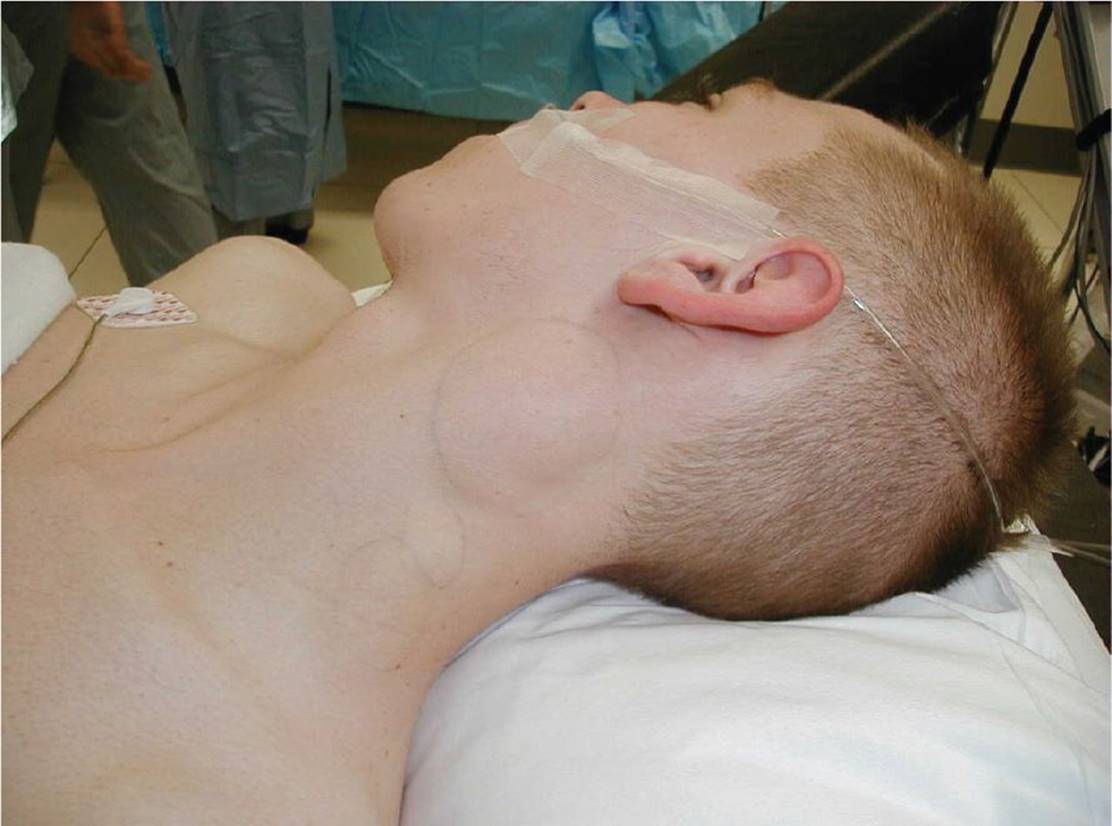
Figure 31-14 Hodgkin lymphoma. Large, fixed cervical masses in a 14-year-old adolescent with weight loss. (Courtesy of Mary L. Brandt, MD.)

Figure 31-15 Thyroid lymphoma. (Courtesy of Eden Palmer and Jonathan Perkins, DO.)
DIFFERENTIAL DIAGNOSES


Other Diagnoses to Consider
• Less common causes of infection/lymphadenopathy:
• Viral: VZV, HSV, HIV, measles, and mumps
• Bacterial: anaerobes, enteric bacteria (Acinetobacter, E. coli, Proteus, Salmonella, Shigella), zoonoses (Brucella, Francisella tularensis, Yersinia pestis, Yersinia enterocolitica), spirochetes, Rickettsiae, and leptospirosis
• Mycobacterial: tuberculosis
• Fungal: Aspergillus, Histoplasma, Cryptococcus, and coccidiomycosis
• Protozoa: Toxoplasma, Leishmania, and Trypanosoma
• Immunodeficiencies (Hyper IgE, leukocyte adhesion deficiency, chronic granulomatous disease)
• Pyriform sinus
• Other vascular anomalies (e.g., fast-flow AV malformations)
• Benign tumors: lipoma, fibroma, benign thyroid nodules, and neurofibromas
• Other malignant neoplasms
• Neuroblastoma, thyroid carcinoma, melanoma, neurogenic tumors, and rhabdomyosarcoma
• Cervical rib
• Kawasaki disease
• Ectopic thyroid
• Connective tissue/autoimmune diseases (lupus, juvenile idiopathic arthritis, and dermatomyositis)
• Serum sickness
When to Consider Further Evaluation or Treatment
• Enlargement of supraclavicular or posterior cervical nodes
• Systemic symptoms concerning for malignancy (e.g., night sweats, weight loss)
• Painless, firm, immobile nodes
• Generally, a node larger than 2 cm
• A single, enlarged node (>1 cm) that does not improve/resolve within 4 to 6 weeks with appropriate antibiotic therapy
SUGGESTED READINGS
Abramowicz S. Padwa BL. Vascular anomalies in children. Oral Maxillofac Surg Clin N Am. 2012;24(3):443–455.
American Academy of Pediatrics. Red Book: 2012 Report of the Committee on Infectious Diseases. 29th ed. Elk Grove Village, IL: American Academy of Pediatrics; 2012:269–271, 736–767.
Dickson PV, Davidoff AM. Malignant neoplasms of the head and neck. Sem Pediatr Surg. 2006;15(2):92–98.
Friedmann AM. Evaluation and management of lymphadenopathy in children. Pediatr Rev. 2008;29(2):53–60.
Gross E, Sichel JY. Congenital neck lesions. Surg Clin North Am. 2006;86(2):383–392.
Gujar S, Gandhi D, Mukherji SK. Pediatric head and neck masses. Topics Magn Resonan Imag. 2004;15(2):95–101.
Kliegman R, Behrman R, Jenson H, et al. Nontuberculous mycobacteria. In: Nelson Textbook of Pediatrics. 19th ed. 2011:1011–1016.
Kliegman R, et al. Cat-scratch disease (Bartonella henselae). In: Nelson Textbook of Pediatrics. 19th ed. 2011:983–986.
Koch BL. Cystic malformations of the neck in children. Pediatr Radiol. 2005;35(5):463–477.