
Approach to the Problem
An abdominal mass in an infant or child has a long differential diagnosis and includes a spectrum of conditions. These conditions range from those that are benign and self-resolving to those that are life threatening and require urgent intervention. These illnesses involve the many organ systems contained in the abdominal cavity, and the pathophysiology of each disease is diverse.
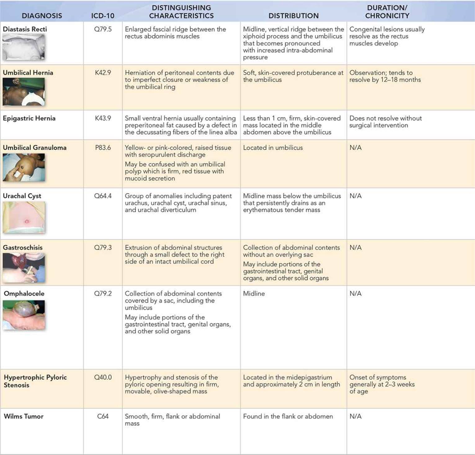
The etiology of some bulges is related to normal processes that occur during fetal development that fail to complete. For example, omphaloceles and gastroschisis can result from the normal process of gastrointestinal development that begins outside the fetus and then reenters the abdomen. Urachal cysts are remnants of the allantois that forms the umbilicus in fetal development. Diastasis recti and umbilical hernias are due to failure of abdominal wall musculature to fully close. In the majority of cases, these midline bulges resolve spontaneously.
Key Points in the History
• Diastasis recti is common in newborns.
• Umbilical hernias are more prevalent in preterm (up to 75% of <1,500 g birth weight) and African American infants (20% vs 3% of Caucasian neonates).
• Omphaloceles are often associated with chromosomal disorders, including trisomy 13, 18, and 21, and with Beckwith–Wiedemann syndrome, whereas gastroschisis often occurs in isolation.
• Hypertrophic pyloric stenosis presents at about 2 to 6 weeks of life with projectile nonbilious vomiting.
• Persistent urachus and other urachal anomalies can present with a persistently draining umbilical cord.
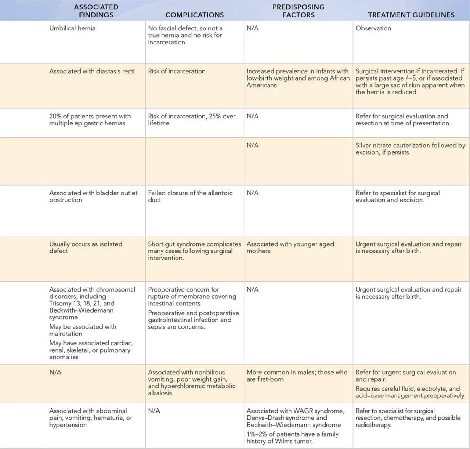
• About 1% to 2% of patients have a positive family history in patients with Wilms tumor.
Key Points in the Physical Examination
• Diastasis recti presents as a midline, vertical ridge that can extend between the xiphoid process and the umbilicus, when an infant cries.
• An epigastric hernia is often located in the middle abdomen above the umbilicus and may occur in multiples.
• Umbilical granulomas can have a seropurulent discharge on yellow- or pink-colored tissue and are distinguished from umbilical polyps in that they usually resolve following treatment with silver nitrate.
• The classic finding for hypertrophic pyloric stenosis is a firm, movable, olive-shaped mass in the midepigastrium about 2 cm in length.
• Persistent urachus may have urine or stool in the drainage, if present.
|
PHOTOGRAPHS OF SELECTED DIAGNOSES |
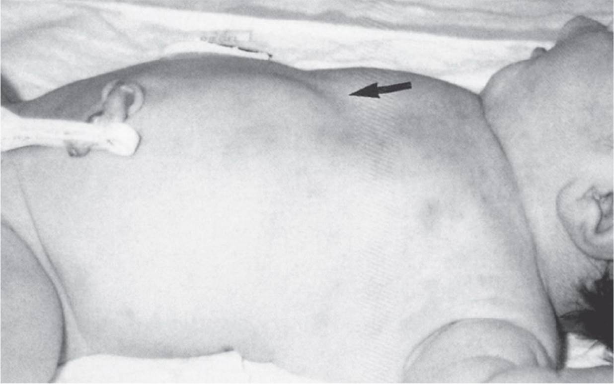
Figure 35-1 Diastasis recti. (Used with permission from Fletcher MA. Physical Diagnosis in Neonatology. Philadelphia, PA: Lippincott–Raven Publishers; 1998:357.)
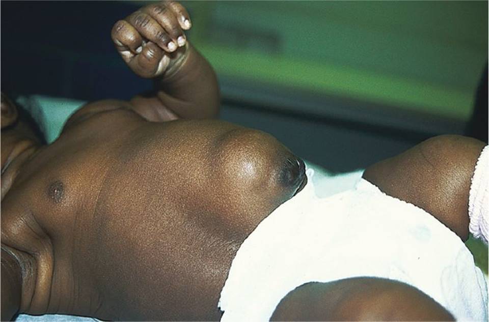
Figure 35-2 Umbilical hernia and diastasis recti. (Courtesy of George A. Datto, III, MD.)
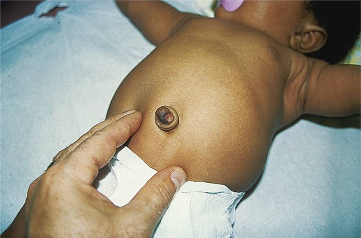
Figure 35-3 Umbilical granuloma. Pink granulation tissue at the base of the umbilicus. (Courtesy of George A. Datto, III, MD.)

Figure 35-4 Umbilical granuloma following treatment with silver nitrate. (Courtesy of George A. Datto, III, MD.)

Figure 35-5 Infected urachal cyst. Infected urachal cyst in a 3-month old infant. (Courtesy of Ben Alouf, MD.)

Figure 35-6 Gastroschisis. (Courtesy of Douglas Katz, MD.)
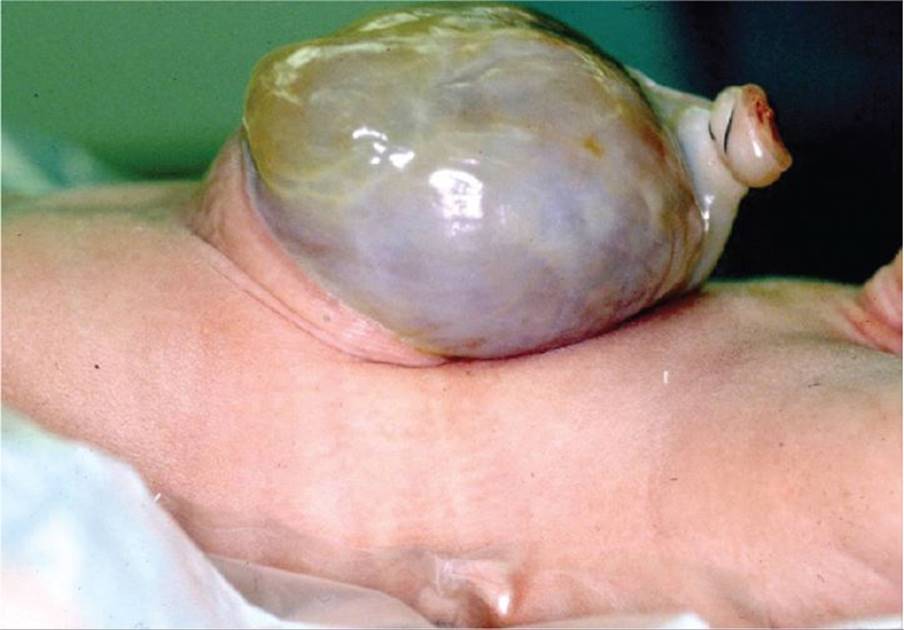
Figure 35-7 Omphalocele. (Courtesy of Douglas Katz, MD.)
DIFFERENTIAL DIAGNOSIS
Other Diagnoses to Consider
• Intestinal duplication
• Polycystic kidney disease
• Neuroblastoma
• Omphalomesenteric cyst or fistula
• Hepatic masses (hepatic hemangioma, hepatic mesenchymal hamartoma, hepatoblastoma, and choledochal cyst)
• Hydrocolpos/hydrometrocolpos, fused hymen resulting in the formation of a mass after menarche
• Ovarian mass, when large, may be palpable in the midline
When to Consider Further Evaluation or Treatment
• Epigastric hernias are at high risk of incarceration. Patients with these should be referred to pediatric surgery for further evaluation.
• Umbilical hernias have a 5% risk of strangulation, incarceration, or evisceration. Hernias smaller than 1.5 cm in diameter are twice as likely to become incarcerated.
• Umbilical hernias that present with symptoms, are very large, or persist beyond 4 to 5 years of age and are generally repaired.
• Silver nitrate can be used to cauterize an umbilical granuloma. If it persists, surgical resection can be considered.
• Ultrasound can be used to confirm the diagnosis of pyloric stenosis. In pyloric stenosis, the loss of electrolytes requires fluid and electrolyte resuscitation and acid–base management preoperatively.
• Hepatic, flank, and pelvic masses often require multidisciplinary approaches to diagnosis and management.
SUGGESTED READINGS
Brodeur AE, Brodeur GM. Abdominal masses in children: Neuroblastoma, Wilms’ tumor, and other considerations. Pediatr Rev. 1991;12:196–206.
Geller E, Kochan PS. Renal neoplasms of childhood. Radiol Clin North Am. 2011;49(4):689–709.
Ipp LS, Taubel D, Walters H, et al. Adolescent with an abdominal mass. Adolesc Med State Art Rev. 2012;23(2):285–289.
Kelly KB, Ponsky TA. Pediatric abdominal wall defects. Surg Clin North Am. 2013;93(5):1255–1267.
Ladino-Torres MF, Strouse PJ. Gastrointestinal tumors in children. Radiol Clin North Am. 2011;49(4):665–677.
Pomeranz A. Anomalies, abnormalities, and care of the umbilicus. Pediatr Clin North Am. 2004;51(3):819–827.
Salameh JR. Primary and unusual abdominal wall hernias. Surg Clin North Am. 2008;88(1):45–60.
Schwartz MZ, Shaul DB. Abdominal masses in the newborn. Pediatr Rev. 1989;11:172–179.