
Approach to the Problem
An increase in the girth of a child’s abdomen, abdominal distension, may be physiologic (e.g., swallowed air, lumbar lordosis in toddlers) or pathologic (e.g., intestinal obstruction, functional or mechanical). It occurs at all ages and in healthy, acutely ill, and chronically ill children. Abdominal distension can be the result of accidental and nonaccidental trauma. For example, a pneumomediastinum may occur in a child on a ventilator, or a pneumoperitoneum may be the result of blunt trauma. The mechanisms that result in abdominal distension are intraluminal or extraluminal accumulation of air or fluid, intra-abdominal mass, organomegaly, ascites, and abdominal wall hypotonia. Rapid recognition of pathologic abdominal distension is essential to reducing morbidity and mortality. Imaging studies and laboratory tests often have a role in determining the etiology of a child’s enlarged abdomen.
Key Points in the History
• A child typically complains of minimal pain with an ileus, but significant pain with an intestinal obstruction.
• Assume bilious vomiting is secondary to intestinal obstruction until proven otherwise.
• Delayed passage of meconium (after 48 hours of life) in the newborn period is highly concerning for Hirschsprung disease, gastrointestinal structural abnormality, cystic fibrosis, and hypothyroidism.
• Hematemesis, melena, and jaundice are clinical features of portal hypertension.
• Failure to thrive, rapid weight loss or gain, fever, fatigue, irritability, and bone pain suggests malignancy.
• Absence of historical details in an ill child with abdominal distension is a red flag for nonaccidental trauma.
• Gastrointestinal infections, pneumonia, and peritonitis with recent history of surgery may be associated with paralytic ileus.
• Ovulation may precede onset of menses, and therefore pregnancy should be considered in all pubertal females with lower abdominal distension. Cyclical distension, with or without abdominal pain, may represent hemato(metro)colpos.
• Pica-induced bezoars should be considered in neurologically or psychologically impaired children.
• Children with hemolytic disease are at risk for distension from splenomegaly.
• A prenatal history of oligohydramnios may result in distal urinary obstruction in the newborn, whereas polyhydramnios is associated with upper intestinal obstruction.
• History of abdominal surgery puts a child at risk for adhesions and small-bowel obstruction.
• Family history should include asking about metabolic diseases, early infant death among relatives, polycystic kidney disease, and cystic fibrosis.
• Constipation and ileus can be caused by misuse of medications (e.g., tricyclic antidepressants, antihistamines, antidiarrheal agents), ingestion of herbal products contaminated with belladonna alkaloids (e.g., teas, meat seasonings, stews), and exposure to anticholinergic substances through recreational drug use (e.g., smoking jimsonweed, use of jimsonweed-laced heroin).
• A history of greasy, foul-smelling stools in the setting of abdominal distension suggests malabsorption.
Key Points in the Physical Examination
• Ascites, flatus, and ileus present with symmetrical abdominal distension.
• Malignancy, constipation, pregnancy, and organomegaly may present with more localized abdominal distension.
• Visible peristaltic waves may be seen with intestinal obstruction.
• To detect abdominal organomegaly, it is important to begin palpation in the lowest part of the abdomen and then proceed upward toward the chest.
• Bowel sounds are decreased or absent in ileus and hyperactive in intestinal obstruction.
• Consider a rectal examination in the evaluation of abdominal distension as the presence of hard stool is suggestive of functional constipation or a pelvic mass. An empty rectal vault in the setting of constipation is suggestive of Hirschsprung disease.
• Pallor and nail clubbing are physical findings that suggest chronic malabsorption.
• Spider nevi, palmar erythema, and jaundice suggest chronic liver disease.
• Prominent abdominal superficial veins may represent portal hypertension or an obstruction in systemic venous return.
|
PHOTOGRAPHS OF SELECTED DIAGNOSES |
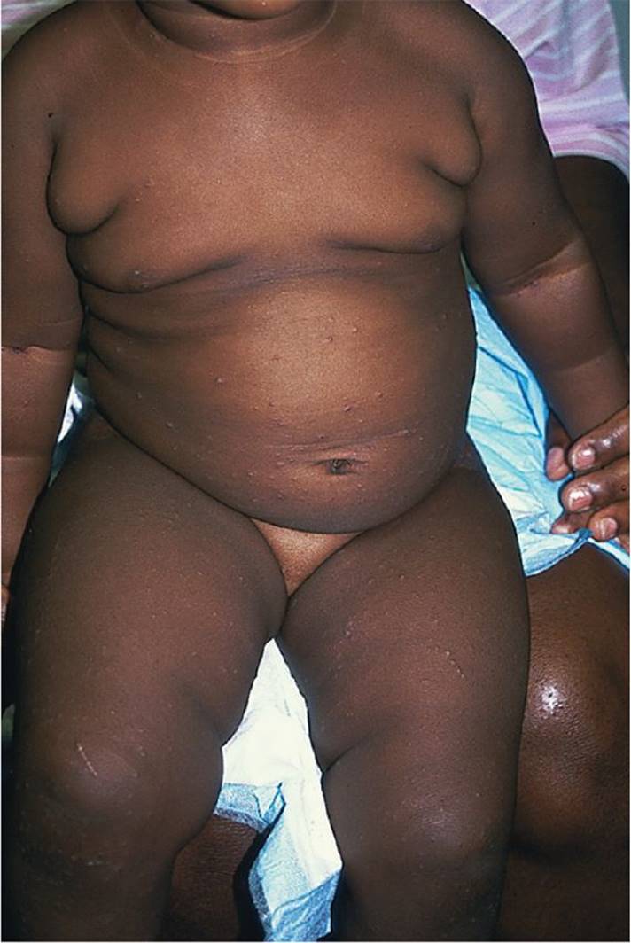
Figure 36-1 Obesity. Enlarged abdomen from increased adiposity. (Courtesy of George A. Datto, III, MD.)
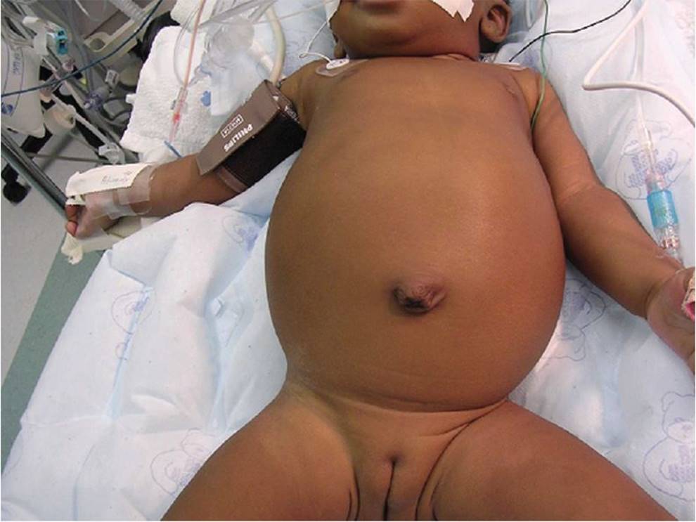
Figure 36-2 Ascites. Large distended abdomen in an infant. (Courtesy of Vani V. Gopalareddy, MD.)
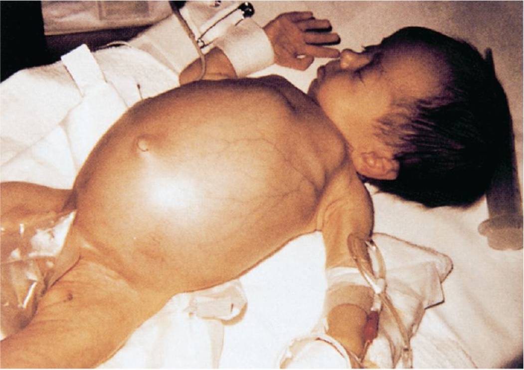
Figure 36-3 Abdominal distension resulting from hepatomegaly in a child with untreated galactosemia. Note that the distension is more prominent in the upper abdomen. (Used with permission from Fletcher MA. Physical Diagnosis in Neonatology. Philadelphia, PA: Lippincott–Raven Publishers; 1998:353.)
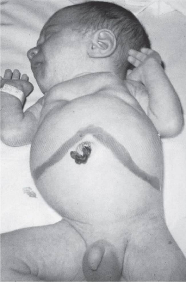
Figure 36-4 Abdominal distension resulting from massive hepatosplenomegaly in an infant with congenital cytomegalovirus infection. Note that the distension is more prominent in the upper abdomen. (Used with permission from Fletcher MA. Physical Diagnosis in Neonatology. Philadelphia, PA: Lippincott–Raven Publishers; 1998:354.)

Figure 36-5 Two young boys suffering from visceral leishmaniasis (Leishmania chagasi) from Brazil. Both have abdominal distension due to hepatosplenomegaly. (Courtesy of WHO. Photograph by Dr. P. Marsden. WHO/TDR/Marsden.)
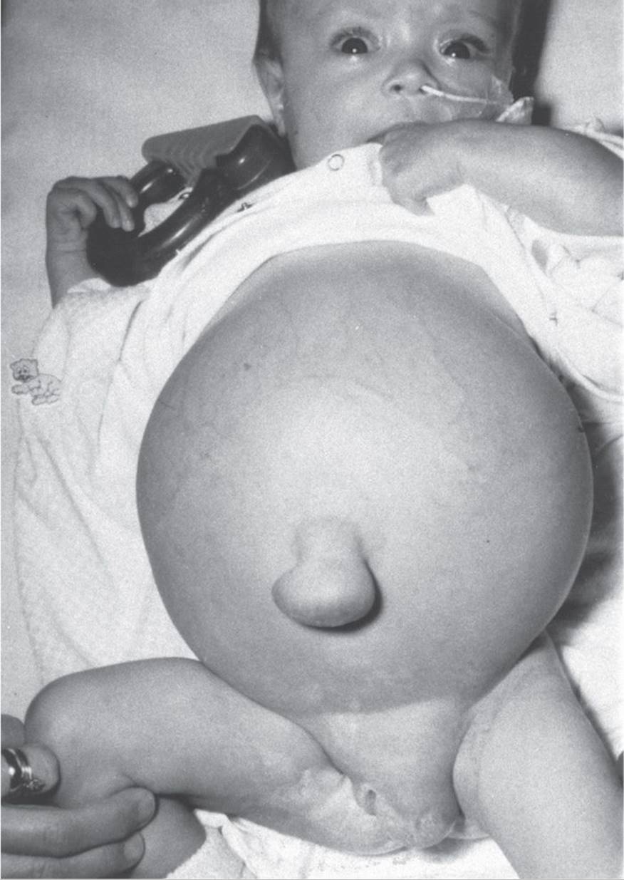
Figure 36-6 Female infant with cirrhosis due to biliary atresia. Note abdominal distension, umbilical hernia, and labial swelling due to massive ascites. McMillan JA, Feigin RD, DeAngelis C, et al. Oski’s Solution. Philadelphia, PA: Lippincott, Williams & Wilkins; 2006.

Figure 36-7 Abdominal distension in an infant with anasarca. Note that the distension is more prominent in the flanks. (Used with permission from Fletcher MA. Physical Diagnosis in Neonatology. Philadelphia, PA: Lippincott-Raven Publishers; 1998:355.)

Figure 36-8 Abdominal distension at the flanks in an infant with prune belly syndrome. (Used with permission from Fletcher MA. Physical Diagnosis in Neonatology. Philadelphia, PA: Lippincott-Raven Publishers; 1998:355.)
DIFFERENTIAL DIAGNOSIS

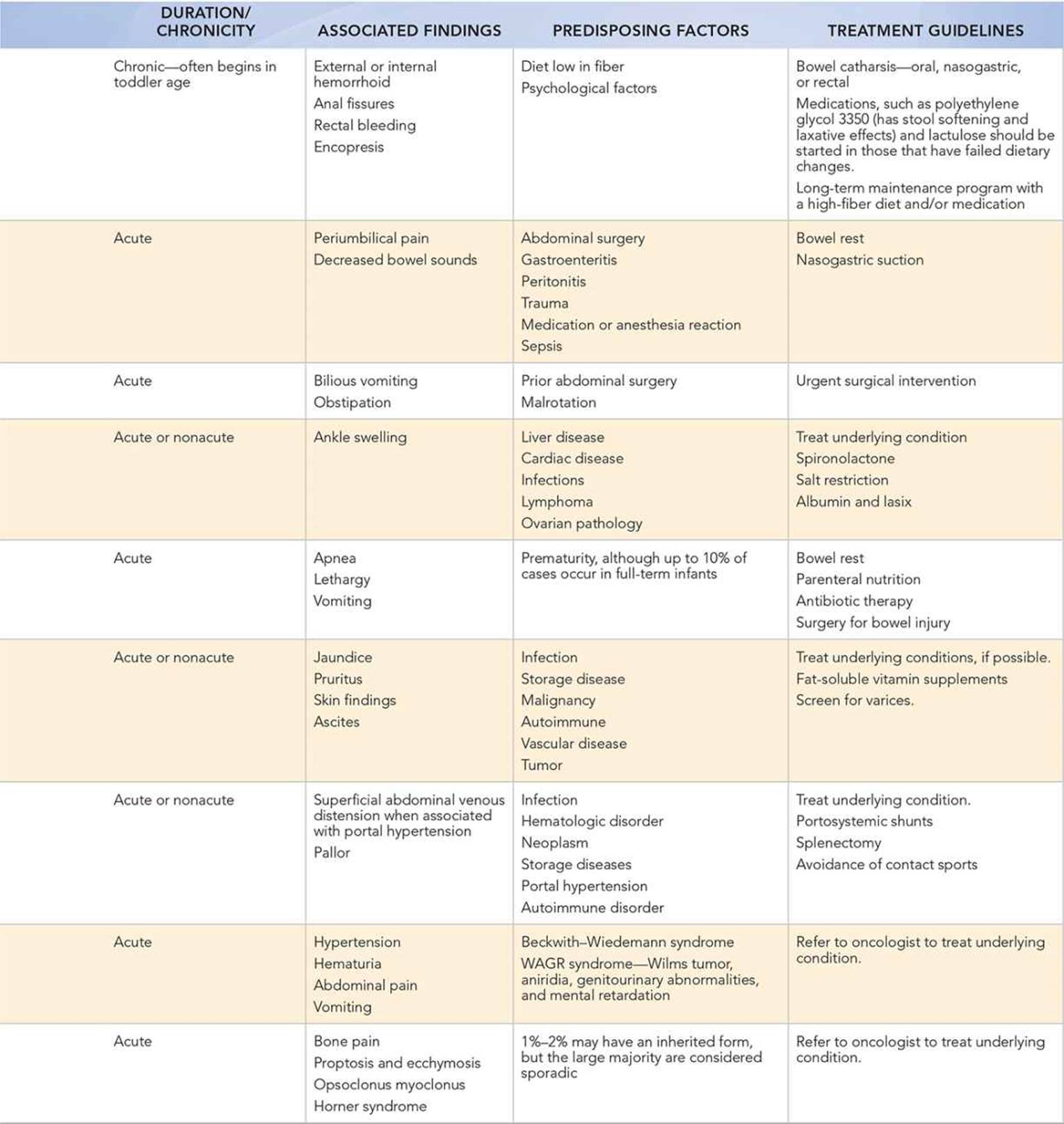
Other Diagnoses to Consider
• Ruptured appendicitis
• Intussusception
• Hirschsprung disease, toxic megacolon
• Malrotation with volvulus
• Incarcerated hernia
• Surgical adhesions
• Obstructed bladder
• Polycystic kidney disease
• Hydronephrosis
• Ovarian mass, cyst
• Pregnancy
• Hematocolpos
• Neoplasms, lymphoma
• Trauma
• Pulmonary hyperinflation
• Bronchiolitis
• Asthma exacerbation
• Anasarca
When to Consider Further Evaluation or Treatment
• Abdominal distension in a newborn accompanied by bilious vomiting is a surgical emergency. An upper gastrointestinal series must be emergently performed to rule out malrotation with or without volvulus.
• If intussusception is suspected, an obstruction series should be performed to rule out perforation, and then an ultrasound and/or air enema should be obtained to make the diagnosis.
• The presence of ascites warrants consultation with pediatric gastroenterology and an ultrasound to identify the cause. A diagnostic paracentesis may be necessary.
• CT scan may be required when history, physical exam, laboratory studies, and other imaging have failed to identify the etiology of abdominal distension.
SUGGESTED READINGS
Avner JR. Abdominal distention. In: Fleischer GR, Ludwig S, et al., eds. Textbook of Pediatric Emergency Medicine. 6th ed. Philadelphia, PA: Lippincott Williams & Wilkins; 2010:153–159.
Belamarich PF. Abdominal distention. In: McInerny TK, Adam HM, Campbell DE, et al., eds. American Academy of Pediatrics Textbook of Pediatric Care. Elk Grove Village, IL: American Academy of Pediatrics; 2008:1369–1376.
Juang D, Snyder CL. Neonatal bowel obstruction. Surg Clin North Am. 2012;92:658–711.
Lambert SM. Pediatric urological emergencies. Pediatr Clin North Am. 2012;59:965–976.
Pepper VK, Stanfill AB, Pearl RH. Diagnosis and management of pediatric appendicitis, intussusception, and Meckel diverticulum. Surg Clin North Am. 2012;92:505–526.
Schonfeld D, Lee LK. Blunt abdominal trauma in children. Curr Opin Pediatr. 2012;24:314–318.