
Pediatricians encounter a vast array of nail disorders in clinical practice. Basic knowledge of the common nail disorders is pivotal for assessment, diagnosis, and referral when necessary. Nail pathology can result from focal or systemic abnormalities. The nail can be directly affected by localized infection, inflammation, mechanical forces, or trauma. Alternatively, nail pathology can result from nutritional deficits, genetic abnormalities, systemic disease, and medication or toxin exposures.
Nails are functional, structural, and cosmetic components of human anatomy. The nail plate is a translucent arrangement of compressed, keratinized dead cells. Nail folds are specialized epithelium that provide structure and protect the nail plate and matrix. The lateral nail fold is the paronychium, and the proximal nail fold is called the eponychium or cuticle. Underlying the eponychium is the nail matrix that contains the germinal cells. It takes 20 days for the fingernail and 80 days for the toenail to emerge from the matrix. Matrix to free edge growth for the fingernail is close to 6 months and 12 to 18 months for the toenail. This growth interval is important to consider for the timing of a potential insult and deciding the treatment course.
Key Points in the History
• Cyanotic congenital heart defects, chronic lung disease, and gastrointestinal disorders are associated with digital clubbing.
• Dermatological disorders such as psoriasis, eczema, alopecia areata, and lichen planus are associated with nail pitting and dystrophy.
• Disease states and certain medications, such as Kawasaki disease, hand–foot–mouth disease, pneumonia, prolonged high fevers, and chemotherapy, can cause a temporary growth arrest in the nail that is later manifested as Beau lines.
• There are benign familial forms of digital clubbing and koilonychias.
• Given the time it takes for nails to emerge from the matrix and grow, a history of medication or toxin exposure should be retrospective and encompassing.
• Nails are porous, and cosmetic products and frequent washing can strip and weaken the nail.
• Nail salons can be the source of fungal and bacterial nail infections.
• Koilonychia is associated with iron deficiency anemia in the adult population.
• Repeated local trauma can impact the growth and integrity of the nail.
Key Points in the Physical Examination
• In digital clubbing, assess for cardiopulmonary pathology such as murmurs or wheezing.
• Lovibond angle, the angle of eponychium to the nail plate, is normally 160 degrees. Clubbing induces angles of greater than 180 degrees.
• Schamroth window is the normal space that is seen when the dorsal aspects of opposing fingers are aligned. This diamond-shaped space is absent in clubbing.
• Dermatologic disorders such as psoriasis, eczema, and lichen planus are associated with nail pitting and dystrophy.
• Inspect the shape, color, texture, and thickness of the nail. Thickened, yellowed nails should raise the suspicion for onychomycosis.
• The nail fold capillaries are dilated in rheumatoid disorders.
• Splinter hemorrhages can be the sign of repeated nail trauma, such as in sports, or more ominously in bacterial endocarditis.
• Local growths, lesions, or cysts can impact the growth and appearance of the nail.
|
PHOTOGRAPHS OF SELECTED DIAGNOSES |

Figure 39-1 Nail clubbing. (Left) The angle between the nail and the digit is normally about 20 degrees in a child. (Center) Flattened angle represents early stage of clubbing. (Right) In advanced clubbing, the nail is rounded over the end of the finger. Note also that the distal phalanx is bulbous and of greater depth than the proximal portion of the finger (interphalangeal depth). (Used with permission from Pillitteri A., Maternal and Child Nursing. 4th ed. Philadelphia, PA: Lippincott, Williams & Wilkins; 2003.)

Figure 39-2 Beau lines. Beau lines of a severe illness in past. Illness was 5 weeks ago, 5 mm distal to the lunula (plate grows 1 mm/week). (Used with permission from Berg D, Worzala K. Atlas of Adult Physical Diagnosis. Philadelphia, PA: Lippincott Williams & Wilkins; 2006.)
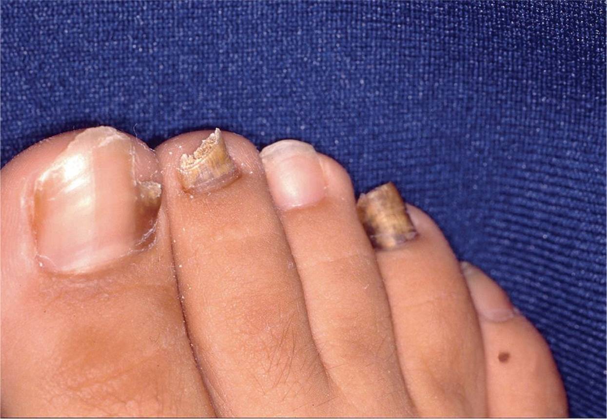
Figure 39-3 Onychomycosis. Note the yellow discoloration and thickening of the nail bed. (Courtesy of Denise W. Metry, MD.)

Figure 39-4 Nail pitting. (Courtesy of Dr. Barankin Dermatology Collection.)

Figure 39-5 Onycholysis. Psoriasis. Pitting and onycholysis are evident in this nail. (Used with permission from Goodheart HP. Goodheart’s Photoguide of Common Skin Disorders. 2nd ed. Philadelphia, PA: Lippincott Williams & Wilkins; 2003.)

Figure 39-6 Koilonychia/nail spooning. Note the concave shape of the nail bed. (Courtesy of Moise L. Levy, MD.)

Figure 39-7 Subungual splinter hemorrhages. (Used with permission from Gold DH, Weingeist TA. Color Atlas of the Eye in Systemic Disease. Baltimore, MD: Lippincott Williams & Wilkins; 2001.)
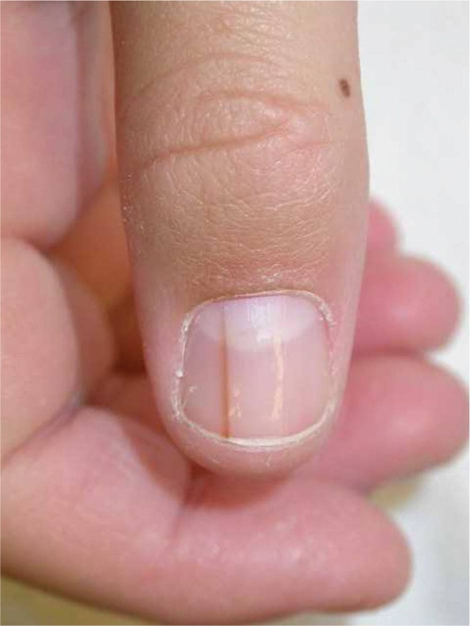
Figure 39-8 Longitudinal melanonychia. Note the longitudinal band of hyperpigmentation with no extension onto surrounding skin. (Courtesy of Moise L. Levy, MD.)
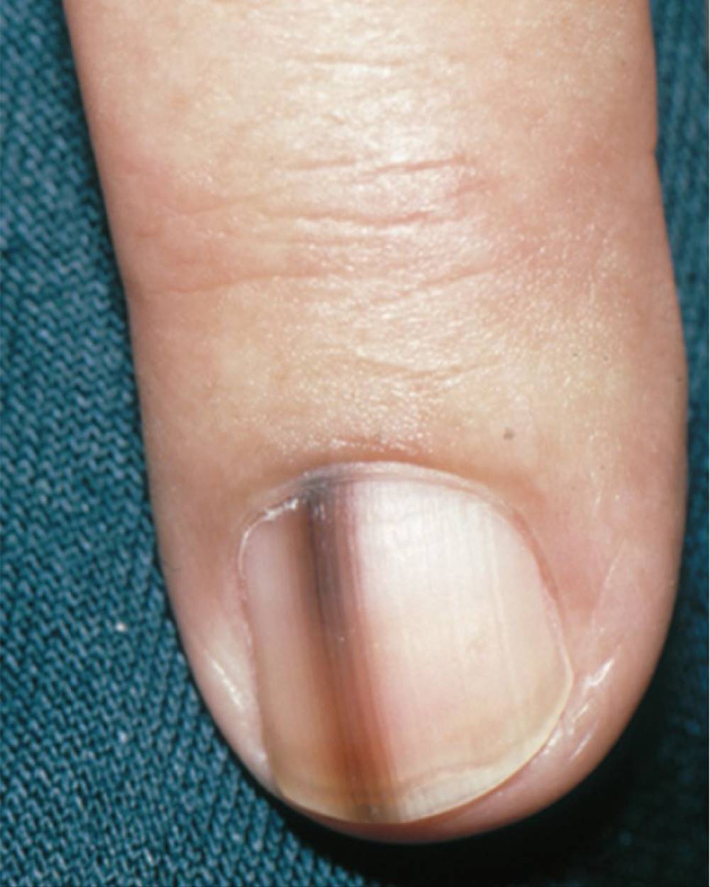
Figure 39-9 Junctional nevus. (Used with permission from Goodheart HP. Goodheart’s Photoguide to Common Skin Disorders. 2nd ed. Philadelphia, PA: Lippincott Williams & Wilkins; 2003:356.)
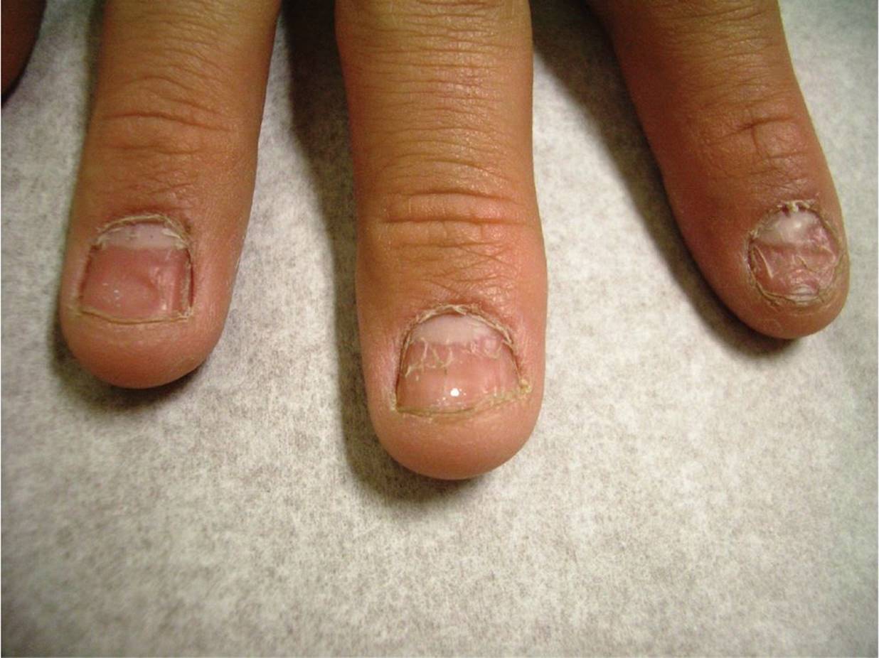
Figure 39-10 Nail dystrophy. A teenager with scaling of the nail suggestive of nail dystrophy. (Courtesy of Paul S. Matz, MD.)

Figure 39-11 Longitudinal ridging. This normal variant is characterized by ridging in all of the nails. (Used with permission from Goodheart HP. Goodheart’s Photoguide to Common Skin Disorders. 2nd ed. Philadelphia, PA: Lippincott Williams & Wilkins; 2003:233.)
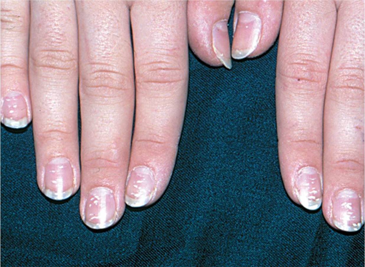
Figure 39-12 Leukonychia striata. (Used with permission from Goodheart HP. Goodheart’s Photoguide to Common Skin Disorders. 2nd ed. Philadelphia, PA: Lippincott Williams & Wilkins; 2003:240.)
DIFFERENTIAL DIAGNOSIS


Other Diagnoses to Consider
• Nail psoriasis
• Pachyonychia congenital (rare genetic disorder characterized by hypertrophic nail dystrophy)
• Onychomadesis (nail dystrophy)
• Leukonychia
• Muehrcke lines
When to Consider Further Evaluation or Treatment
• Children presenting with nail clubbing should be assessed for potential cardiac, pulmonary, or gastrointestinal issues.
• Immunodeficiency should be considered in cases of severe onychomycosis that are unresponsive to antifungal agents.
• Endocarditis should be considered in cases of painful splinter hemorrhages seen in conjunction with Osler nodes.
• Subungual masses should be carefully evaluated. Consider diagnostic imaging, biopsy, and referral to a dermatologist.
• Fungal cultures should be done in cases of suspected onychomycosis.
SUGGESTED READINGS
Piraccini BM, Iorizzo M. Drug reactions affecting the nail unit: diagnosis and management. Dermatol Clinic. 2007;25:215–221.
Piraccini BM, Starace M, Bruni F. Onychomycosis in children. Expert Rev Dermatol. 2012;7:569–578.
Shah KN, Rubin AI. Nail disorders as signs of pediatric systemic disease. Curr Probl Pediatr Adolesc Health Care. 2012;42:204–211.
Tosti A, Daniel R, Piraccini BM, et al. Color Atlas of Nails. Heidelberg: Springer-Verlag; 2010:31–38, 87–88, 103–111.
Zaidi Z, Lanigan SW. Nail disorders. Dermatology in Clinical Practice. London: Springer-Verlag, 2010:381–393.