
Approach to the Problem
Congenital midline back lesions such as sacral dimples, skin tags, hairy patches, hemangiomas, dermal sinuses, and lipomas are present in approximately 5% to 7% of infants. It is important to recognize and diagnose midline back lesions as they may correlate with a spinal dysraphism. Spinal dysraphisms are characterized as open or closed. Open lesions involve protrusion of the spinal cord or nerves through defects in the meninges or vertebral column. Examples of open lesions are spina bifida with meningocele or spina bifida with myelomeningocele. Closed lesions (occult dysraphisms) are improperly formed nervous system structures covered by skin. Examples of closed lesions are spina bifida occulta or isolated tethered cord. Closed lesions may have overlying cutaneous stigmata that signal an underlying defect. Spinal dysraphisms, such as tethered cord or dermal sinus, are important to identify early in order to prevent serious neurologic and infectious complications. Acquired midline back lesions, such as pilonidal cysts, occur in adolescence and may only be identified after infection.
Key Points in the History
• Midline back lesions are typically present at birth.
• Back lesions combined with the presence of neurologic symptoms such as bowel or bladder incontinence, extremity weakness, parasthesias, or gait abnormalities strongly suggest spinal dysraphism.
• Back lesions without the presence of neurologic symptoms do not rule out an underlying spinal defect.
• Long-standing tethered cord may result in asymmetric lower extremity growth and neurologic sequelae.
• Erythema, warmth, and tenderness surrounding a midline back lesion in an adolescent may suggest an infected pilonidal cyst.
• An infected congenital dermal sinus tract may be the cause of recurrent meningitis.
Key Points in the Physical Examination
• Simple sacral dimples are located <2.5 cm from the anus and are within the gluteal cleft.
• Atypical sacral dimples are >2.5 cm from the anus, have a diameter >5 mm, and may be associated with other cutaneous markers.
• Vascular nevi—salmon patches, nevus flammeus—are patches of pink to red discolorations of the skin.
• Hemangiomas on the back are soft masses of vascular endothelial overgrowth that may have a blue or reddish discoloration.
• Lumbosacral hypertrichosis or “hairy patch” is often V-shaped and poorly circumscribed. Hair can be dark or light with a silky texture. This must be distinguished from the normal sacral hair seen in newborns.
• Subcutaneous lipomas on the back are palpated as homogeneous, soft masses that are flesh colored.
• Skin tags are small outgrowths of skin that may be located on the back.
• Dermoid sinuses present as small skin openings that lead to a narrow duct. Protruding hair, hairy patches, or vascular nevi may be present nearby.
• Deviation of the gluteal cleft suggests an underlying mass, such as a lipoma or myelomeningocele.
|
PHOTOGRAPHS OF SELECTED DIAGNOSES |
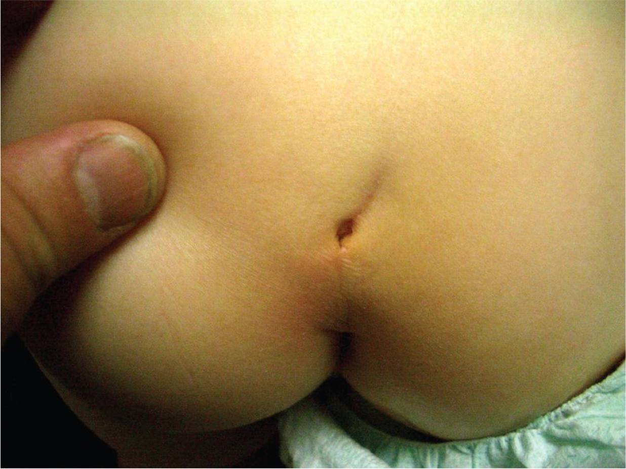
Figure 38-1 Sacral dimple. Shallow dimple visible within the gluteal fold. (Courtesy of Paul S. Matz, MD.)
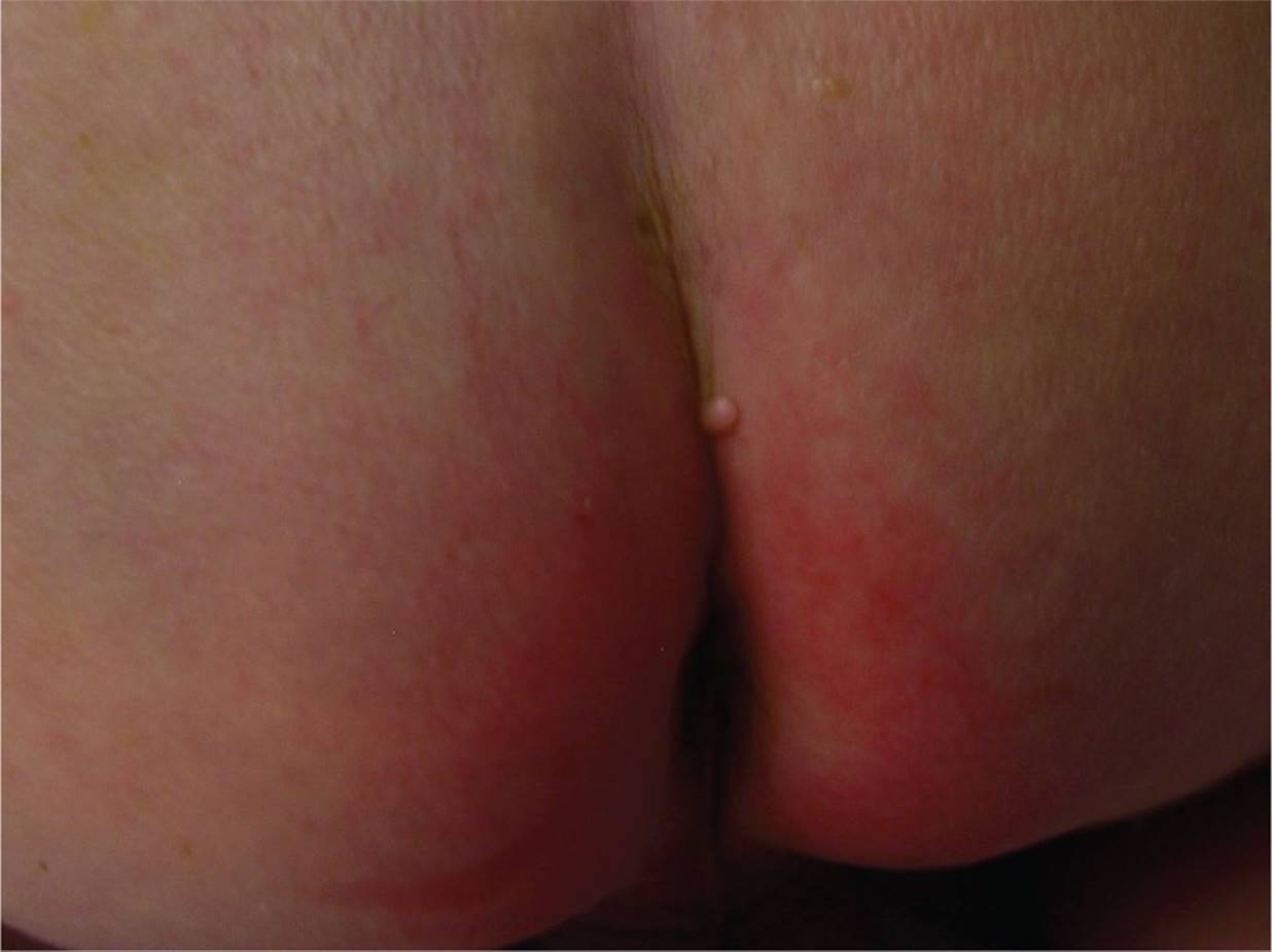
Figure 38-2 Sacral skin tag with no underlying spinal pathology. Note the isolated nodule within the gluteal fold. (Courtesy of Esther K. Chung, MD, MPH.)
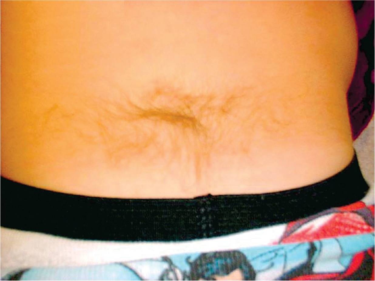
Figure 38-3 Hair tuft. (Courtesy of Joseph Piatt, MD.)

Figure 38-4 Infected dermal sinus connected to intramedullary dermoid cyst. Thoracic sinus associated with purulent discharge. (Courtesy of Joseph Piatt, MD.)
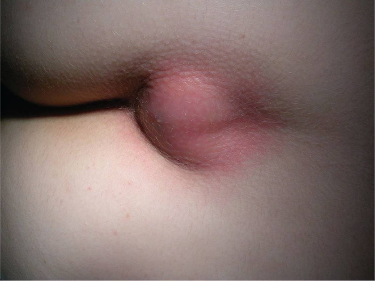
Figure 38-5 Infected pilonidal cyst. A large erythematous fluctuant mass visible at the superior portion of the gluteal fold. (Courtesy of Scott VanDuzer, MD.)
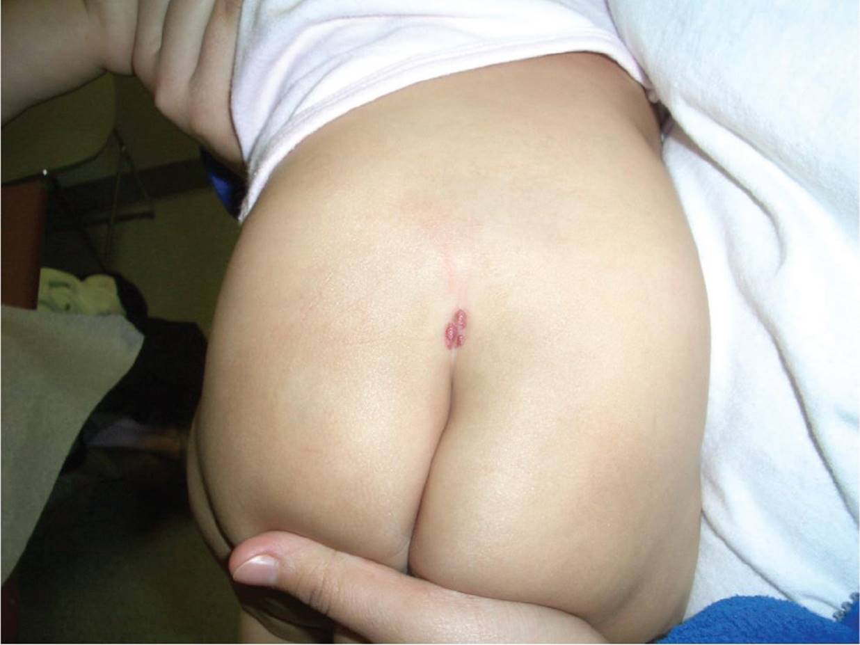
Figure 38-6 Sacral hemangioma. Vascular malformation visible overlying sacral spine. (Courtesy of Paul S. Matz, MD.)
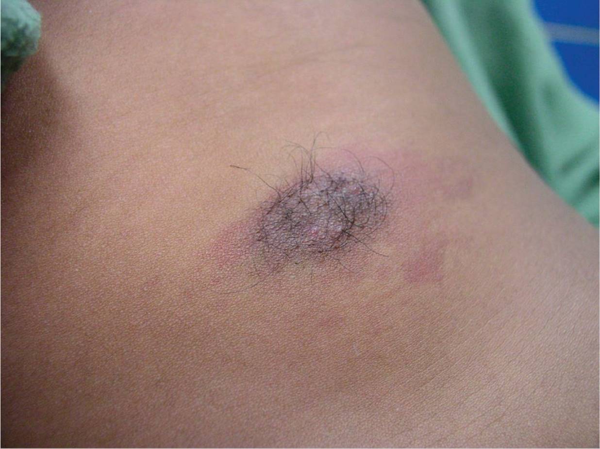
Figure 38-7 Lumbar hemangioma. This midline lesion was associated with a dermal sinus and an underlying tethered cord. (Courtesy of Esther K. Chung, MD, MPH.)
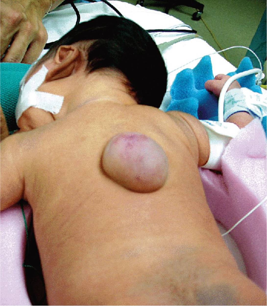
Figure 38-8 Thoracic meningocele. Large skin-covered thoracic mass in an infant. (Courtesy of Joseph Piatt, MD.)
DIFFERENTIAL DIAGNOSIS
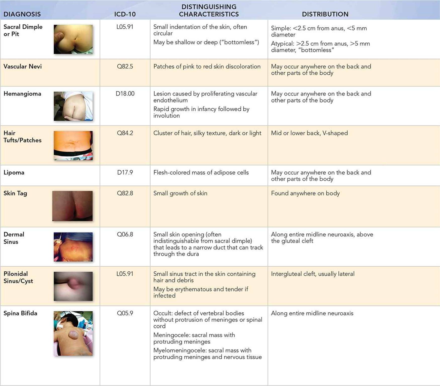
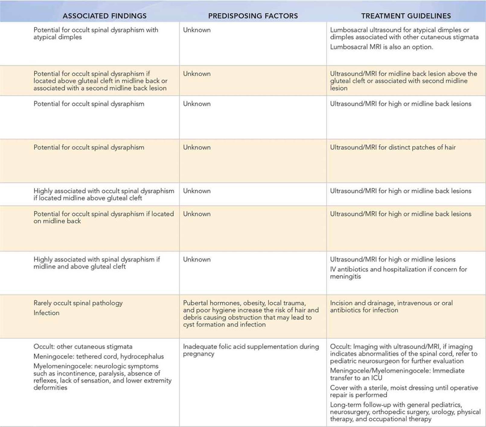
When to Consider Further Evaluation or Treatment
• Two or more congenital midline back lesions are strong indicators of spinal dysraphism.
• Atypical sacral dimples warrant further imaging.
• Simple sacral dimples with a visible base do not warrant further imaging.
• Midline back hairy patches, lipomas, skin tags, and hemangiomas may be indicative of occult spinal dysraphism and should be further evaluated with imaging.
• Vascular nevi that are located above the gluteal cleft in the midline back or associated with a second midline back lesion should be imaged.
• Tracts of a dermoid sinus may pass through the dura, increasing the risk for cerebrospinal infection.
• Any child with the presence of neurologic signs and symptoms in combination with a back lesion should be referred immediately for further imaging.
• Infants with spina bifida with meningocele or myelomeningocele should have coordinated care involving general pediatrics, neurosurgery, orthopedic surgery, urology, and physical and occupational therapy.
SUGGESTED READINGS
Ackerman LL, Menezes AH. Spinal congenital dermal sinuses: a 30-year experience. Pediatrics. 2003;112:641–647.
Drolet BA. Cutaneous signs of neural tube dysraphism. Pediatr Clin North Am. 2000;47(4):813–823.
Guggisberg D, Hadj-Rabia S, Viney C, et al. Skin markers of occult spinal dysraphism in children. Arch Dermatol. 2004;140:1109–1115.
Kinsman SL, Johnston MV. Spina bifida occulta. In: Kliegman RM, Stanton BF, St. Geme JW, et al., eds. Nelson Textbook of Pediatrics. 19th ed. Philadelphia, PA: Saunders Elsevier; 2011.
Medina LS, Kerry Crone K, Kuntz KM. Newborns with suspected occult spinal dysraphism: a cost-effectiveness analysis of diagnostic strategies. Pediatrics. 2001;108:e101.
Sardana K, Gupta R, Garg V, et al. A prospective study of cutaneous manifestations of spinal dysraphism from India. Pediatr Dermatol. 2009;26(6):688–695.
Schropp C, Sorensen N, Collman H, et al. Cutaneous lesions in occult spinal dysraphism—correlation with intraspinal findings. Childs Nerv Syst. 2006;22:125–131.
Zywicke HA, Rozelle CJ. Sacral dimples. Pediatr Rev. 2011;22(3):109–113.