
Approach to the Problem
Arm swelling can be a common manifestation of musculoskeletal, infectious, or rheumatologic diseases. The first consideration in a patient presenting with arm swelling is inquiry regarding a previous history of trauma. Traumatic events can result in injuries such as hematomas, sprains, and fractures. A clinician must also consider nonaccidental trauma in immobile children and in children presenting without a history to support a traumatic injury. Other potential diagnoses in patients presenting with arm swelling are infection and tumors. If more than one joint presents with swelling, consideration can be given to associated systemic diseases such as systemic lupus erythematosus, juvenile idiopathic arthritis, or inflammatory bowel disease. It is also very important to differentiate between conditions that might require urgent medical intervention.
Key Points in the History
• Presence of precipitating factors such as trauma or recent illness.
• For infants or patients presenting with an inconsistent history, nonaccidental trauma should be considered.
• Younger children who are not able to express pain may have a history of decreased use of the affected arm.
• Osteomyelitis, septic arthritis, and soft-tissue infections should be considered in patients who also present with subacute arm swelling, erythema, decreased arm movement, and fever. The most common bacteria in bone infections are Staphylococcus aureus and Group A streptococcus.
• Predisposing factors for soft-tissue infections include breaks in the skin from bites, abrasions, or preexisting skin conditions.
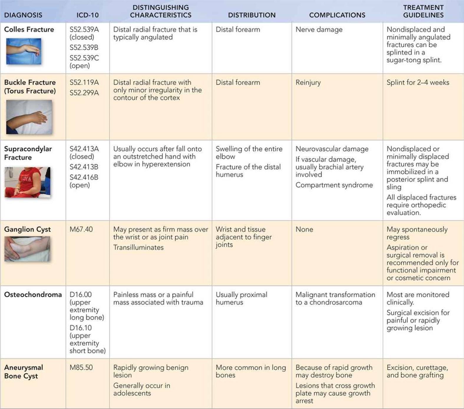
• It is common for distal radial, ulnar (Colles), greenstick, and buckle (torus) fractures to occur following trauma, such as falling on an outstretched hand, when a child is trying to protect himself/herself from a fall.
• Supracondylar fractures in children can be secondary to high-velocity injuries such as falling from a bicycle or playground equipment, such as monkey bars.
• Younger children usually sustain supracondylar fractures after shorter falls, such as fall from a bed or sofa.
• A ganglion cyst is a cystic swelling that can present as a nontender wrist mass. However, if it occurs near a joint, it can be painful.
• In the arm, osteochondromas are common benign, slow-growing, nonpainful, bone tumors that usually occur at the proximal humerus. Osteochondromas usually stop growing once the child reaches skeletal maturity.
• Children with a history of a bleeding disorder, such as hemophilia, may develop hemarthrosis or hematoma with minimal trauma.
• In a child with a history of a central venous line who presents with acute arm swelling, venous thromboembolism should be considered.
Key Points in the Physical Examination
• If a history of known trauma or nonaccidental trauma is suspected, the practitioner should perform a complete examination looking for other areas of trauma.
• In evaluating a child presenting with arm swelling, it is important to perform a complete neurovascular examination.
• Supracondylar fractures are at risk for nerve damage, so it is important to assess the median, radial, and ulnar nerves. The median nerve can be assessed by asking the patient to make the “OK” sign. The radial nerve can be assessed by asking the patient to make the “thumbs-up” sign. The ulnar nerve can be assessed by asking the patient to abduct all fingers against resistance.
• In distal forearm fractures, clinicians should also evaluate the elbow and wrist for associated fractures.
• Increased pain on passive extension of the fingers, cold hands, and excessive swelling can be suggestive of compartment syndrome in patients with supracondylar fractures.
• Ganglion cysts transilluminate and are typically firm, nontender, mobile nodules over the wrist.
• Osteochondromas and other bone tumors usually present as a discrete, painless, nonmobile mass.
|
PHOTOGRAPHS OF SELECTED DIAGNOSES |

Figure 41-1 Colles fracture. Wrist swelling with obvious deformity. (Courtesy of William Phillips, MD.)

Figure 41-2 Colles fracture. Distal radial fracture with dorsal angulation. (Courtesy of William Phillips, MD.)
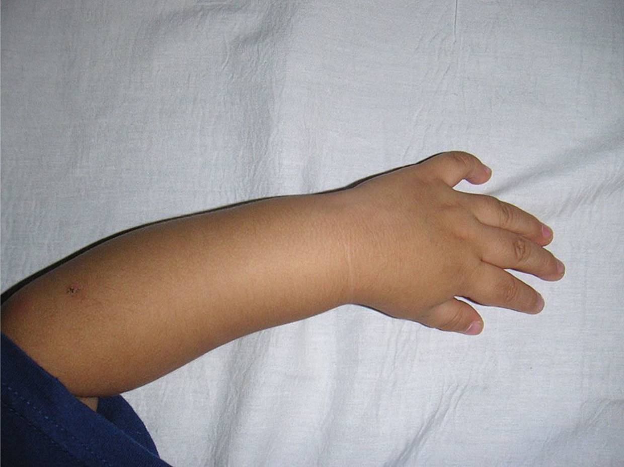
Figure 41-3 Torus fracture. (Courtesy of Michael C. Distefano, MD.)

Figure 41-4 Torus fracture. (Courtesy of Michael C. Distefano, MD.)

Figure 41-5 Supracondylar fracture. Note the swelling of the left elbow. (Courtesy of Michael C. Distefano, MD.)
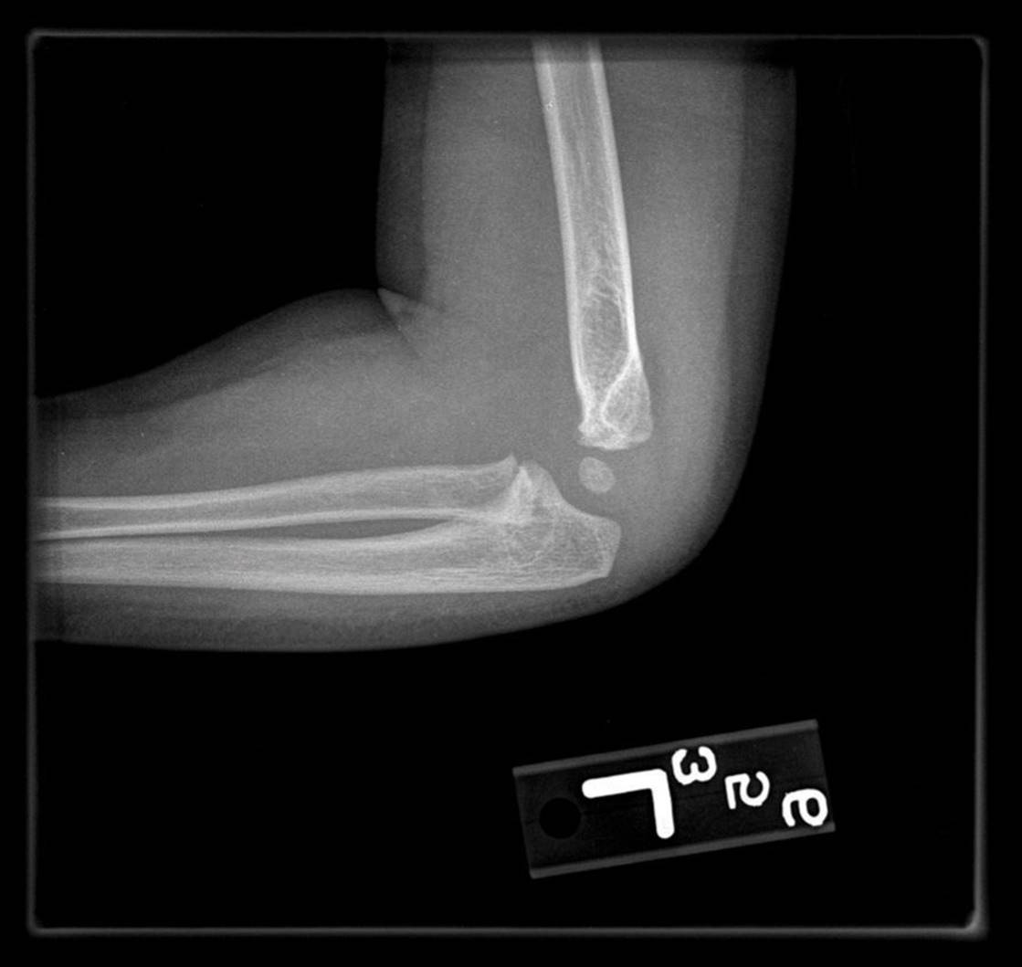
Figure 41-6 Supracondylar fracture. Left grade 1 supracondylar fracture (nondisplaced) with associated posterior fat pad. No other radiographic evidence of fracture. (Courtesy of Michael C. Distefano, MD.)

Figure 41-7 Ganglion cyst. Localized swelling over the volar surface of the wrist of a school-aged child. (Courtesy of Mary L. Brandt, MD.)

Figure 41-8 Ganglion cyst. Localized swelling over the volar surface of the wrist of an infant. (Courtesy of Mary L. Brandt, MD.)

Figure 41-9 Large hemangioma of right forearm of an infant. Swelling with raised areas of vascular prominence is present. (Courtesy of Moise L. Levy, MD.)
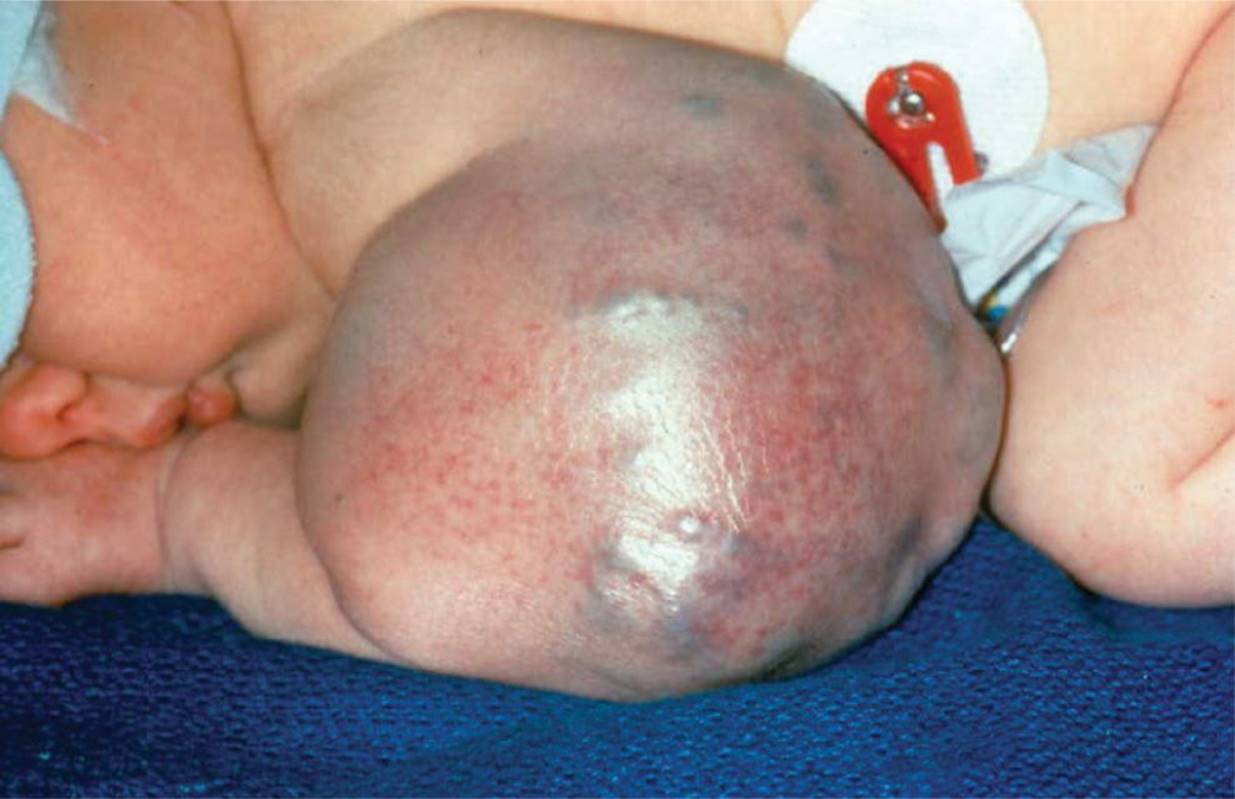
Figure 41-10 Cavernous hemangioma. (Courtesy of Jan Edwin Drutz, MD.)

Figure 41-11 Rickets. Bilateral wrist swelling secondary to vitamin D deficiency rickets. (Courtesy of Tom Thacher, MD.)
DIFFERENTIAL DIAGNOSIS
Other Diagnoses to Consider
• Septic arthritis
• Osteomyelitis
• Systemic lupus erythematosus
• Juvenile idiopathic arthritis
• Postinfectious arthritis
• Rickets
• Sickle cell vasooclusive crisis
• Rickets
• Venous thromboembolism
• Klippel–Trenaunay syndrome
• Hemangioma
• Bone tumor
When to Consider Further Evaluation or Treatment
• Recent fever with monoarticular disease can be suggestive of a joint or bone infection and should be evaluated rapidly.
• Fractures in stages of healing that are not consistent with the clinical history, or multiple fractures with different stages of healing, should raise suspicion for child abuse.
• In fractures that are open, or show evidence of neurovascular compromise or compartment syndrome, a prompt evaluation by an orthopedic surgeon is indicated.
• Most ganglion cysts resolve spontaneously. A ganglion cyst that causes significant pain or decreased function may be referred to an interventional radiologist for aspiration or a surgeon for excision.
• Wrist sprains that have not improved with rest, ice, and range of motion exercises should be referred to an orthopedic surgeon.
SUGGESTED READINGS
Chasm RM, Swencki SA. Pediatric orthopedic emergencies. Emerg Med Clin North Am. 2010;28(4):907–926.
Cramer CE, Sheri SA, eds. Pediatrics: Orthopedic Surgery Essentials. Philadelphia, PA: Lippincott Williams & Wilkins; 2003:104–135.
Gereige R, Kumar M. Bone lesions: Benign and malignant. Pediatr Rev. 2010;31:355–363.
Plint AC, Perry JJ, Correll R, et al. A randomized, controlled trial of removable splinting versus casting for wrist buckle fractures in children. Pediatrics. 2006;117(3):691–697.