
Approach to the Problem
Intoeing is a lower extremity rotational abnormality. It is one of the most common reasons why children are referred to an orthopedic surgeon. Concerned parents perceive that their child will have lasting structural, cosmetic, and/or functional issues. Concomitantly, many pediatricians feel ill-equipped to deal with or address orthopedic issues. The pediatric practitioner can effectively address intoeing by knowing the normal lower extremity developmental process, normal variations, key historical red flags, and a few basic physical examination maneuvers. The vast majority of cases of intoeing resolve with time. It is only a minority of cases that will necessitate referral and/or surgical intervention.
Intoeing is the manifestation of one of three likely underlying processes: metatarsus adductus, internal tibial torsion, or femoral anteversion. Rotational variation is a more concise term to describe these processes. As a result of intrauterine crowding, many infants are born with varying degrees of femoral anteversion, internal tibial torsion, or angulation of the feet. The normal maturational process of the lower extremity involves external rotation of the tibia and femur as the child grows. The foot also rotates to assume the normal position. Thus, these counteracting forces result in the resolution of most cases of intoeing as the child grows.
Key Points in the History
• It is essential to obtain a thorough birth history, including prenatal complications, Apgar scores, gestational age, and the nursery or NICU course. Cerebral palsy should be part of the differential diagnosis for complicated births.
• Multiple gestation births, due to intrauterine crowding, have higher rates of rotational issues.
• Genetic disorders such as achondroplasia and vitamin D resistant rickets predispose children to lower extremity rotational and structural issues.
• Clubfoot is associated with syndromes such as arthrogryposis, Down syndrome, and myelodysplasia. Children with these syndromes will likely require corrective surgery.
• Pain, progression, and worsening of the disorder warrant further evaluation.
• Children with neuromuscular and/or developmental disorders warrant a more in-depth and encompassing assessment.
• Ascertain if there is a family history of lower extremity rotational disorders. There are familial and ethnic tendencies for rotational issues.
Key Points in the Physical Examination
• Assess the child for any sign of dysmorphism associated with an underlying genetic disorder that could predispose to rotational abnormalities.
• The child with disproportionate or severe short stature should be evaluated for skeletal dysplasia.
• In metatarsus adductus, the medial aspect of the foot is convex, and the lateral aspect is concave, giving the foot a “C” or kidney bean appearance. There might also be a medial foot crease.
• An outline or photocopy of the feet is helpful in assessing the resolution of metatarsus adductus.
• The heel bisector is a line drawn bisecting the heel and extending to the forefoot. The normal point of bisection is the second interspace. Metatarsus adductus is mild if the line intersects the third ray, moderate if it intersects the fourth ray, and severe if it bisects the fifth ray.
• Clubfoot is a rigid deformity of the foot.
• Children with femoral anteversion tend to sit in the “W” position and run with an “egg-beater” gait.
• Femoral anteversion is marked by medial thigh rotation greater than 60 to 65 degrees.
• Tibial torsion is assessed with the thigh-foot angle. The normal angle is 10 to 15 degrees; children with internal tibial torsion have negative thigh-foot angles.
|
PHOTOGRAPHS OF SELECTED DIAGNOSES |
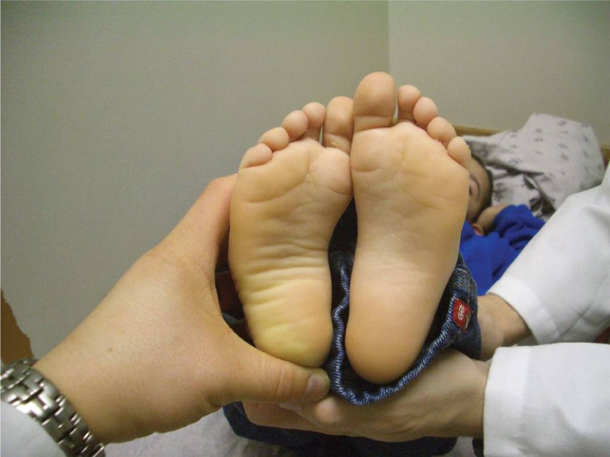
Figure 47-1 Metatarsus adductus. The convex (“C”) shape of the child’s right foot suggests metatarsus adductus. (Courtesy of Paul S. Matz, MD.)
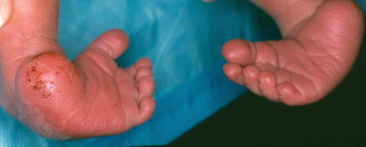
Figure 47-2 Clubfeet. Bilateral clubfeet in an infant with notable metatarsus adductus. (Courtesy of Gerardo Cabrera-Meza, MD.)

Figure 47-3 Femoral torsion. A 5-year-old girl with increased medial rotation of the hips because of femoral torsion. (Courtesy of Julie A. Boom, MD.)
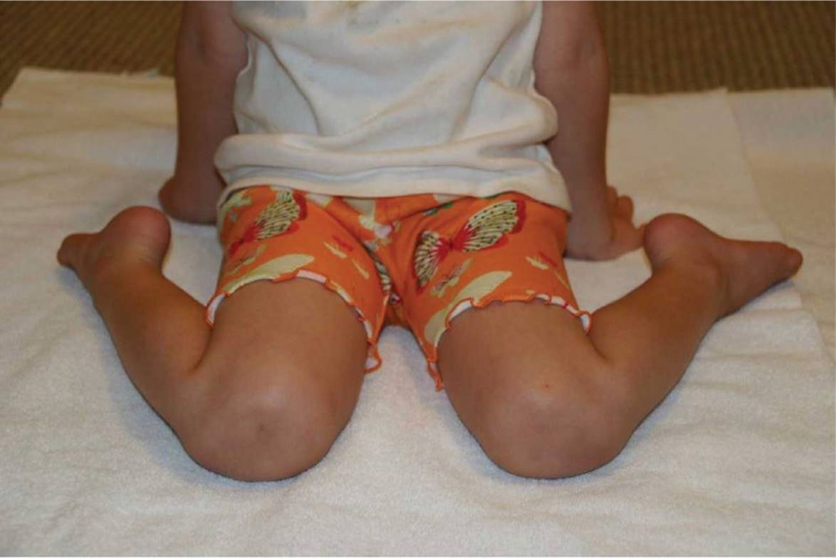
Figure 47-4 Femoral torsion. A 5-year-old girl comfortably “W” sitting. (Courtesy of Julie A. Boom, MD.)

Figure 47-5 Femoral torsion. Limited lateral hip rotation in a 5-year-old girl with femoral torsion. (Courtesy of Julie A. Boom, MD.)

Figure 47-6 Tibial torsion. A 4-year-old child with a negative thigh-foot angle. (Courtesy of Julie A. Boom, MD.)
DIFFERENTIAL DIAGNOSIS

Other Diagnoses to Consider
• Metatarsus primus varus
• Skew foot
• Dynamic hallux abductus
• Neuromuscular disorders
• Genetic syndromes
• Skeletal dysplasia
When to Consider Further Evaluation or Treatment
• Severe or marked femoral anteversion or internal tibial torsion that results in functional issues and/or fails to resolve by the age of 8
• Patients with rigid metatarsus adductus or clubfoot
• Children with underlying genetic, developmental, or neuromuscular disorders
• Progression of the abnormality instead of the expected resolution
• Metatarsus adductus that persists beyond the ages of 4 to 5 years
SUGGESTED READINGS
Craig CL, Goldberg MJ. Foot and leg problems. Pediatr Rev. 1993;14:395–400.
Lincoln TL, Suen PW. Common rotational variation in children. J Am Acad Orthop Surg. 2003;11:312–320.
Sass P, Hassan G. Lower extremity abnormalities in children. Am Fam Physician. 2003;68:461–468.
Scherl SA. Common lower extremity problems in children. Pediatr Rev. 2004;25:52–61.
Smith BG. Lower extremity disorders in children and adolescents. Pediatr Rev. 2009;30:287–294.