
Approach to the Problem
Knee swelling in the pediatric patient suggests a broad differential diagnosis, including musculoskeletal, rheumatic, and infectious processes. Knees are large joints commonly involved in juvenile idiopathic arthritis (JIA) (previously known as juvenile rheumatoid arthritis). Because there are many soft-tissue structures within it, the knee may be injured in almost every type of sport. The knee is involved in approximately 90% of cases of joint swelling associated with Lyme disease. Knee swelling in children can be subtle or obvious. A thorough history and complete physical examination cannot be overemphasized. Knowing the age, ethnicity, gender, and geographic location of the affected individual is vital to narrowing the differential diagnosis. Specifically, gonococcal arthritis may be seen in an adolescent, Lyme disease should be considered in a patient from Connecticut, girls more than boys are diagnosed with pauciarticular arthritis, osteosarcoma is more predominant in whites than in African Americans, and sports injuries are common in young athletes.
Knee swelling can be approached by acuity of presentation and the presence or absence of pain, recurrence (as with tuberculosis), and localized or systemic disease. Identifying the etiology also depends on the history: recent or past trauma, symptoms of infection, presence of inflammation, a family history of rheumatic disease or cancer, recent travel, and so forth. A child with septic arthritis, osteomyelitis, systemic onset JIA, or acute leukemia may be ill-appearing. On the other hand, a child with exostosis may be well-appearing. Swelling of the knee joint may occur as a result of surrounding skin and soft-tissue infection, a knee effusion with blood (as with bone and growth plate fractures and an anterior cartilage ligament tear) or nonbloody fluid (as with a meniscal tear and ligament sprain), synovial thickening, a cyst, or bursitis.
Key Points in the History
• An acute onset of knee swelling suggests trauma, septic arthritis, rheumatic fever, or Lyme disease.
• Chronic knee swelling suggests JIA, tuberculosis, or malignancy.
• The mechanism of trauma may provide clues to the most likely structures injured. For example, a history of knee hyperextension may suggest an anterior cruciate ligament (ACL) injury.
• An audible pop may be concerning for a serious ligamentous injury or fracture.
• Knee instability or “giving way” may indicate a ruptured ACL or patellar instability.
• Knee locking with limited extension may indicate a torn meniscus, avulsed cruciate ligament, or bony fragment.
• Extremely painful migratory polyarthritis, involving the knees, elbows, wrists, and ankles, and a prior Group A streptococcal pharyngitis warrant the consideration of rheumatic fever.
• Antecedent diarrheal illness can be a clue for reactive arthritis.
• Pain in septic arthritis is constant and generally worsens over time.
• Morning joint stiffness is seen in JIA or systemic lupus erythematosus (SLE).
• Fever may be suggestive of an infectious or rheumatologic process; high intermittent fevers (≥39.5°C) that occur once or twice daily may indicate systemic onset JIA.
• An evanescent rash (small, pale red macules with central clearing) that occurs during periods of temperature elevation is suggestive of systemic onset JIA.
• Consider Lyme disease when knee arthritis with an erythema chronicum migrans rash follows a tick bite or an exposure to a Lyme-endemic area.
• Individuals with hemophilia are susceptible to hemarthrosis.
• A growing painless bony lump in the arm or leg of a 10- to 20-year-old could be exostosis.
• A painful soft-tissue mass growing over months may be osteosarcoma; fever, weight loss, and malaise are generally absent.
• Consider risk factors such as exposure to tuberculosis or sexually transmitted infections in monoarticular arthritis.
Key Points in the Physical Examination
• The site of bruising may provide a clue to the direction of force that caused the swelling.
• An effusion, indicated by asymmetry of the suprapatellar pouches, may indicate synovitis—the hallmark of late Lyme disease.
• Effusion immediately following trauma may suggest acute bleeding into the knee.
• Edema over the lower pole of the patella suggests a prepatellar bursitis.
• A mass palpated in the popliteal area that gets bigger upon standing suggests the presence of a popliteal cyst (Baker cyst).
• The discovery of a new heart murmur, especially one consistent with mitral or aortic insufficiency, may suggest acute rheumatic fever.
• Excruciating knee pain with bright erythema or dramatic warmth is characteristic of acute rheumatic fever or septic arthritis.
• Knee swelling accompanied by urticaria, erythematous maculopapules, or purpura involving primarily the lower extremities with gastrointestinal symptoms suggests Henoch–Schönlein purpura.
• A malar rash with painful knee swelling can be SLE.
• Pain with palpation over the tibial tuberosity suggests Osgood–Schlatter disease.
|
PHOTOGRAPHS OF SELECTED DIAGNOSES |
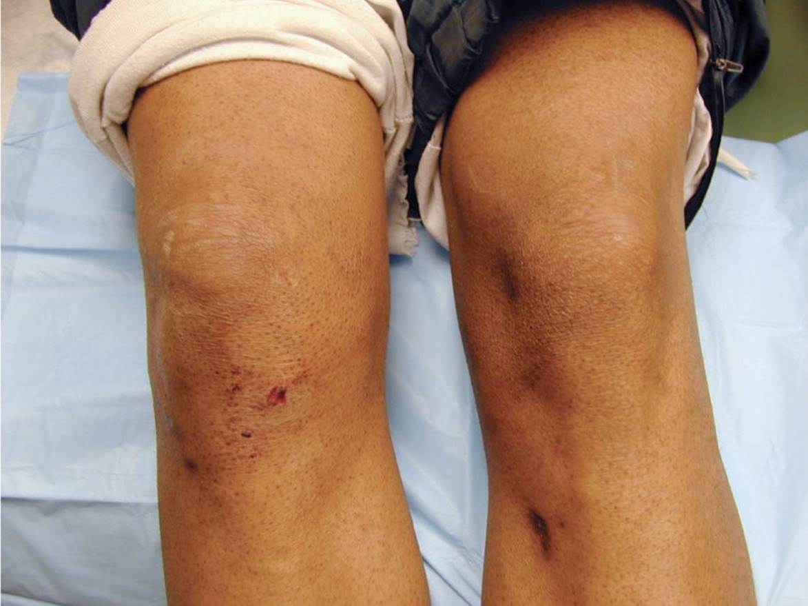
Figure 48-1 Knee effusion. Clinically obvious effusion of the right knee. (Used with permission from Fuchs MA. Hemarthrosis. In: Greenberg MI, ed. Greenberg’s Atlas of Emergency Medicine. Philadelphia, PA: Lippincott Williams & Wilkins; 2005:525.)

Figure 48-2 Knee cellulitis. Localized erythema suggestive of cellulitis overlying the knee. (Used with permission from Fleisher GR, Ludwig S, Baskin MN, eds. Atlas of Pediatric Emergency Medicine. Philadelphia, PA: Lippincott Williams & Wilkins; 2004:202.)

Figure 48-3 Juvenile idiopathic arthritis. Unilateral swelling of the right knee in a young girl with JIA. (Courtesy of Shriners Hospitals for Children, Houston, Texas.)
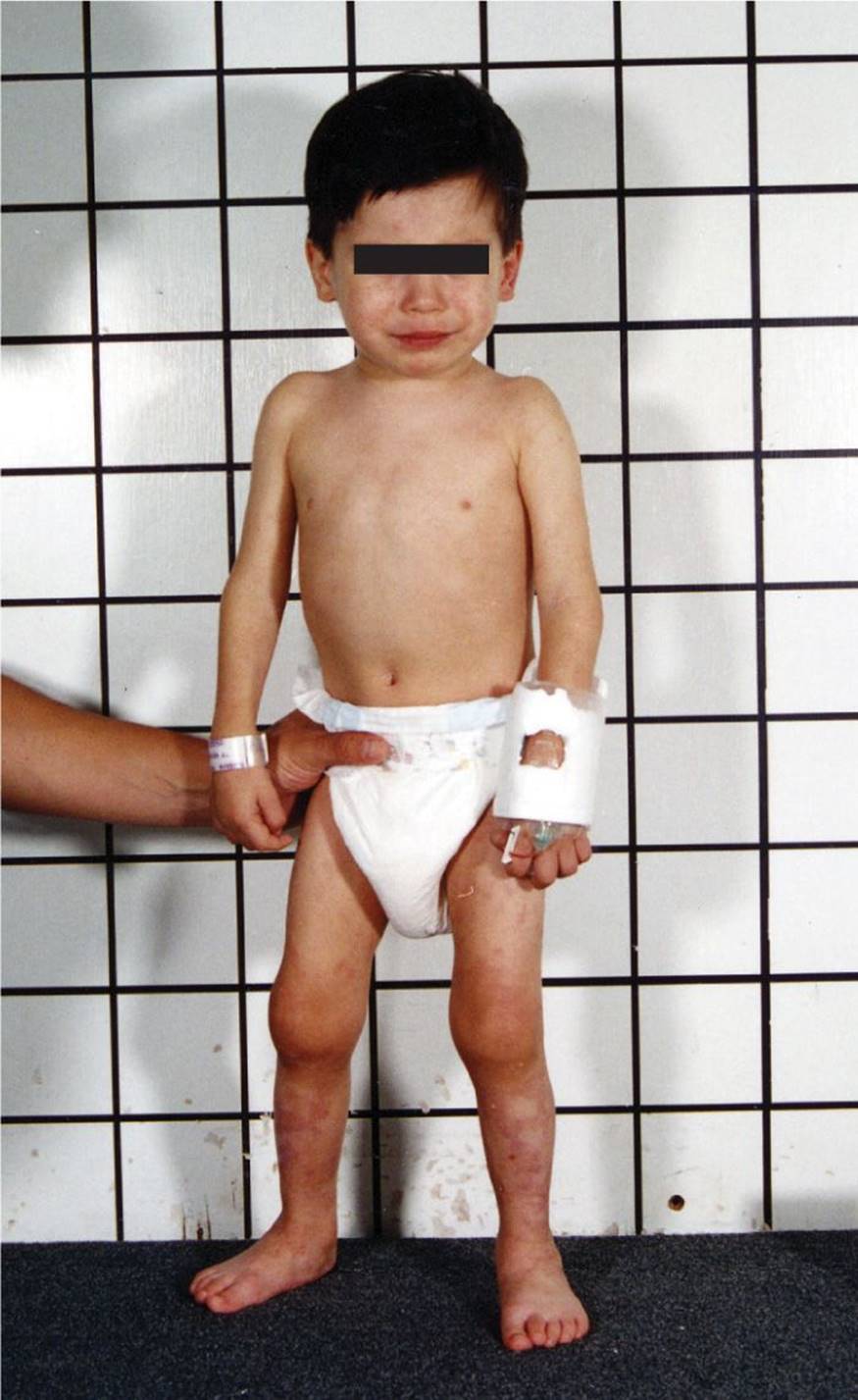
Figure 48-4 Juvenile idiopathic arthritis. A toddler with bilateral knee swelling due to JIA. (Courtesy of Shriners Hospitals for Children, Houston, Texas.)

Figure 48-5 Osgood–Schlatter disease. Lateral view demonstrating prominence of the tibial tuberosity. (Courtesy of Julie A. Boom, MD.)
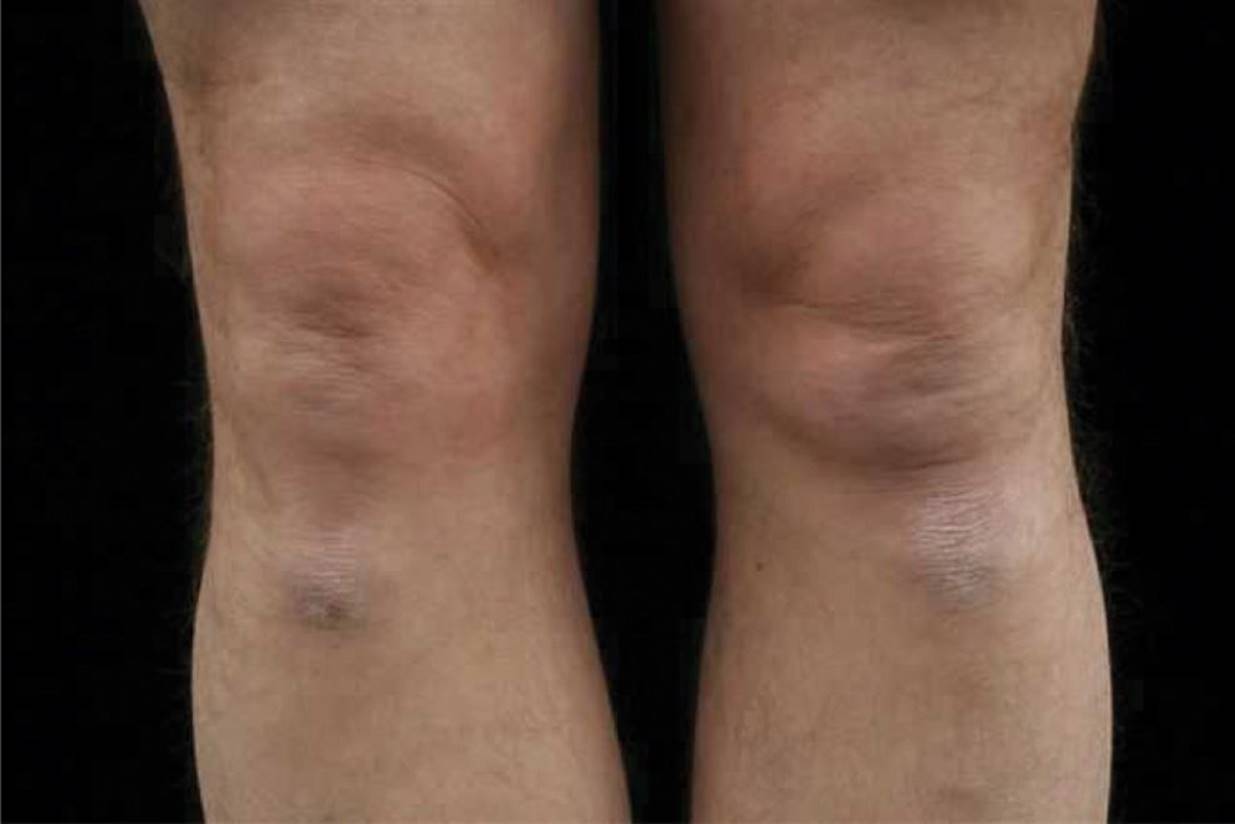
Figure 48-6 Osgood–Schlatter disease. Pain with palpation over the tibial tuberosity is suggestive of Osgood–Schlatter disease. (Courtesy of Julie A. Boom, MD.)
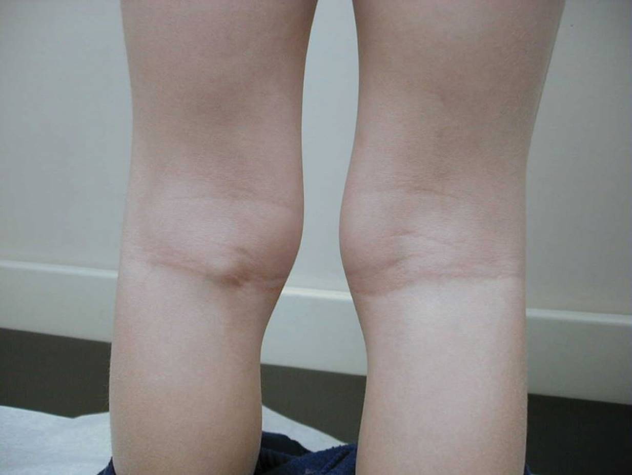
Figure 48-7 Baker cyst. Discrete swelling in the left popliteal fossa without overlying erythema. (Courtesy of Mary L. Brandt, MD.)
DIFFERENTIAL DIAGNOSIS


Other Diagnoses to Consider
• Osteochondritis dissecans
• Osgood–Schlatter disease
• Pigmented villonodular synovitis (rare—painless chronic knee swelling in an adolescent)
• Serum sickness
• SLE
• Osteomyelitis
• Viral arthritis associated with parvovirus, adenovirus, Epstein–Barr virus, and rubella, mumps, varicella, and hepatitis B virus infection
• Malignancy including leukemia, neuroblastoma, lymphoma, Hodgkin disease, malignant histiocytosis, rhabdomyosarcoma, osteosarcoma, and Ewing sarcoma
• Immunodeficiency and inflammatory bowel disease-associated arthritis
• Rickets secondary to vitamin D deficiency
When to Consider Further Evaluation or Treatment
• Neurovascular injury to the popliteal artery and peroneal nerve occurs most commonly in knee dislocations and displaced fractures. Immediate vascular surgery consultation is warranted if popliteal artery compromise is suspected.
• According to the Ottawa Knee Rules, radiographs of the knee should be obtained after an acute injury in children who meet one of the following criteria: isolated tenderness of the patella, tenderness at the head of the fibula, inability to flex the knee to 90 degrees, inability to bear weight immediately and in the emergency department for four steps, regardless of limp.
• The most serious complication of pauciarticular JIA is the development of uveitis or iridocyclitis, which occurs most commonly in the subgroup of children below 6 years of age who are ANA positive. A prompt ophthalmology evaluation and routine screening are necessary.
• Bleeding disorders should be considered when hemarthrosis of the knee occurs with minimal or no recollected trauma. Initial laboratory tests include a complete blood count, prothrombin time, and activated partial thromboplastin time. Joint bleeding is typically seen in deficiencies in coagulation factors, such as hemophilia.
• Recurrent painful monoarticular hemarthrosis of the knee may be caused by a synovial hemangioma, which occurs more commonly in children. MRI is the preferred mode of imaging. Refer to orthopedic surgery for treatment, which includes embolization, local steroid injection, and surgical excision.
SUGGESTED READINGS
Baskin MN. Injury—knee. In: Fleisher GR, Ludwig S, Henretig FM, eds. Textbook of Pediatric Emergency Medicine. 6th ed. Philadelphia, PA: Lippincott Williams & Wilkins; 2010:345–352.
Fleisher GR, Ludwig S, Baskin MN, eds. Atlas of Pediatric Emergency Medicine. Philadelphia, PA: Lippincott Williams & Wilkins; 2004:202.
Greenberg MI, ed. Greenberg’s Atlas of Emergency Medicine. Philadelphia, PA: Lippincott Williams & Wilkins; 2004:525.
Mirkinson L. The diagnosis of rheumatic fever. Pediatr Rev. 1998;19:310–311.
Schaller JG. Juvenile rheumatoid arthritis. Pediatr Rev. 1997;18:337–349.
Schwartz MW, Bell LM, Bingham P, et al., eds. The 5-Minute Pediatric Consult. 5th ed. Philadelphia, PA: Lippincott Williams & Wilkins; 2008:58–59, 724–725, 760–761.