
Approach to the Problem
Rashes on the face can be an isolated local phenomenon or associated with systemic disease. Clues to the etiology of the rash are found in the anatomic location, distribution, color, type, and the shape of the involved lesions. Many patients and their families are often concerned about the impact of the facial rash on the child’s appearance, self-esteem, and interpersonal interactions.
Key Points in the History
• When acne lesions appear in a child under 7, consider an evaluation for endocrinologic abnormalities.
• In a female with acne and oligomenorrhea or hirsutism, consider evaluation for polycystic ovarian syndrome.
• The lesions of impetigo may be distinguished from contact dermatitis in that impetigo lesions are generally painful, whereas contact dermatitis lesions are pruritic.
• Children with eczema frequently have family members with a history of eczema, allergies, or asthma.
• During winter, when the air has lower humidity, facial rashes exacerbated by dry skin such as eczema, pityriasis alba, and lip-licking dermatitis are often worse.
• Autoimmune diseases can present with a chronic erythematous facial rash. Associated fever and arthritis raise the suspicion for these disorders.
Key Points in the Physical Examination
• Focal rashes on the face are seen with impetigo and tinea corporis.
• Symmetric lesions are seen with infectious processes, such as erythema infectiosum or scarlet fever.
• When evaluating acne, it is important to assess the type of lesions (comedone vs inflammatory), the number and distribution of lesions, and the presence of scarring in order to guide therapy.
• In severe cases of eczema, lesions can be found throughout the body; however, lesions in the axilla or groin area should prompt consideration of other diagnoses, such as psoriasis. If pustules are present, consider superinfection with Staphylococcus aureus.
• Contact dermatitis presents as a localized skin lesion exposed to an irritant, such as nickel-containing clothing snaps or jewelry.
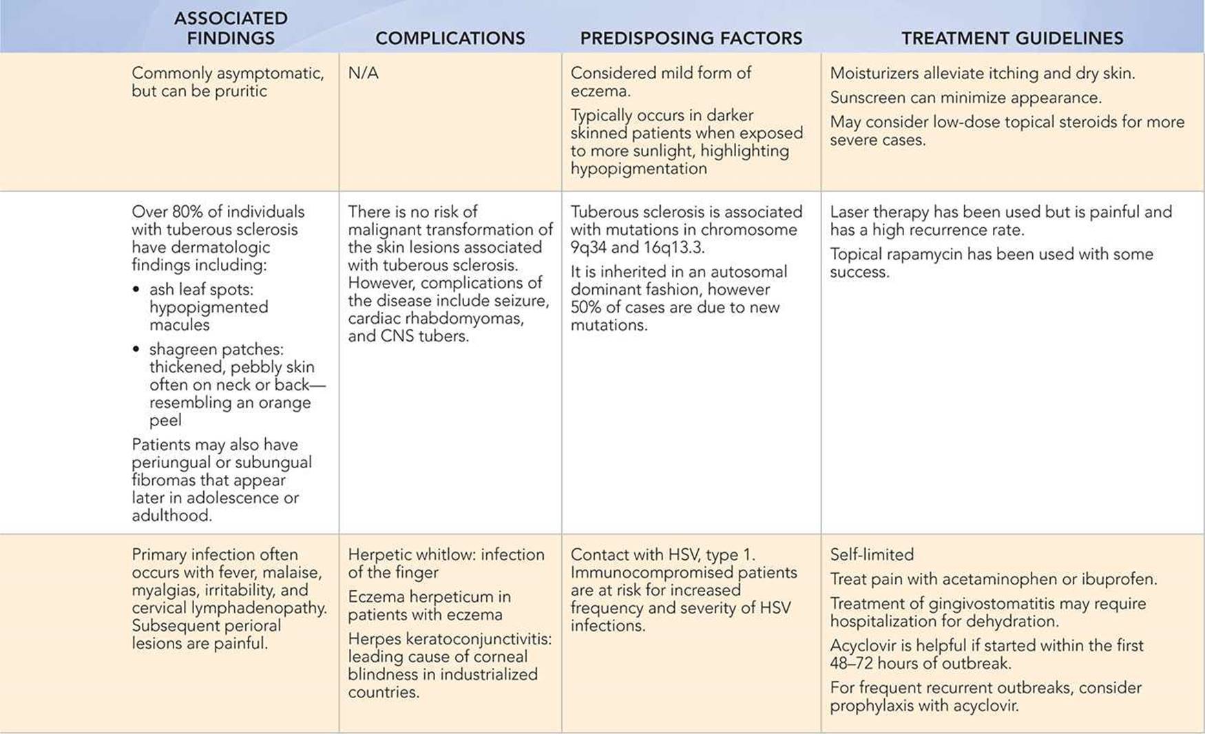
• Vesicular lesions occur in herpes simplex virus (HSV) and coxsackievirus infections. In herpes infections, the vesicles are grouped or dermatomal.
• Rhus dermatitis and some cases of eczema are associated with vesicular lesions.
• Acne presents with comedones.
• Pearl-like lesions with central umbilication are seen in molluscum contagiosum.
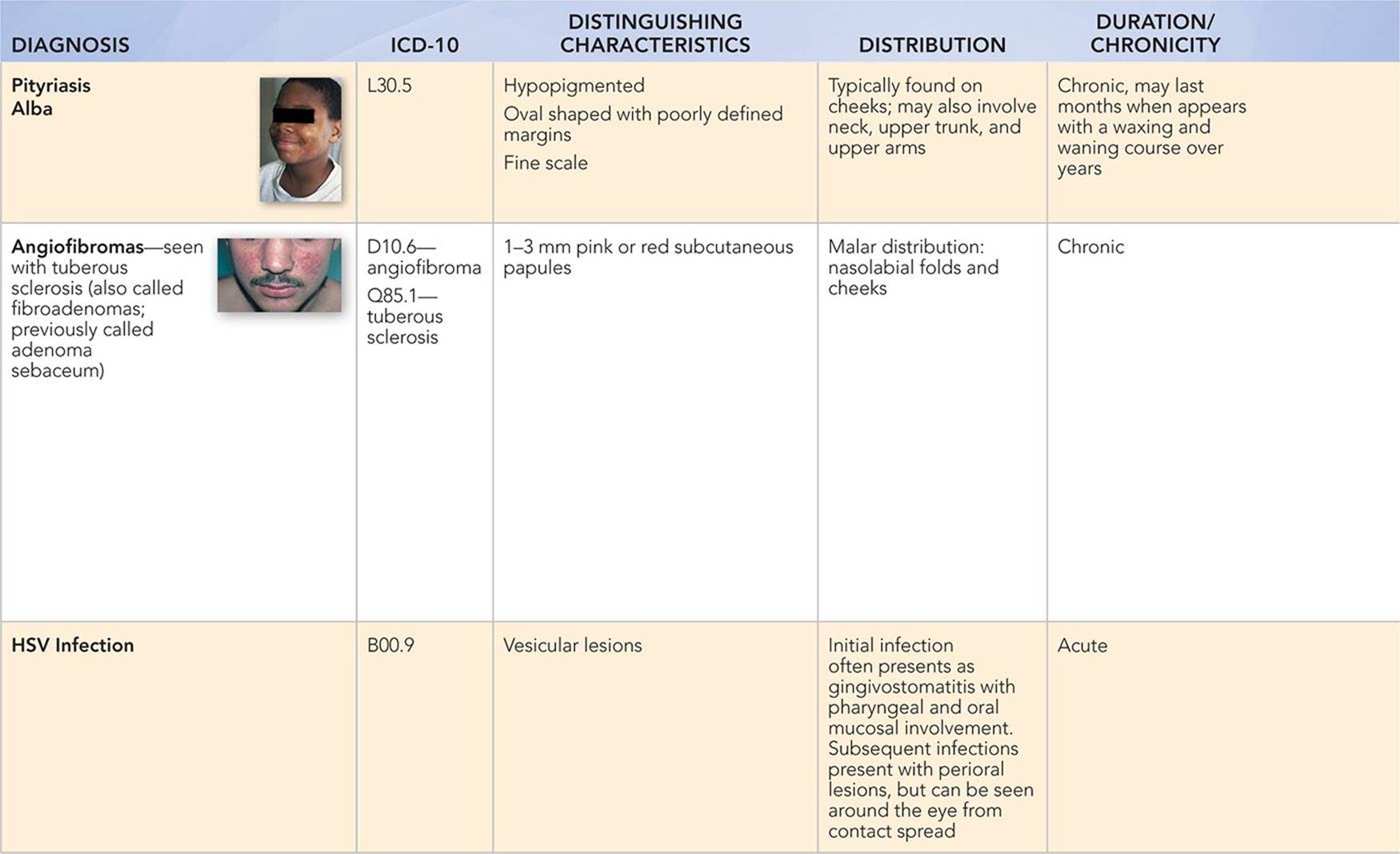
• The facial lesions of tuberous sclerosis are very hard subcutaneous papules. They are often misdiagnosed as acne; look for other dermatologic findings of tuberous sclerosis, including ash leaf spots and shagreen patches.
• Individuals with scaly rashes such as eczema, seborrhea, psoriasis, and pityriasis alba often have skin changes present in other parts of their body.
• Seborrhea classically has a greasy scale, whereas psoriasis has a silvery scale.
• Erythematous plaques sparing the nasolabial folds are characteristic of the malar rash seen in systemic lupus erythematosus.
|
PHOTOGRAPHS OF SELECTED DIAGNOSES |
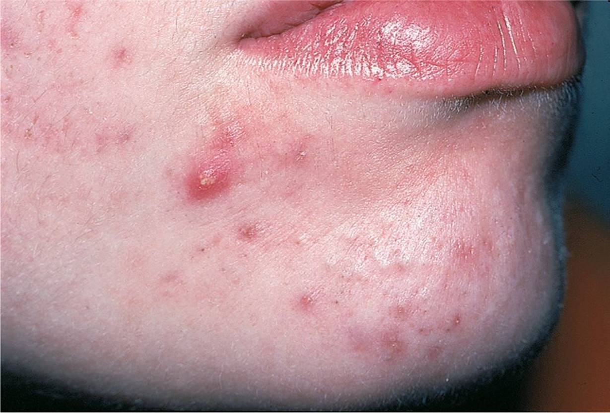
Figure 62-1 Inflammatory acne. Erythematous papules and pustules on chin. (Used with permission from Goodheart HP. Goodheart’s Photoguide to Common Skin Disorders. 2nd ed. Philadelphia, PA: Lippincott Williams & Wilkins; 2003:14.)

Figure 62-2 Eczema. Symmetric bilateral scaly rash on cheeks. (Used with permission from Goodheart HP. Goodheart’s Photoguide to Common Skin Disorders. 2nd ed. Philadelphia, PA: Lippincott Williams & Wilkins; 2003:46.)
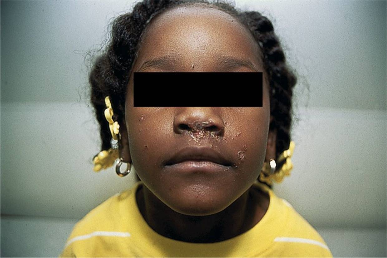
Figure 62-3 Impetigo. Honey-crusted lesions at base of nares that are self-inoculated onto other parts of the face. (Courtesy of George A. Datto, III, MD.)
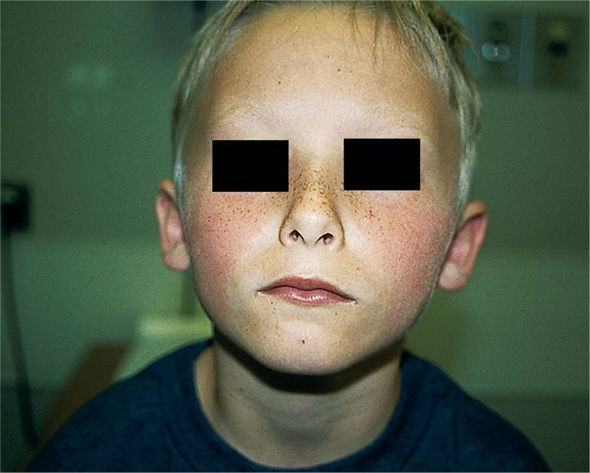
Figure 62-4 Erythema infectiosum. Bilateral erythematous macular rash on cheeks—“slapped cheeks.” (Courtesy of George A. Datto, III, MD.)

Figure 62-5 Molluscum contagiosum. Umbilicated papules on face of child. (Used with permission from Goodheart HP. Goodheart’s Photoguide to Common Skin Disorders. 2nd ed. Philadelphia, PA: Lippincott Williams & Wilkins; 2003:138.)

Figure 62-6 Pityriasis alba. Hypopigmented scaly macules on cheeks. (Courtesy of George A. Datto, III, MD.)
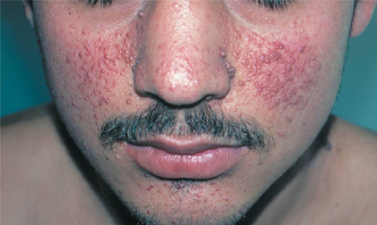
Figure 62-7 Tuberous sclerosis. Adenoma sebaceum (angiofibroma). (Used with permission from Goodheart HP. Goodheart’s Photoguide to Common Skin Disorders. 2nd ed. Philadelphia, PA: Lippincott Williams & Wilkins; 2003:388.)
DIFFERENTIAL DIAGNOSIS


Other Diagnoses to Consider
• Systemic lupus erythematosus
• Dermatomyositis
• Scarlet fever
• Hemangiomas
• Psoriasis
• Petechiae/Purpura
When to Consider Further Evaluation or Treatment
• Because acne scars may be irreversible, their presence should prompt the clinician to be aggressive in the selection of therapeutic agents and to consider referral to a dermatologist.
• Severe eczema is often infected with S. aureus, especially with pustular or eroded lesions.
• For severe seborrheic dermatitis, consider an alternate diagnosis including psoriasis or HIV infection.
• Special precautions should be taken with pregnant women exposed to parvovirus B19. Due to the effect of the virus on red cell production, complications include miscarriage, intrauterine fetal demise, and hydrops fetalis.
SUGGESTED READINGS
Gollnick H, Cunliffe W, Berson D, et al. Management of acne: a report from a global alliance to improve outcomes in acne. J Am Acad Dermatol. 2003;49:S1.
Goodheart HP. Goodheart’s Photoguide to Common Skin Disorders. 3rd ed. Philadelphia, PA: Lippincott Williams & Wilkins; 2008: Chapters 1, 2, 5, 14, and 16.
Krakowski AC, Eichenfield LF, Dohil MA. Management of atopic dermatitis in the pediatric population. Pediatrics. 2008;122(4):812.
Krueger DA, Franz DN. Current management of tuberous sclerosis complex. Paediatr Drugs. 2008;10(5):299–313.
Naldi L, Rebora A. Clinical practice. Seborrheic dermatitis. N Engl J Med. 2009;360(4):387–396.
Paller AS, Mancini AJ, eds. Hurwitz Clinical Pediatric Dermatology: A Textbook of Skin Disorders of Childhood and Adolescence. 4th ed. Philadelphia, PA: Elsevier Saunders; 2011:Chapters 3, 4, 14, 15, and 19.
Silverberg N. Pediatric molluscum contagiosum: optimal treatment strategies. Paediatr Drugs. 2003;5:505–512.