
Approach to the Problem
When faced with a concerning cutaneous injury, it is important to consider not only child abuse but also other causes. Age and developmental ability of the patient, as well as the location and type of the lesion(s), help in determining the likelihood of an abusive or nonaccidental cause of cutaneous findings. Common cutaneous injuries seen in child physical abuse include bruises, pattern injuries, and burns. It is important to match the history (or mechanism) of how the findings appeared with the actual observed finding(s). Try to determine whether the mechanism is plausible, based on the child’s developmental ability. Photo-documentation can be important for the investigative process, for review by experts, and to document changes. For example, if a suspected bruise does not resolve with time, then it is not a bruise and could be a birthmark.
Key Points in the History
• Obtain a detailed history of the alleged cause of the cutaneous findings, if available.
• Sometimes no history is given, or only potential mechanisms are offered. For example, a caretaker may not have witnessed an actual event, but may believe that something specific happened to explain the findings.
• A vague history such as “fell from chair” is not adequate to match an injury to a mechanism; therefore, it is essential to gather details related to the fall.
• A changing or discrepant history is concerning for abuse.
• Height of the fall, specific details about the fall (i.e., Was it straight down or in an arc? Was there twisting or rotational motion? Were there multiple strikes with the child falling down stairs?), surface onto which the fall occurred, whether there were objects on the floor, and other such information can help evaluate whether the explanation fits the injury.
• Consider detailed questions about the environment, witnesses, and the child’s response to the event or injury.
• Delay in seeking care is potentially a concerning factor; an explanation for a delay in seeking care should be elicited and can be evaluated as being a reasonable explanation or not.
• For scald injuries, a temperature of the water should be obtained at the scene by investigators.
• Family history of bleeding disorders or other underlying conditions should be obtained.
• Medications, such as recent anticoagulant use, should be assessed.
• Previous accidents resulting in injuries such as fractures, burns, and head trauma can suggest a pattern of abuse and should raise suspicion for child physical abuse.
• Include in the medical record any explanation given by the child or adolescent for the injury, using their exact words, when possible.
Key Points in the Physical Examination
• Corroborate the child’s developmental ability.
• Location is a key determinant of suspicion for abuse: buttocks, trunk, neck, ears, genitalia, and upper arms are concerning locations for bruises or other injuries.
• Examine the entire body. Subtle bruising or pattern marks can easily be missed.
• Pattern marks can be suspicious. Linear or other pattern marks should be measured, documented, and photographed.
• Scald burns with sharply demarcated lines, without splash marks, are suspicious for forced immersion.
• Examine behind the ears, as there may be bruises from inflicted trauma.
• Examine the mouth carefully; injuries to the frenulum can result from forced bottle feeding or smothering of a crying baby.
• Perform a genital examination on every child with suspected physical abuse.
• Penile bruises may result from pinching and may reflect physical punishment or an accidental toilet seat injury.
• Photo-documentation can be important for follow-up, expert review, and investigating agencies.
|
PHOTOGRAPHS OF SELECTED DIAGNOSES |
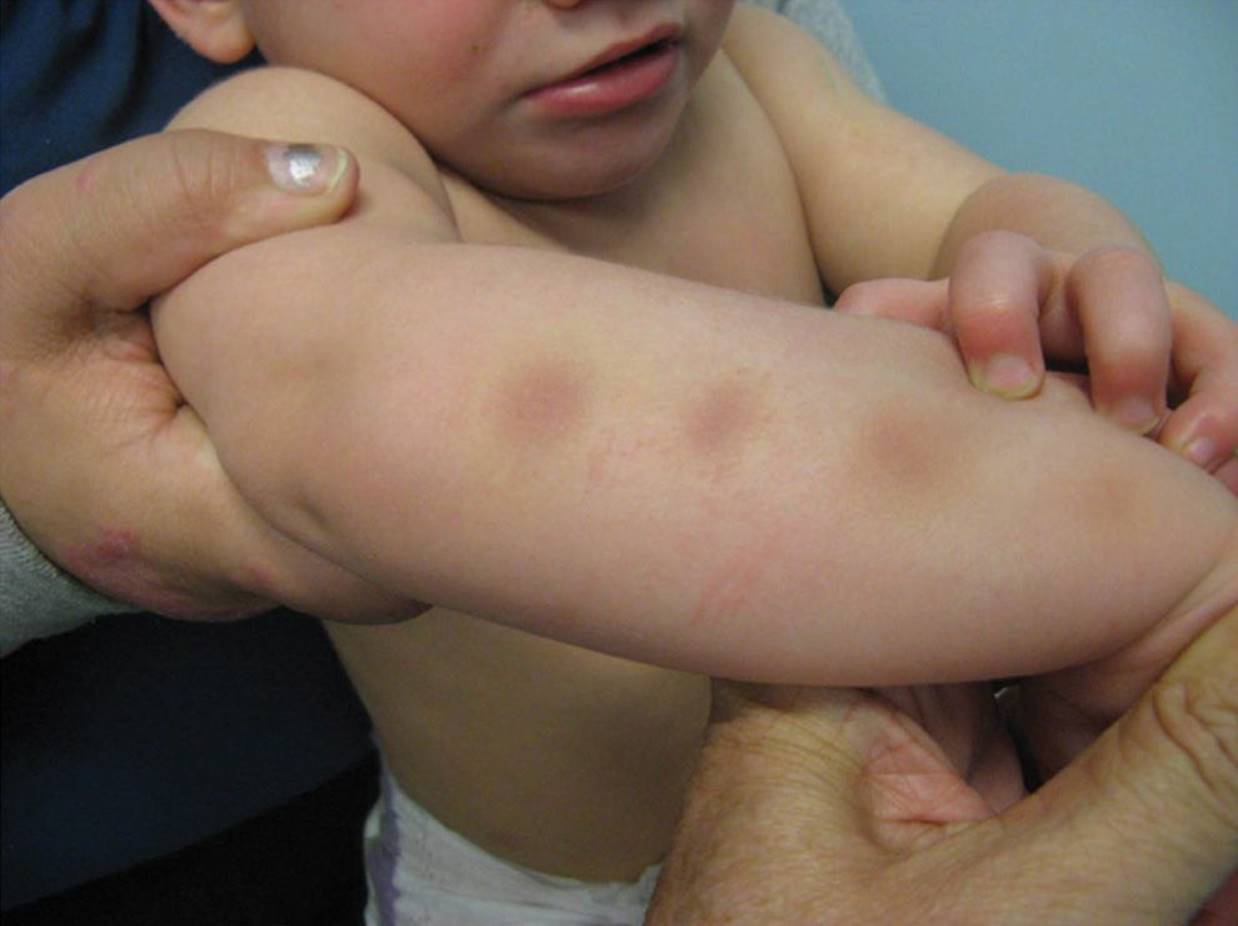
Figure 61-1 Circular bruises. These are typical of fingertip marks from squeezing, shown here on an infant’s forearm. (Courtesy of Dr. Jean Labbé.)

Figure 61-2 Bruise of the external ear. Ear injuries are highly concerning for child physical abuse. (Courtesy of Dr. Jean Labbé.)
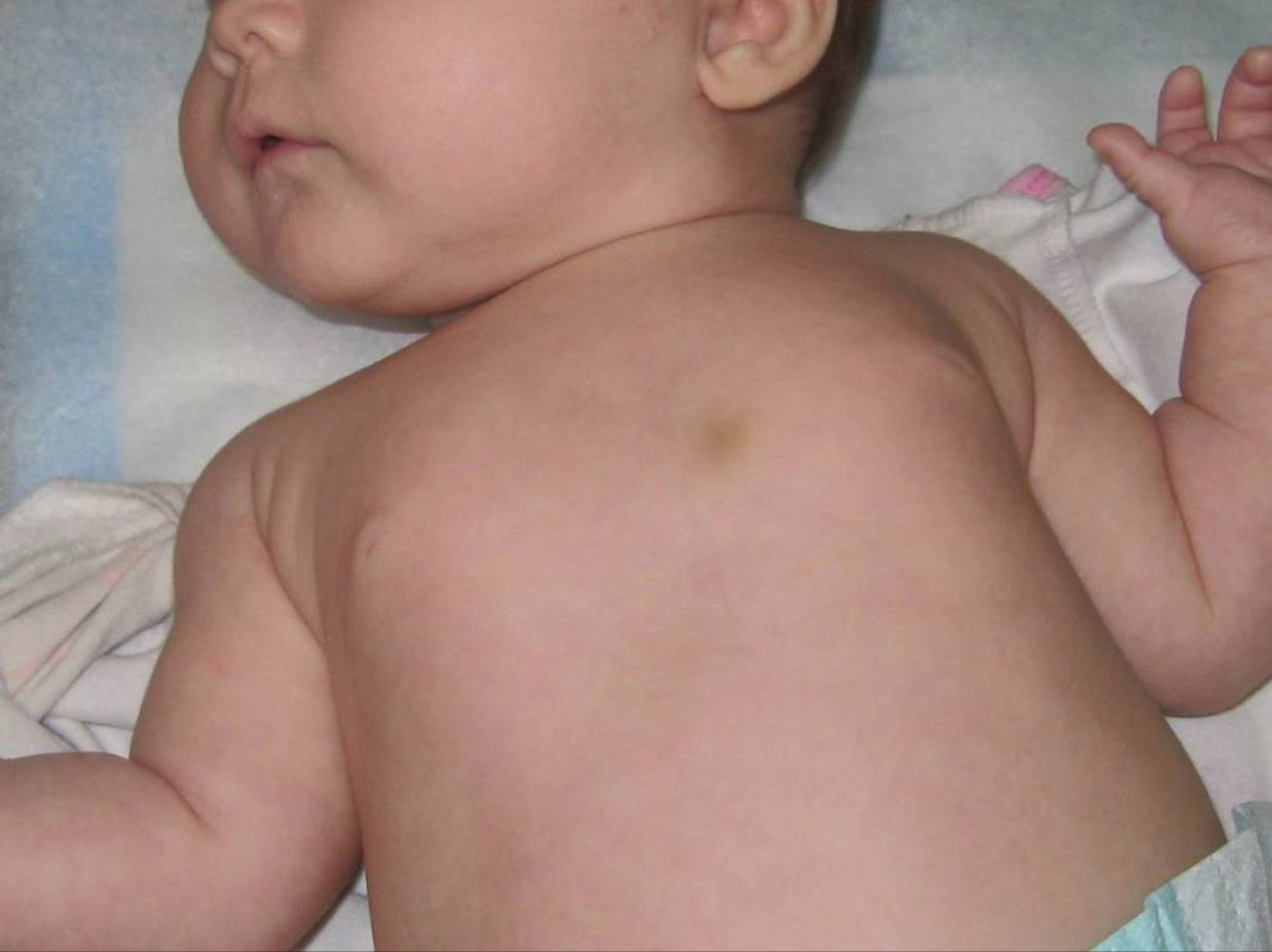
Figure 61-3 Bruise on infant’s chest. Both the location of the bruise, and the fact that it is an infant make this less likely to be from an accident. (Courtesy of Dr. Jean Labbé)
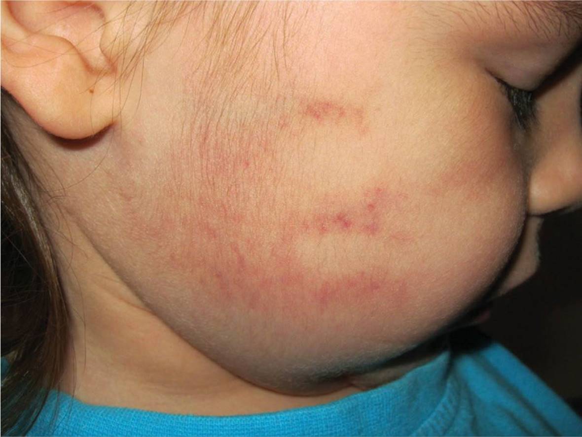
Figure 61-4 Slap mark on child’s cheek. Petechial lines or an outline of a hand may be observed in slaps when blood is pushed through the space between the fingers of the slapping hand. (Courtesy of Dr. Jean Labbé.)

Figure 61-5 Pattern mark on a child’s arm (A) from being struck by a wooden spoon. Wooden spoon (B) matching the mark on child’s arm seen in (A). (Courtesy of Dr. Jean Labbé.)

Figure 61-6 Dermal melanocytosis on child’s back. These lesions may be mistaken for bruises. Bruises should resolve, however, over days to weeks. (Courtesy of Dr. Jean Labbé.)
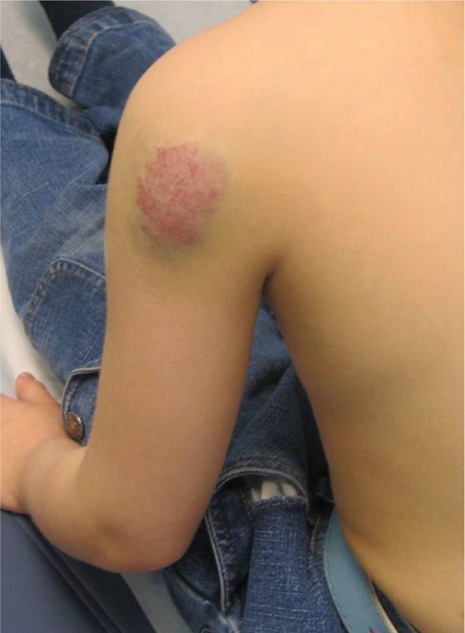
Figure 61-7 Cavernous hemangioma on child’s shoulder. This type of lesion is sometimes mistaken for a bruise from an inflicted injury. (Courtesy of Dr. Jean Labbé.)

Figure 61-8 Henoch–Schönlein purpura (HSP). The purpuric lesions of HSP may be mistaken for bruises from inflicted injury. (Courtesy of Dr. Jean Labbé.)

Figure 61-9 Cigarette burn (A). Close-up of cigarette burn showing a typical measurement of approximately 8 mm (B). (Courtesy of Dr. Jean Labbé.)
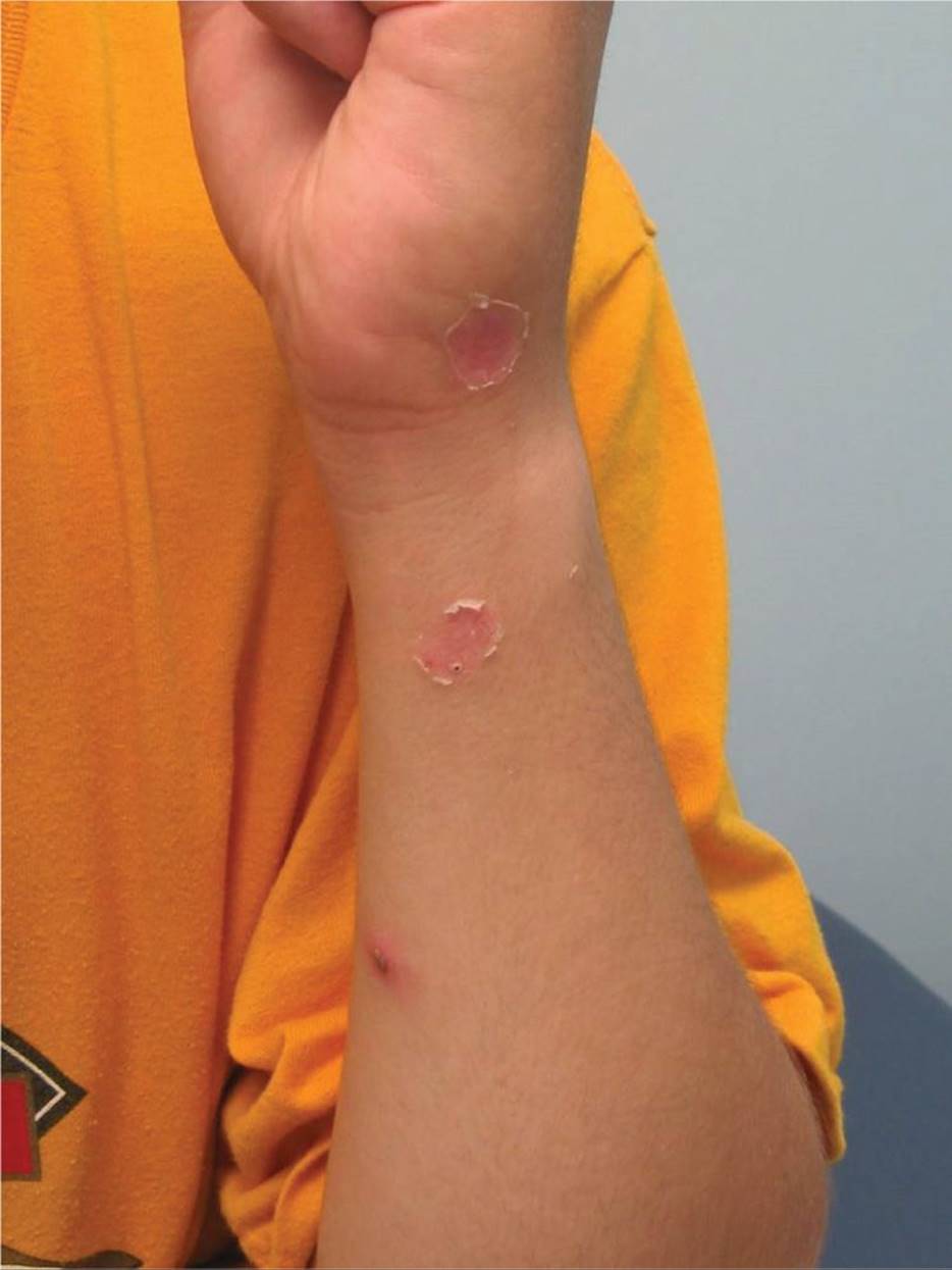
Figure 61-10 Impetigo. This entity is sometimes mistaken for a cigarette burn. (Courtesy of Dr. Jean Labbé.)
DIFFERENTIAL DIAGNOSIS

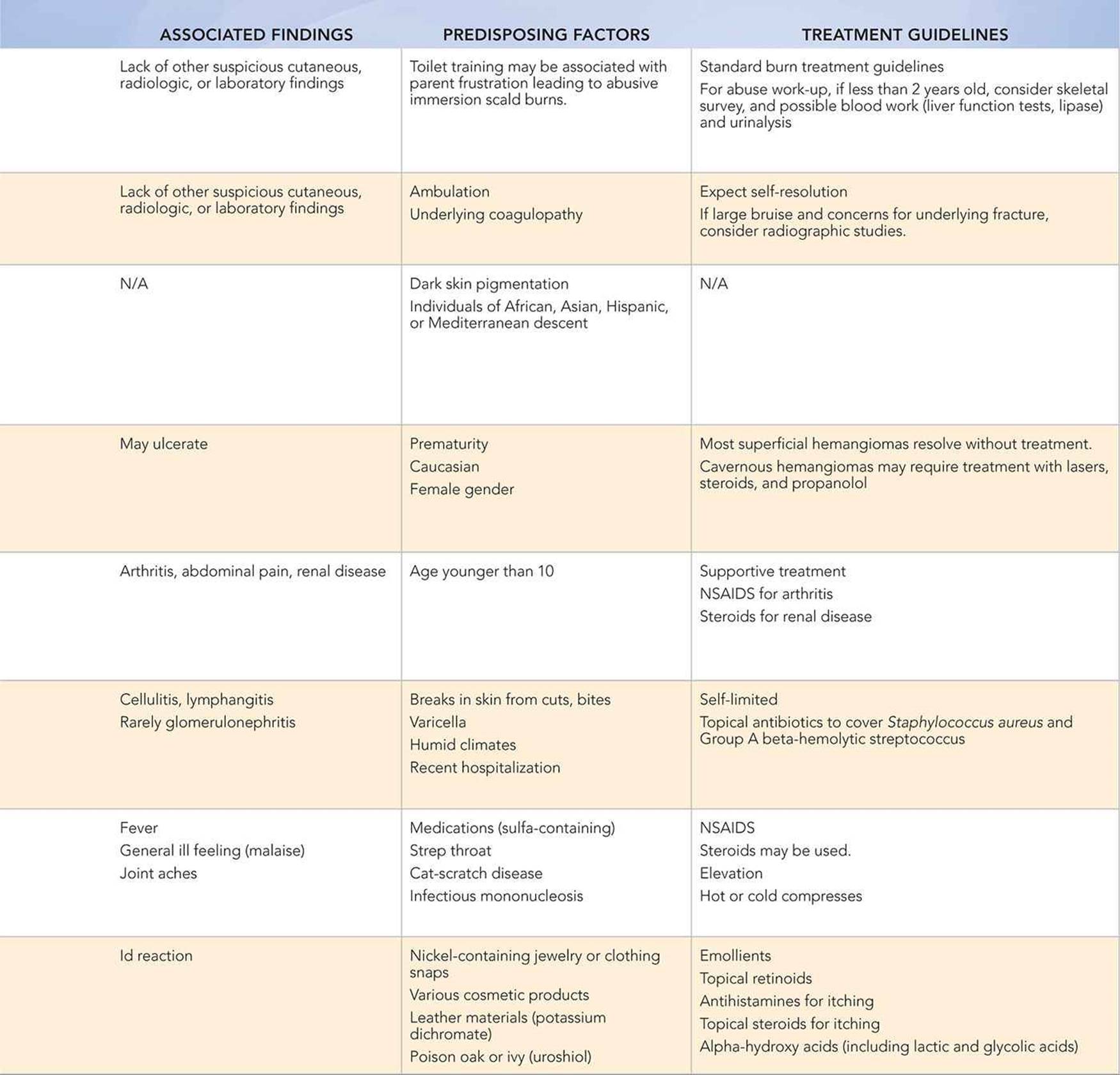
Other Diagnoses to Consider
Bruising
• Dermal melanosis, slate grey nevi, mongolian spots
• Hemangiomas
• Henoch–Schönlein purpura
• Cupping, spooning, coining
• Leukemia
• Von Willebrand disease
• Idiopathic thrombocytopenic purpura
• Hemophilia
• Erythema nodosum
Burns
• Epidermolysis bullosa
• Staphylococcal scalded skin syndrome
• (Bullous) impetigo
• Contact dermatitis
• Accidental scald or contact burns
• Phytophotodermatitis
• Chemical burns from home remedies like laxatives
When to Consider Further Evaluation or Treatment
• In general, whenever there is bruising suspicious for child abuse, a complete blood count and coagulation panel (prothrombin time, partial thromboplastin time, and international normalized ratio) should be obtained to rule out malignancy or bleeding disorder.
• Consider referral to a dermatologist if a medical condition is being considered.
• In children less than 2 years old with concerning cutaneous findings, a skeletal survey should generally be done to look for fractures specific for abuse.
• Consider head imaging and fundoscopic examination in infants or children less than a year of age if suspicious injuries.
• Suspicious injuries should be referred to a child abuse specialist.
• Child protective services or the police should be contacted when appropriate, and may be responsible for scene investigation.
SUGGESTED READINGS
Harris TS. Bruises in children: normal or child abuse? J Pediatr Health Care. 2010;24(4):216–221.
Hobbs CJ. ABC of child abuse. Burns and scalds. BMJ. 1989;298(6683):1302–1305.
Kos L, Shwayder T. Cutaneous manifestations of child abuse. Pediatr Dermatol. 2006;23(4):311–320.
Sugar NF, Taylor JA, Feldman KW. Bruises in infants and toddlers: those who don’t cruise rarely bruise. Arch Pediatr Adolesc Med. 1999;153(4):399–403.
Swerdlin A, Berkowitz C, Craft N. Cutaneous signs of child abuse. J Am Acad Dermatol. 2007;57(3):371–392.