
Approach to the Problem
The key to successful diagnosis of a red rash or swelling is a careful history and physical examination while keeping a broad list of differential diagnoses in mind. Timing, appearance, and progression of the rash; presence of associated signs and symptoms; and recent exposures and activities aid in identifying the etiology and diagnosis. The distribution, appearance, and feel of the skin are also helpful in determining the etiology.
Key Points in the History
• Lesions that change in location, shape, and size over minutes to hours are likely urticarial. They can be caused by an exposure to a new antigen (detergent, perfume, food), but are more commonly caused by viral infections.
• Cellulitis and erysipelas are characterized by erythema and tender swelling surrounding a break in the skin, and may be accompanied by fever.
• Insect bites can be discrete or clustered and are more frequent on exposed surfaces of the skin. Time spent outdoors, near a pet, or with other similarly affected individuals is suggestive.
• A hemangioma may start off as a flat area of telangiectasia with a surrounding ring of pallor. A hemangioma typically increases in size over the child’s first months of life prior to involution.
• Hematoma should be suspected with a suggestive history or whenever there is concern for nonaccidental trauma.
• Contact dermatitis is an allergic hypersensitivity response to an irritant or allergen that is usually limited to the area where the exposure occurred and is frequently pruritic.
• Erythema infectiosum is often accompanied by fever, rhinorrhea, and/or arthralgias.
• Pityriasis rosea presents initially as a single patch (the “herald patch”) that evolves into a diffuse macular eruption. It is sometimes pruritic. Systemic symptoms are rare.
• Erythema nodosum often occurs in the setting of infection but can also be due to a wide variety of immunologic and oncologic conditions.
Key Points in the Physical Examination
• Noting whether the patient has multiple, clustered, or discrete lesions can help to narrow the differential diagnosis.
• Urticarial lesions have a wheal and flare appearance that can evolve during examination.
• Cellulitis and erysipelas are typically warm and tender. Erysipelas is raised, with well-defined edges, whereas cellulitis lacks defined edges. The presence of fluctuance suggests an underlying abscess.
• Insect bites frequently have a central punctum with surrounding erythema. If multiple bites are present, they are usually grouped rather than randomly scattered.
• A subtle cluster of blanching telangiectasias with a ring of pallor is suggestive of an early hemangioma. They later develop into nonblanching cherry red macules or papules. Bluish discoloration suggests deeper involvement.
• Hematomas may vary in size, shape, and distribution. They are usually tender and not well demarcated. They may change in color from reddish to shades of purple, blue, and green over time.
• Contact dermatitis may present as papular, papulovesicular, or eczematous lesions. Location may implicate the offending stimulus.
• Erythema infectiosum is suggested by a nontender “slapped cheek” erythema, which spares the nasal bridge and periorbital areas, and is typically associated with a lacy or reticular rash that may be seen on the trunk and the extremities.
• Pityriasis rosea initially presents as a pink, scaly, oval patch, followed by the eruption of oval macules with a collarette of scale. Pityriasis rosea often has a “Christmas-tree” distribution, with lesions following skin cleavage lines (Langer lines).
• Erythema nodosum is characterized by tender, raised, reddish blue lesions found on the shins. These lesions may also appear on the buttocks, calves, ankles, thighs, and arms.
|
PHOTOGRAPHS OF SELECTED DIAGNOSES |
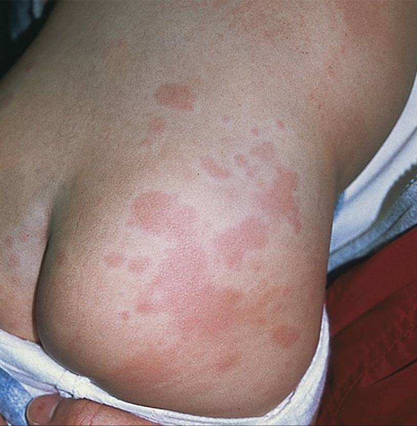
Figure 64-1 Urticaria. Erythematous wheals on buttocks of child. (Courtesy of George A. Datto, III, MD.)

Figure 64-2 Cellulitis. Poorly defined erythematous lesion on hand that developed after skin abrasion. (Courtesy of George A. Datto, III, MD.)
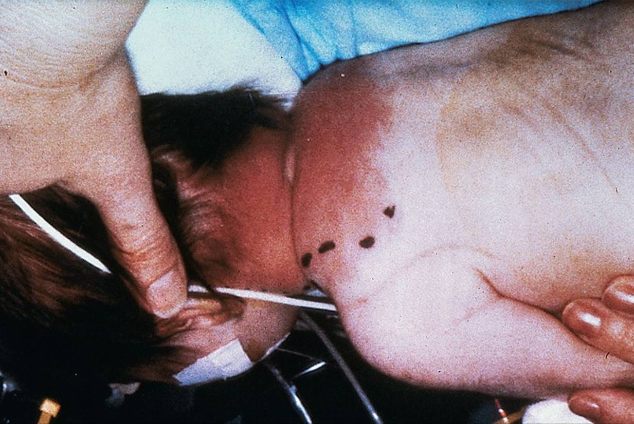
Figure 64-3 Erysipelas. Very erythematous rash on neck with sharply demarcated borders. (Courtesy of George A. Datto, III, MD.)

Figure 64-4 Insect bites. These fleabites are grouped in a characteristic nonfollicular, “breakfast, lunch, and dinner” pattern. (Used with permission from Goodheart HP, MD. Goodheart’s Photoguide of Common Skin Disorders. 2nd ed. Philadelphia, PA: Lippincott Williams & Wilkins; 2003.)

Figure 64-5 Erythema chronicum migrans. Note the central punctum following a tick bite and the ring-like appearance (Courtesy of Paul S. Matz, MD.)

Figure 64-6 Mixed hemangiomas. (Used with permission from Stedman’s.)
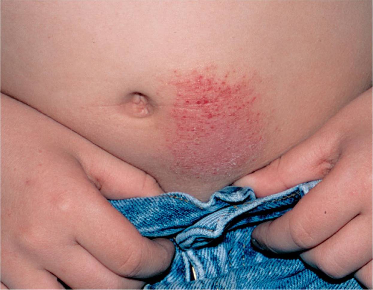
Figure 64-7 Allergic contact dermatitis. This boy developed an eczematous eruption at the site where the nickel snap on his blue jeans contacted his skin. (Used with permission from Goodheart HP, MD. Goodheart’s Photoguide of Common Skin Disorders. 2nd ed. Philadelphia, PA: Lippincott Williams & Wilkins; 2003.)

Figure 64-8 Allergic contact dermatitis. This patient reacted to the rubber in the elastic waistband of her underpants. Note sparing at the sites where the garment did not come into constant contact with the skin. (Used with permission from Goodheart HP, MD. Goodheart’s Photoguide of Common Skin Disorders. 2nd ed. Philadelphia, PA: Lippincott Williams & Wilkins; 2003.)

Figure 64-9 Erythema nodosum. Tender erythematous nodules on extensor aspects of lower legs. (Courtesy of George A. Datto, III, MD.)
DIFFERENTIAL DIAGNOSIS


Other Diagnoses to Consider
• Erythema multiforme
• Stevens–Johnsons syndrome
• Dermatographia
• Tinea cruris
• Tinea corporis
• Rubeola
• Langerhans cell histiocytosis
When to Consider Further Evaluation or Treatment
• Urticaria with another sign of anaphylaxis (angioedema, wheezing, hypotension, or vomiting) warrants emergent epinephrine administration. Urticaria after exposure to a new food or medication may represent an allergy. Recommend future avoidance of the offending agent.
• Cellulitis that does not improve within 72 hours of starting systemic antibiotics should be reevaluated. Consider broader spectrum antibiotics or admission for intravenous antibiotics and observation. Abscesses often require incision and drainage.
• Insect bites that are associated with other systemic symptoms should prompt additional workup and treatment for superinfection or anaphylaxis. If concern for venomous spider bite, prompt hospitalization and possible antivenom administration are indicated.
• Hemangiomas on the face should prompt a referral to a pediatric dermatologist as treatment may be indicated to prevent disfigurement. Hemangiomas near the eye can obstruct vision and lead to amblyopia. The presence of a very large hemangioma or multiple hemangiomas should raise suspicion for a genetic syndrome and/or internal lesions as seen with hemangiomatosis.
• Large hematomas may overlie boney fractures. Multiple hematomas, especially in unusual sites should prompt consideration of child physical abuse.
SUGGESTED READINGS
Bisno A. Current concepts: streptococcal infections of the skin and soft tissues. N Engl J Med. 1996;334(4):240–245.
Hartzell LD, Buckmiller LM. Current management of infantile hemangiomas and their common associated conditions. Otolaryngol Clin N Am. 2012;45(3):545–556.
Kakourou T, Drosatou P, Psychou F, et al. Erythema nodosum in children: a prospective study. J Am Acad Dermatol. 2001;44(1):17–21.
Kelly BP. Superficial fungal infections. Pediatr Rev. 2012;33(4):e22–e37.
Khangura S, Wallace J, Kissoon N, et al. Management of cellulitis in a pediatric emergency department. Pediatr Emerg Care. 2007;23:805–811.
Scott LA, Stone MS. Viral exanthems. Dermatol Online J. 2003;9(3):4.
Sicherer S, Leung D. Advances in allergic skin disease, anaphylaxis, and hypersensitivity reactions to foods, drugs and insects in 2007. J Allergy Clin Immunol. 2008;121:1351–1358.