
Approach to the Problem
Raised red rashes are common in pediatrics and can be concerning to parents and practitioners. The majority of raised red rashes, however, are not indicative of serious illness. Many red rashes have associated symptoms that may be helpful in making a final diagnosis. For example, symptoms of fatigue, fever, and lymphadenopathy suggest infectious mononucleosis. Complications can occur in some individuals with certain red rashes. For example, exposure of a pregnant woman to parvovirus may place her fetus at risk. At times, typical eruptions may not follow a predicted pattern—the distribution may be atypical, the season may not fit, or the age may be unusual. These diagnostic challenges emphasize the importance of a detailed history and astute observation.
Key Points in the History
• High fever for 3 to 5 days followed by acute defervescence that precedes the rash eruption is characteristic of roseola.
• Classic characteristics in the history aid in the diagnosis. For example, a history of “slapped cheeks” indicates Fifth disease; the presence of Koplik spots (clustered white papules in the buccal mucosa opposite the second molars) denotes measles.
• Individuals acutely affected by infectious mononucleosis are at risk for rash development following exposure to penicillins; however, those with infectious mononucleosis may also develop a rash in the absence of penicillin exposure.
• Antibiotic exposure is associated with a drug rash and erythema multiforme.
• Pruritus is typical in erythema multiforme, varicella, hot tub folliculitis, and scabies.
• Seasonal occurrence can provide clues to the diagnosis: late summer and fall would be the season for coxsackievirus, for example; spring and fall is when erythema multiforme, for example, may be seen.
• Family members with a similar rash may suggest scabies.
• The location of rash origin is important. For example, a red rash that begins on the scalp and travels downward is characteristic of measles.
Key Points in the Physical Examination
• Erythema may be more apparent in light-skinned children.
• The size and types of papules may support specific diagnoses: fine micropapules indicate scarlet fever; target lesions denote erythema multiforme.
• Papules and vesicles in combination may indicate scabies or impetigo.
• The color of the lesions aids in the diagnosis of the rash: rose-pink lesions are seen in roseola; red maculopapular lesions, in measles; and brownish lesions on the palms and soles, in syphilis.
• Red lesions on the palms and soles are present in syphilis, measles, erythema multiforme, scabies, and Gianotti–Crosti syndrome.
• Symmetric lesions may be noted in erythema multiforme, syphilis, and Gianotti–Crosti syndrome.
• Diffuse mucosal inflammation (i.e., urethritis, conjunctivitis, pharyngitis) is seen in Kawasaki disease.
• There are often oral mucous membrane findings in infectious mononucleosis, erythema multiforme, roseola, Kawasaki disease, and measles.
• Conjunctivitis is seen in Kawasaki disease and measles.
• In syphilis, the rash follows lines of cleavage.
• Periorbital edema is associated with roseola and infectious mononucleosis.
• Measles presents with Koplik spots (gray-white papules) on the buccal mucosa and dark red macules and papules that start on the head and spread caudally.
|
PHOTOGRAPHS OF SELECTED DIAGNOSES |

Figure 67-1 Roseola. Rose pink-colored rash on the trunk of an infant. (Courtesy of John Loiselle, MD.)

Figure 67-2 Scarlet fever. Fine, sandpapery rash on the trunk and neck. (Courtesy of George A. Datto, III, MD.)

Figure 67-3 Erythema infectiosum. Erythematous “slapped” cheeks along with erythematous rash on extensor surfaces of arms. (Courtesy of Philip Siu, MD.)
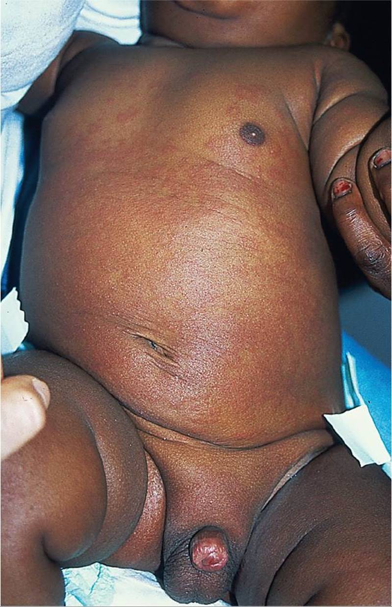
Figure 67-4 Kawasaki disease. Erythematous maculopapular rash that started in the groin and spread onto the trunk. (Courtesy of George A. Datto, III, MD.)

Figure 67-5 Infectious mononucleosis. (Courtesy of Kathleen Cronan, MD.)

Figure 67-6 Gianotti–Crosti syndrome. Note the reddish-brown papular lesions on the extremities. (Courtesy of John Loiselle, MD.)
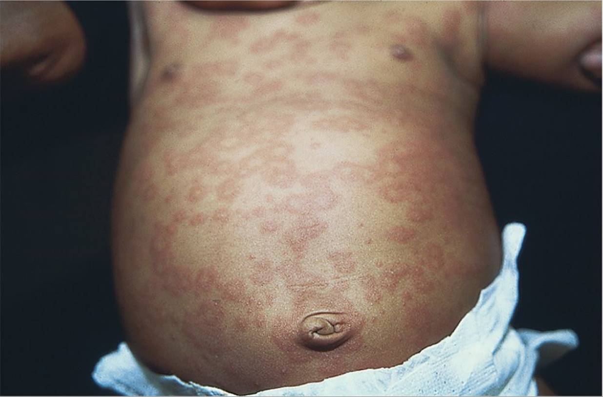
Figure 67-7 Erythema multiforme. Target-shaped lesions in an infant. (Courtesy of George A. Datto, III, MD.)

Figure 67-8 Scabies. Note the lesions in the axilla of a child. (Courtesy of George A. Datto, III, MD.)
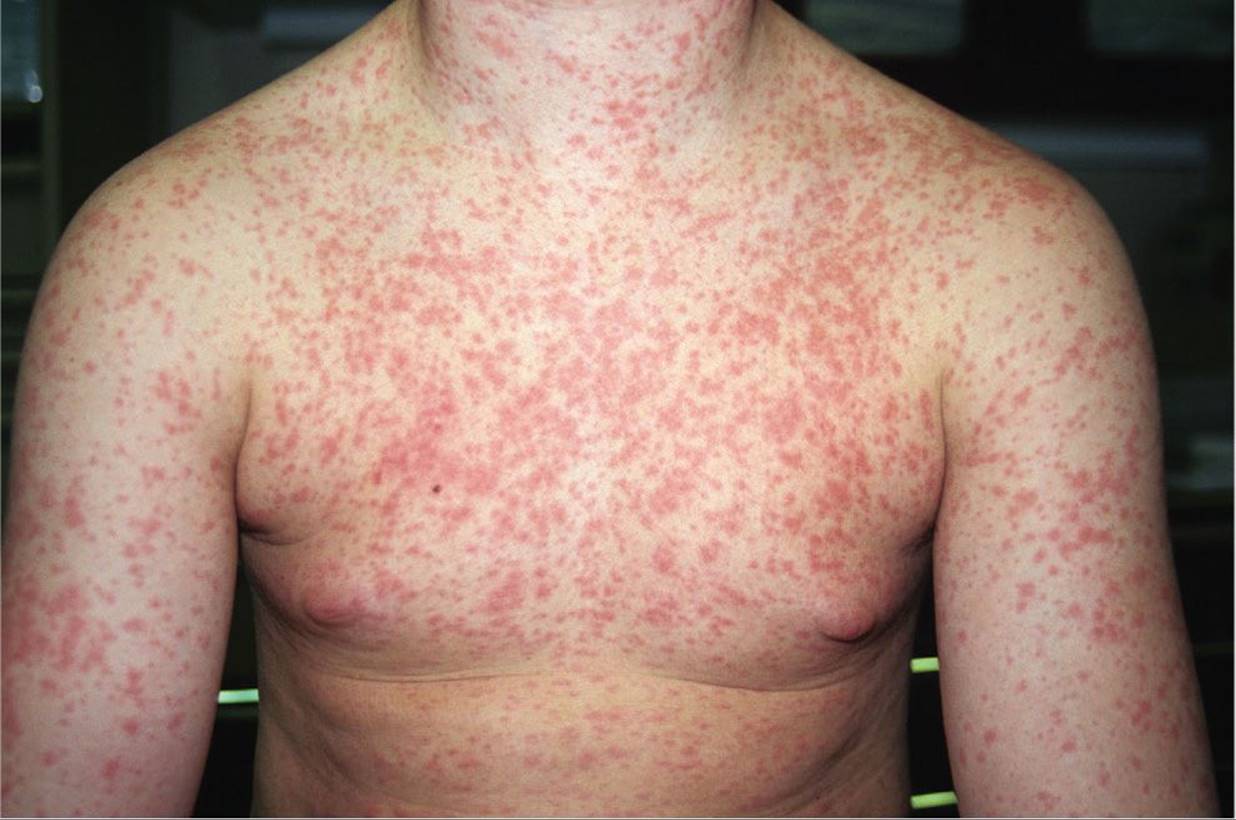
Figure 67-9 Measles. Lesions typically start on the head and travel downward. (Courtesy of Kathleen Cronan, MD.)
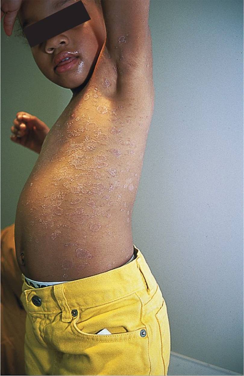
Figure 67-10 Unilateral laterothoracic exanthem. Pink, scaly rash involving the axilla and trunk on the right side of this child. (Courtesy of George A. Datto, III, MD.)
DIFFERENTIAL DIAGNOSIS


Other Diagnoses to Consider
• Drug eruptions
• Urticaria
• Contact dermatitis
• Henoch–Schönlein purpura
• Meningococcal disease
• Varicella (particularly in children who have received varicella vaccine for whom the rash may be atypical)
• Other viral exanthem
When to Consider Further Evaluation or Treatment
• A second course of permethrin 5% cream is often necessary 1 week following the initial dose in the treatment of scabies. Family members should also be treated, and bedding and clothing should be washed in the hottest water possible or dry-cleaned.
• In patients with diagnosed measles and poor nutrition or vitamin A deficiency, vitamin A supplementation is recommended. Measles is associated with significant complications, including pneumonia, encephalitis, myocarditis, and the late-occurring subacute sclerosing panencephalitis. If these are suspected, appropriate referrals and inpatient management should be sought.
• Fever and petechiae or purpura should always be considered as high risk for meningococcemia, and appropriate testing and treatment should be instituted promptly.
• Patients with suspected Kawasaki disease should be evaluated for coronary artery aneurysm formation.
• Acute abdominal pain in a child with infectious mononucleosis warrants an evaluation of the spleen for possible rupture.
• Patients with roseola erythema infectiosum should be instructed to avoid contact with pregnant women until the infection subsides due to the risk of nonimmune hydrops in the fetus.
SUGGESTED READINGS
Cherry JD. Roseola infantum (exanthem subitum). In: Feigin RD, Cherry JD, Demmler-Harrison GJ, Kaplan SL. eds. Feigin and Cherry’s Textbook of Pediatric Infectious Diseases. 6th ed. Philadelphia, PA: Saunders; 2009:780.
Currie BJ, McCarthy JS. Permethrin and ivermectin for scabies. N Engl J Med. 2010;362:717–725.
Dyer JA. Childhood viral exanthems. Pediatr Ann. 2007;36(1):21–29.
Kwon NH, Kim JE, Cho BK, et al. Gianotti-Crosti syndrome following novel Influenza A (H1N1) vaccination. Ann Dermatol. 2011;23(4):554–555.
Luzuriaga K, Sullivan JL. Infectious mononucleosis. N Engl J Med. 2010;362:1993–2000.
Paller AS, Mancini AJ, eds. Hurwitz Clinical Pediatric Dermatology: A Textbook of Skin Disorders of Childhood and Adolescence. 4th ed. Philadelphia, PA: Elsevier Saunders; 2011.