
Approach to the Problem
A vesicle is a raised skin lesion filled with clear fluid that is less than 1 cm in diameter. A raised, clear fluid-filled lesion larger than 1 cm is referred to as a bulla. In childhood, there are many diseases that manifest as vesicular rashes, the most familiar of which is the rash seen with herpes simplex virus (HSV) infection. Other viral and bacterial infections also may present with vesiculobullous lesions as may many noninfectious processes, including allergic and immune-mediated diseases, mechanical disorders of the skin, burns, and insect bites.
Vesicular eruptions may be benign and self-limited or may be progressive and life threatening. Early identification of potentially serious disease and prompt attention to complications are critical, particularly in infants and immunocompromised hosts.
Key Points in the History
• Recurrent herpetic skin outbreaks in the same location almost always represent the reactivation of a latent infection rather than a new primary infection.
• Immunocompromised hosts may have disseminated disease due to HSV, varicella zoster virus (VZV), and coxsackievirus infections.
• Primary HSV lesions are often associated with fever and systemic symptoms, whereas secondary lesions or reactivation of HSV lesions are usually not.
• The reactivation of HSV or VZV, known as “shingles,” is typically preceded by a prodrome of pain, tingling, itching, or burning at the site.
• In assessing vesicular rashes in the neonate, a detailed maternal history is necessary to elicit possible HSV exposure.
• Lethargy, poor feeding, temperature instability, jaundice, irritability, or seizures in an infant with vesicular lesions should raise suspicion for neonatal HSV infection.
• Frequently accompanying genital HSV is painful inguinal adenopathy, dysuria, urinary retention, and vaginal discharge. However, most primary genital HSV infections are asymptomatic.
• Primary VZV infection or chickenpox is very contagious; therefore, a history of household or school exposure in a child with characteristic lesions is highly suggestive.
• Children vaccinated against VZV may still develop chickenpox, though the disease course is milder.
• When contact dermatitis is suspected, a detailed environmental exposure history is warranted.
• Symptoms of allergic contact dermatitis may not manifest for 6 to 24 hours after the exposure. Symptoms are often worse with second or subsequent exposures.
• A history of outdoor exposure can indicate rhus dermatitis—poison oak, poison ivy, or poison sumac.
Key Points in the Physical Examination
• Grouped vesicles on an erythematous base are the hallmark of HSV infection; however, in immunocompromised patients, the erythematous base is not always apparent.
• HSV lesions on skin or mucous membranes may appear vesicular or, if they have ruptured, the lesions may appear eroded or ulcerated.
• Lesions of neonatal herpes often appear at 5 to 14 days of life; lesions appearing in the first 2 days of life suggest intrauterine exposure. Intrauterine-acquired HSV may not present with vesicles but rather scarring.
• The oral vesicles and ulcers of HSV tend to form more anteriorly on the gingivae, tongue, and hard palate; whereas, the lesions of hand-foot-and-mouth disease are typically more posterior on the soft palate, tonsillar pillars, and posterior oropharynx.
• Primary HSV infection of the eye may appear as blepharitis or keratoconjunctivitis. Signs include corneal or conjunctival erythema, watery discharge, lid swelling, and preauricular adenopathy.
• Lesions of primary VZV infection or chickenpox progress from papules to vesicles to erosions with crust. They occur in successive crops over 2 to 5 days and are predominant on the trunk, face, and scalp and progress in a centripetal distribution.
• Smallpox lesions spread centrifugally, with lesions concentrated on the extremities and spreading inward. A distinguishing feature of smallpox is the central umbilication of the lesions. Children with this disease are typically very ill.
• The presence of a dermatomal vesicular eruption is consistent with the diagnosis of shingles or reactivation of VZV infection.
• Extensive herpes zoster skin lesions may indicate an underlying immunodeficiency and an increased risk of visceral involvement.
• Lesions of contact dermatitis are typically limited to the area of exposure. The skin is often erythematous and edematous with vesiculation and weeping.
• Papular urticaria is characterized by recurrent crops of pruritic papulovesicles on skin areas exposed to insect bites.
|
PHOTOGRAPHS OF SELECTED DIAGNOSES |

Figure 68-1 Hand-foot-and-mouth disease. Vesicles on palms and soles. (Courtesy of Philip Siu, MD.)

Figure 68-2 Papular urticaria. Vesicular lesion following an insect bite on the lower leg of a child. (Courtesy of Shirley P. Klein, MD, FAAP.)

Figure 68-3 Papular urticaria. Vesiculobullous eruption secondary to insect bites on exposed area. (Courtesy of Ilona J. Frieden, MD.)

Figure 68-4 Breakthrough varicella. Note the “dewdrop-on-a-rose-petal” appearance of this lesion in a child previously immunized against varicella. (Courtesy of Esther K. Chung, MD, MPH.)

Figure 68-5 Varicella. Note the various stages of the lesions: papular, vesicular, and crusted. (Courtesy of Shirley P. Klein, MD, FAAP.)
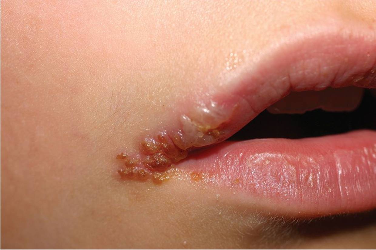
Figure 68-6 Herpes labialis. Grouped vesicles predominantly on one portion of the lip. (Courtesy of Ilona J. Frieden, MD.)

Figure 68-7 Herpetic whitlow. A group of vesicular lesions on the distal phalanx. (Courtesy of Paul S. Matz, MD.)
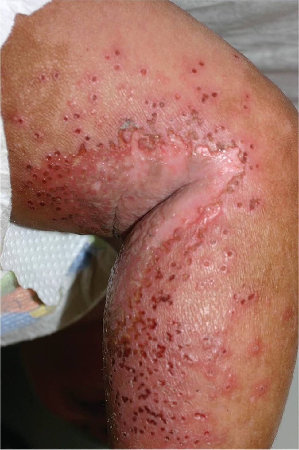
Figure 68-8 Eczema herpeticum. Multiple eroded vesicles with umbilication and crusting overlying a patch of eczematous skin. (Courtesy of Ilona J. Frieden, MD.)
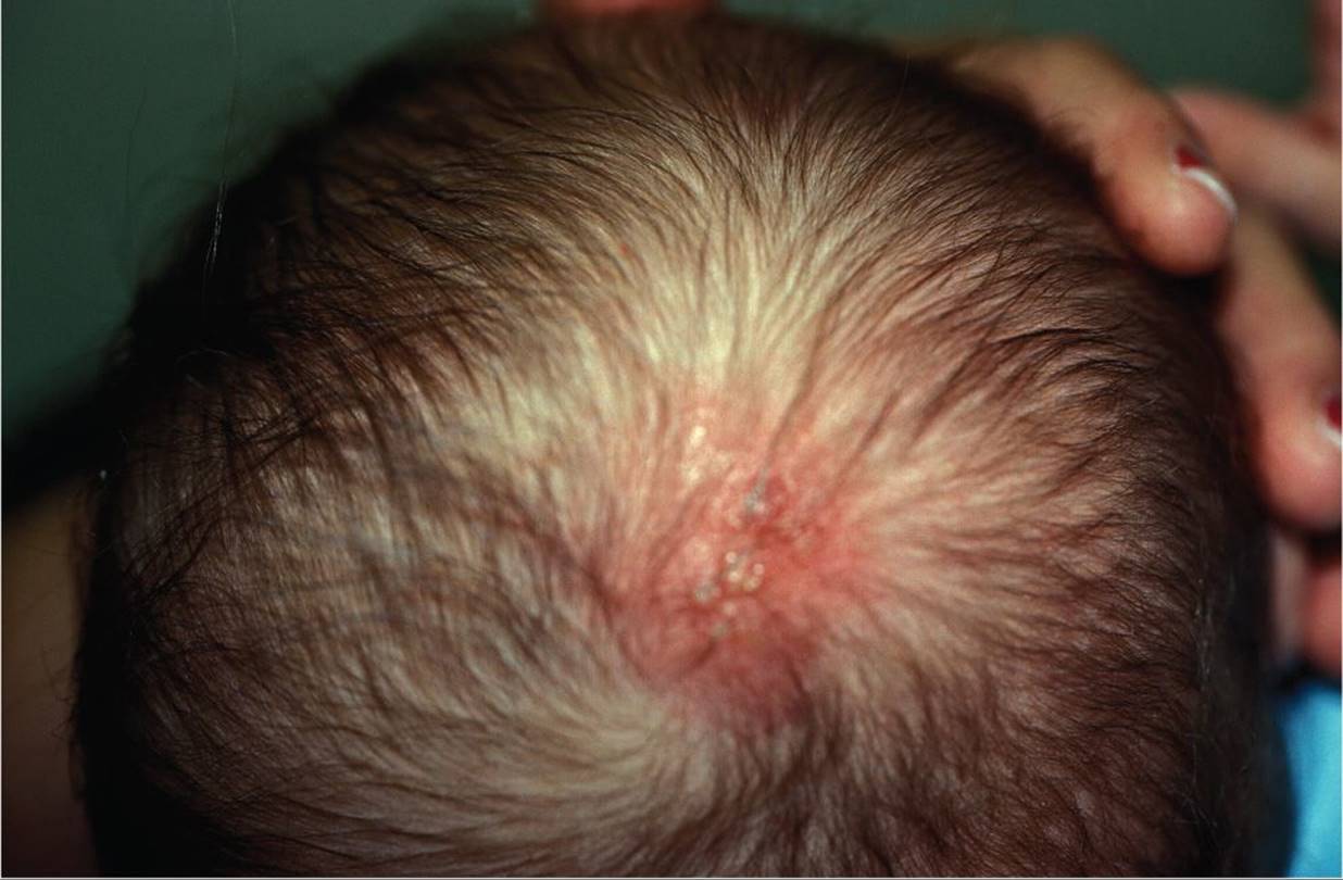
Figure 68-9 Neonatal herpes. Scalp erythema and vesicle at site of scalp electrode. (Courtesy of Shirley P. Klein, MD, FAAP.)

Figure 68-10 Herpes zoster. Grouped vesicles on an erythematous base in a dermatomal distribution. (Courtesy of Hans B. Kersten, MD.)
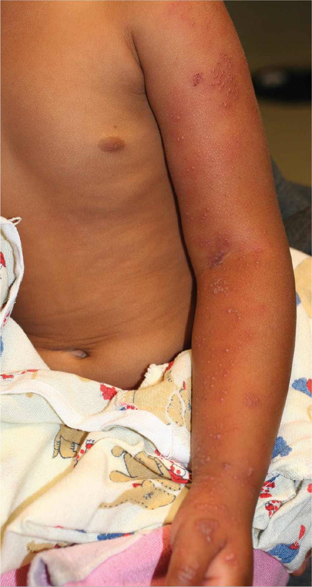
Figure 68-11 Herpes zoster. Grouped vesicles and erosions on an erythematous base in a C6 dermatomal distribution. (Courtesy of Ilona J. Frieden, MD.)
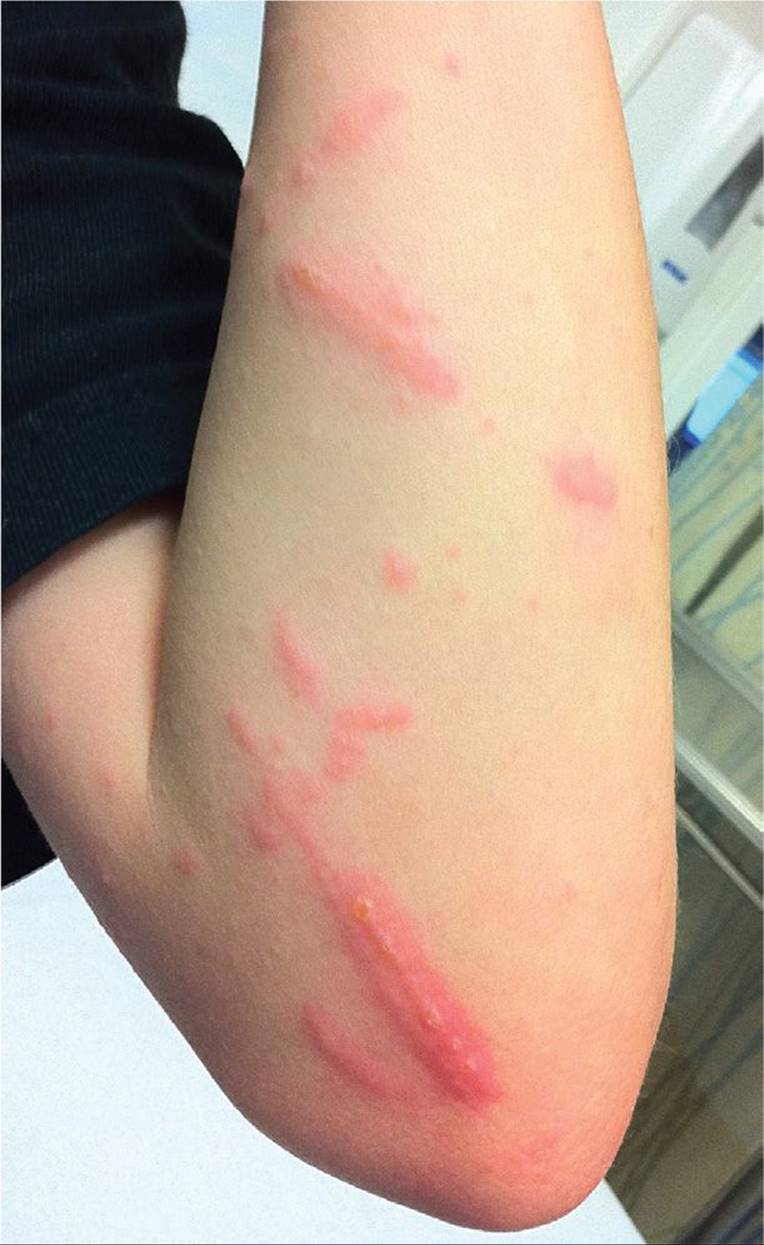
Figure 68-12 Contact dermatitis. Erythema and vesicle formation in a linear pattern characteristic of poison ivy. (Courtesy of Darren Fiore, MD.)
DIFFERENTIAL DIAGNOSIS
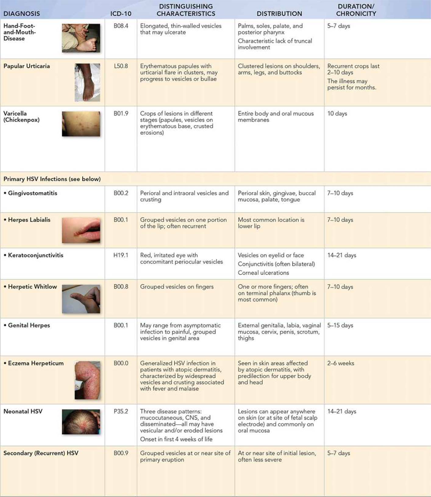


Other Diagnoses to Consider
• Incontinentia pigmenti
• Photosensitivity reactions
• Bullous impetigo
• Eczema herpeticum
• Staphylococcal scalded skin syndrome
• Langerhans cell histiocytosis
• Pemphigus
When to Consider Further Evaluation or Treatment
• HSV infections are often diagnosed clinically; however, diagnostic testing should be performed in uncertain or complex cases. A Tzank smear is a rapid, but not sensitive and not specific test. For more definitive results, consider viral culture, DNA detection, or direct fluorescent antibody testing.
• The use of systemic antiviral therapy in uncomplicated cutaneous HSV infections is not always warranted; treatment may shorten the duration of illness if initiated early in the first 72 hours of symptoms or in cases of severe disease.
• HSV and VZV infections in immunocompromised hosts or neonates may disseminate rapidly, and consultation with an infectious disease specialist and/or a neonatologist is recommended.
• The presence of HSV-2 in young children should raise concerns for child sexual abuse.
• Vesicular lesions may become bacterially superinfected, typically by staphylococci, and may require systemic antibiotics.
• HSV or VZV keratoconjunctivitis should be referred to an ophthalmologist for evaluation and treatment. Similarly, in young patients with periorbital HSV lesions, evaluation by pediatric ophthalmology may be helpful to rule out ocular involvement.
• Recurrent episodes of genital herpes may be treated with episodic or chronic suppressive antiviral medication.
• The last documented case of smallpox in the United States was in 1949, and the last case in the world was in 1977. Any confirmed or suspected case of smallpox must be reported to public health officials.
• Immunocompromised patients exposed to VZV are candidates for VZIG and should be referred to an infectious disease specialist.
• Atypical, recurrent or poorly healing, contact dermatitis should be referred to a dermatologist for further evaluation.
SUGGESTED READINGS
Chayavichitslip P, Buckwalter JV, Krakowski AC, et al. Herpes simplex. Pediatr Rev. 2009;30:119–130.
Cohen B. Pediatric Dermatology. 3rd ed. London: Mosby; 2005:101–120.
Eichenfield LF, Frieden IJ, Esterly NB, eds. Neonatal Dermatology. 2nd ed. Philadelphia, PA: Elsevier; 2008:131–158.
Gnann JW Jr, Whitley RJ. Clinical practice: herpes zoster. N Engl J Med. 2002;347:340–346.
Weston WL, Lane AT, Morelli JG. Color Textbook of Pediatric Dermatology. 4th ed. Philadelphia, PA: Mosby; 2007:127–138, 195–212.