
Approach to the Problem
Fine, bumpy rashes are a common complaint seen in the pediatric outpatient setting. They may be acute or chronic, and may be so subtle and asymptomatic that they are not noted for a period of time. The most classic example of a fine, bumpy rash is the sandpaper rash of scarlet fever, but this category of rashes also includes nonspecific rashes such as viral exanthems, heat rashes, drug reactions, and rarer lichenoid eruptions. Most of these conditions are benign and self-limited, and at times it may not be possible to make the exact diagnosis. It is important to note, however, that there are serious conditions such as toxic shock syndrome (TSS) that may initially present as a mild rash. It is important to have a high clinical suspicion for these serious disorders, particularly when systemic symptoms accompany the rash.
Key Points in the History
• Scarlet fever is generally seen with the accompanying symptoms of fever, headache, sore throat, and abdominal pain; the rash usually appears 24 to 48 hours after the onset of symptoms.
• Folliculitis, which occurs in all skin types, is not always infectious but also may be caused by chemical irritation or physical injury from shaving.
• Keratosis pilaris is often prominent in patients with underlying atopic dermatitis or obesity. Many patients with keratosis pilaris have a family member with this condition.
• Lichen nitidus is usually a chronic, nonpruritic, asymptomatic condition.
• Though usually precipitated by heat, miliaria can occur in the winter in association with fever, overbundling, or the use of certain ointments.
• Fine papular eruptions are the most frequent of all cutaneous drug reactions, classically due to ampicillin or amoxicillin, and are often identical in appearance to viral exanthems.
• Penicillin therapy in patients with Epstein–Barr virus infection may result in a morbilliform (measles-like) exanthem, which is considered to be a drug reaction.
Key Points in the Physical Examination
• Scarlet fever often produces Pastia lines, linear rows of petechiae, in the skin folds, particularly in the antecubital and popliteal fossae and the inguinal area.
• Scarlet fever is most often associated with pharyngitis, but may also be seen with other group A streptococcal skin infections such as cellulitis.
• In patients with more skin pigmentation, the rash of scarlet fever may not readily appear erythematous; therefore, it is important to examine patients in bright lighting.
• The Koebner phenomenon, linear papules along the lines of skin trauma, is a hallmark of lichen nitidus and presents in almost all cases.
• Keratosis pilaris, which is rough in texture, occurs along the extensor surfaces of extremities.
• Folliculitis, which may consist of pustules of varying size, is usually confined to hair-bearing areas such as the scalp, forearms, groin, and legs. Often the hair follicle cannot be seen in folliculitis.
• Miliaria is often found on the face and upper trunk and back.
|
PHOTOGRAPHS OF SELECTED DIAGNOSES |
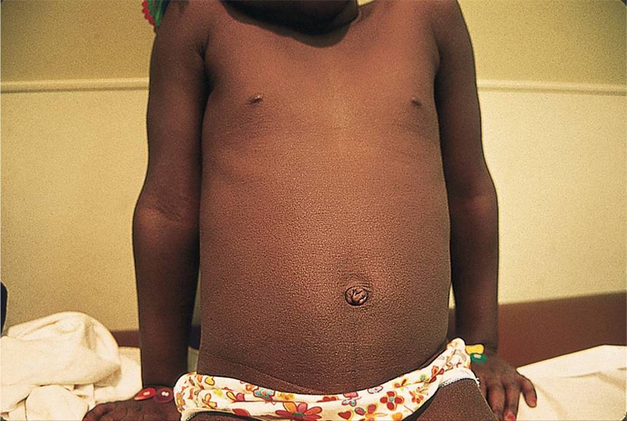
Figure 71-1 Scarlet fever. Fine papules, “sandpaperlike” rash on trunk of child with scarlet fever. (Courtesy of George A. Datto, III, MD.)
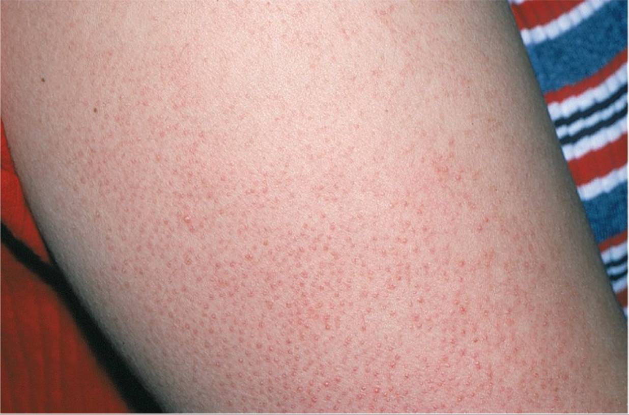
Figure 71-2 Keratosis pilaris. Tiny, rough-textured, follicular papules on lateral upper arms. (Used with permission from Goodheart HP. Goodheart’s Photoguide to Common Skin Disorders. 2nd ed. Philadelphia, PA: Lippincott Williams & Wilkins; 2003:49.)

Figure 71-3 Folliculitis. Erythematous papulovesicular lesions on abdomen after exposure in a hot tub. (Courtesy of Lee R. Atkinson-McEvoy, MD.)

Figure 71-4 Lichen nitidus. Shiny small papules on elbow. (Courtesy of George A. Datto, III, MD.)
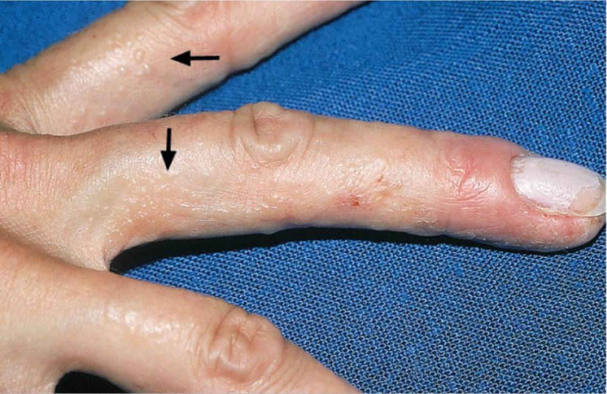
Figure 71-5 Dyshidrotic eczema. Note the fine, fluid-filled bumps on the fingers as depicted by the arrows. (Used with permission from Goodheart HP. Goodheart’s Photoguide to Common Skin Disorders. 2nd ed. Philadelphia, PA: Lippincott Williams & Wilkins; 2003:60.)
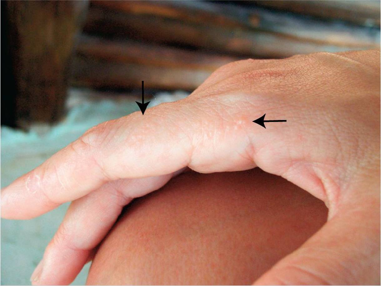
Figure 71-6 Dyshidrotic eczema. Note the fine, fluid-filled bumps on the finger (as depicted by the arrows) and the distal area of peeling. (Courtesy of Esther K. Chung, MD, MPH.)
DIFFERENTIAL DIAGNOSIS


Other Diagnoses to Consider
• Nonspecific viral exanthem
• TSS (early phase)
• Kawasaki disease
• Contact dermatitis (early phase)
• “Id” reaction (autoeczematization to various stimuli, particularly fungi, that results in a symmetrical eczematous, maculopapular, or papulovesicular rash)
• Lichen spinulosus (papular rash with a spine or horn at each hair follicle)
• Keratosis follicularis or Darier disease (hereditary skin disorder where keratotic papules coalesce to form crusty, warty plaques)
• Dyshidrotic eczema
When to Consider Further Evaluation or Treatment
• Most fine, bumpy rashes are benign and self-limited. Any persistent rash or a rash associated with systemic symptoms, such as fever or altered mental status, warrants further evaluation.
• If a rash is bothersome and does not respond to initial therapy, further management should be determined after consultation with a pediatric dermatologist.
• The initial rash of TSS may mimic scarlet fever. The presence of high fever, mental status changes, headache, or early shock-like symptoms should raise suspicion for TSS and warrants immediate evaluation and management.
SUGGESTED READINGS
Goodheart HP. Goodheart’s Photoguide to Common Skin Disorders. 3rd ed. Philadelphia, PA: Lippincott Williams & Wilkins; 2008:60.
Habif T. Clinical Dermatology. 5th ed. St. Louis, MO: Mosby; 2009:263, 351–355, 464–466, 486–488.
Hwang S. Keratosis pilaris: a common follicular hyperkeratosis. Cutis 2008;82:177–180.
Paller AS. Hurwitz Clinical Pediatric Dermatology. 4th ed. Philadelphia, PA: WB Saunders; 2011:106–107.
Shulman ST. Clinical practice guidelines for the diagnosis and management of Group A streptococcal pharyngitis: 2012 update by the Infectious Disease Society of America. Clin Infect Dis. 2012;55:1279–1282.
Tilly JT, Drolet BA. Lichenoid eruptions in children. J Am Acad Dermatol. 2004;51:606–624.