
Approach to the Problem
The most common scaly rash in pediatrics is atopic dermatitis (eczema), which affects 15% to 20% of the pediatric population. While eczema tends to be chronic in nature, some patients have symptoms primarily during cold and dry weather. Other common causes of scaly rash include pityriasis rosea, tinea corporis, and seborrhea. Psoriasis and ichthyosis are less common. Initial lesions of pityriasis may at times be mistaken for tinea corporis, and ichthyosis may at times be mislabeled as severely dry skin. In general, most dry and scaly rashes tend to be pruritic in nature.
Key Points in the History
• The duration of symptoms will help to distinguish acute and subacute rashes, such as tinea corporis, from more chronic conditions, such as eczema.
• A family history of atopy should raise suspicion for eczema.
• Eczema generally spares the groin and diaper areas, whereas seborrhea does not.
• A solitary lesion may suggest tinea corporis or may be the herald patch seen in pityriasis rosea.
• Tinea corporis worsens with topical steroids, whereas eczema generally improves.
• Eczema on the face of young infants may have a circular area of erythema and may be misdiagnosed as tinea corporis.
• Cold weather generally exacerbates eczema, but some patients report worsening in the summer and winter months.
• In pityriasis rosea, the rash often starts as a single isolated lesion, a herald patch, followed by a more generalized rash occurring 5 to 10 days later.
• In the event a child shares a bed with another individual who denies pruritus or rash, a diagnosis of scabies is unlikely.
• Psoriasis affects 1% to 3% of the population, but it is uncommon in African Americans.
Key Points in the Physical Examination
• Patients with eczema often have dry skin, keratosis pilaris, or both.
• Lichenification is pathognomic of chronic atopic dermatitis when it appears in the expected distribution.
• Often, allergic shiners and Dennie–Morgan lines are seen in individuals with atopic dermatitis.
• Seborrhea generally stays within the hairline, whereas psoriasis extends beyond the hairline.
• In ectopic allergic contact dermatitis, the rash may not be in the expected location as can be seen with nail polish (tosylamide/formaldehyde) allergy.
• Lesions associated with tinea corporis tend to be round, whereas the herald patch in pityriasis rosea is oval.
• The generalized rash of pityriasis rosea classically runs parallel to the lines of skin cleavage, in a “Christmas-tree” distribution.
• In some individuals, the scaly lesions of pityriasis may be found in the pubic, inguinal, and axillary areas, and this is referred to as “inverse pityriasis rosea.”
• Postinflammatory hypopigmentation commonly occurs following eczema, pityriasis, and tinea. Hypopigmentation can be distressing to families; therefore, discussing this early in the course of the disease may be helpful.
|
PHOTOGRAPHS OF SELECTED DIAGNOSES |

Figure 70-1 Atopic dermatitis. This lesion shows no evidence of active inflammation. Lichenification and postinflammatory hyperpigmentation are apparent. (Used with permission from Goodheart HP. Goodheart’s Photoguide to Common Skin Disorders. 2nd ed. Philadelphia, PA: Lippincott Williams & Wilkins; 2003:44.)

Figure 70-2 Atopic dermatitis. Note the areas of dryness, hypo- and hyperpigmentation and lichenification in the antecubital fossa of this 5-year-old. (Courtesy of Esther K. Chung, MD.)

Figure 70-3 Nummular eczema. “Coin-shaped” patches and plaques are located on the legs. (Used with permission from Goodheart HP. Goodheart’s Photoguide to Common Skin Disorders. 2nd ed. Philadelphia, PA: Lippincott Williams & Wilkins; 2003:87.)

Figure 70-4 Seborrhea. (Used with permission from Fleisher GR, Ludwig S, Baskin MN. Atlas of Pediatric Emergency Medicine. Philadelphia, PA: Lippincott Williams & Wilkins; 2004:85.)
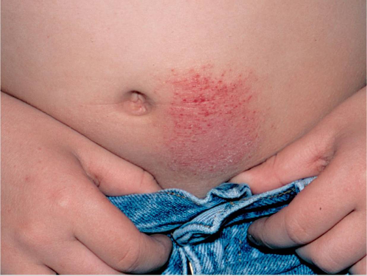
Figure 70-5 Allergic contact dermatitis. This boy developed an eczematous eruption at the site where the nickel snap on his blue jeans contacted his skin. (Used with permission from Goodheart HP. Goodheart’s Photoguide to Common Skin Disorders. 2nd ed. Philadelphia, PA: Lippincott Williams & Wilkins; 2003:67.)

Figure 70-6 Variation in pityriasis rosea. In light skin, a “classic” oval erythematous, scaly herald plaque. (Used with permission from Burkhart C, Morrell D, Goldsmith LA, et al. VisualDx: Essential Pediatric Dermatology.Philadelphia, PA: Lippincott Williams & Wilkins; 2009.)

Figure 70-7 Tinea corporis. Note the large size of this lesion that was made worse by the use of topical steroids. (Courtesy of Esther K. Chung, MD.)
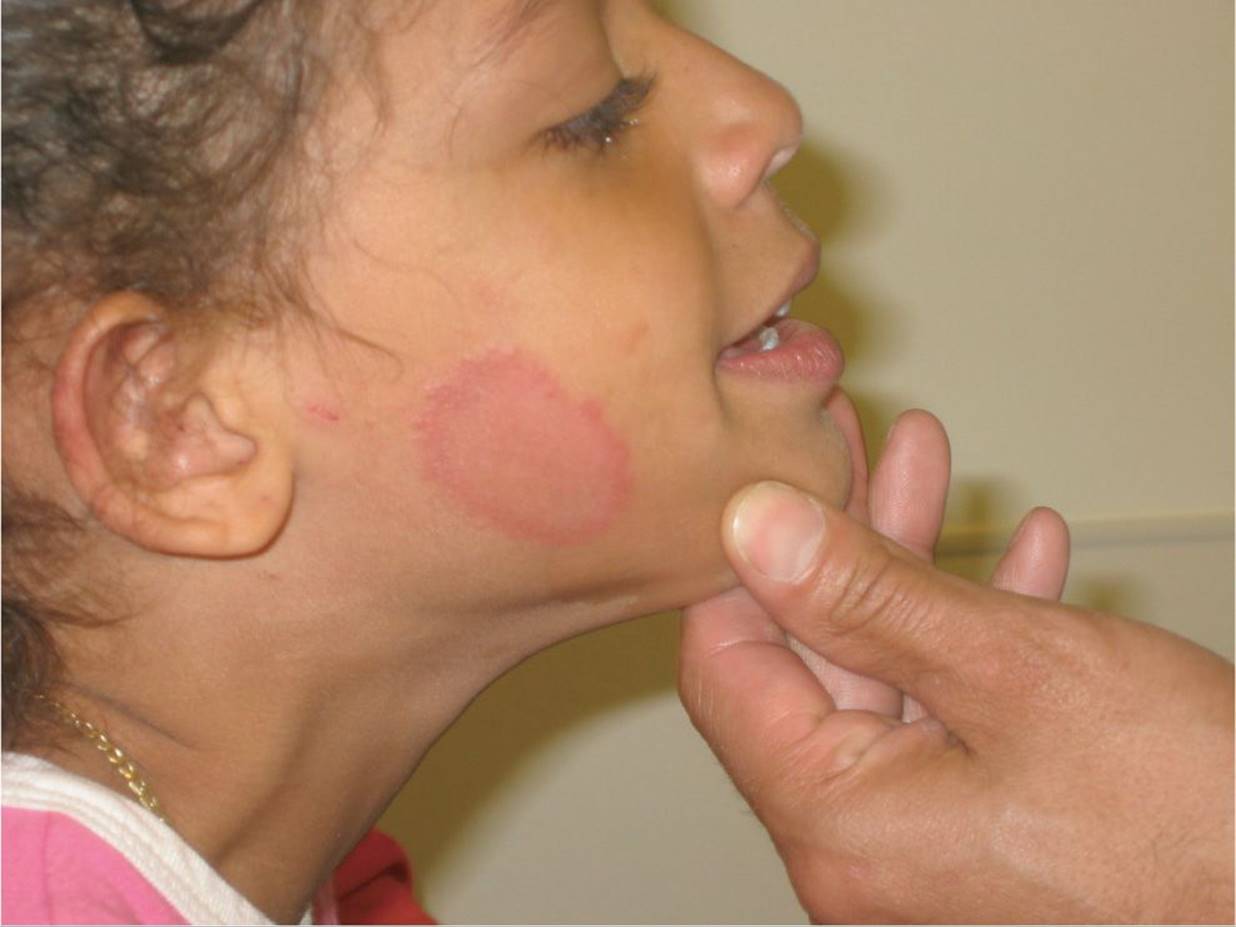
Figure 70-8 Tinea corporis on the face. (Courtesy of George A. Datto, III, MD.)
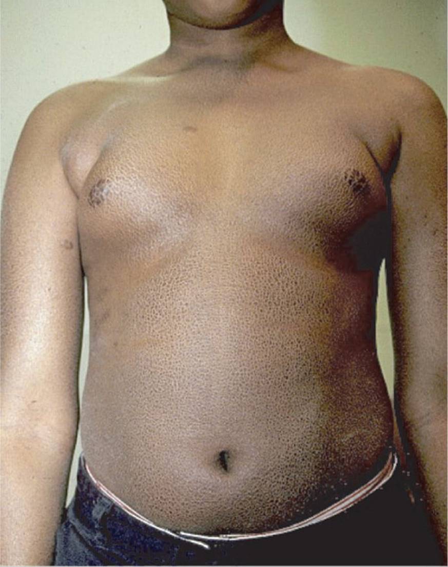
Figure 70-9 Ichthyosis vulgaris. (Courtesy of George A. Datto, III, MD.)
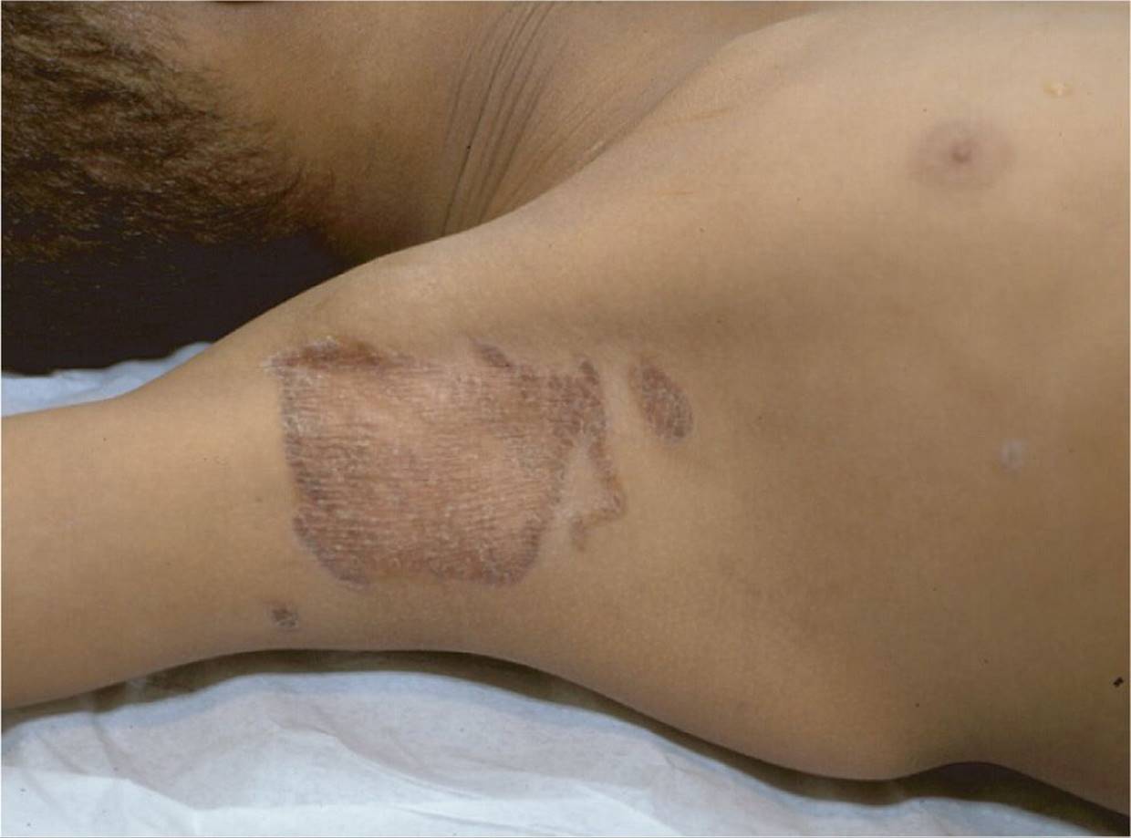
Figure 70-10 Psoriasis. (Courtesy of George A. Datto, III, MD.)
DIFFERENTIAL DIAGNOSIS


Other Diagnoses to Consider
• Scabies
• Letterer–Siwe disease (a form of histiocytosis consisting of lymphadenopathy, hepatosplenomegaly, and a seborrhea-like rash)
• Leiner disease (seborrhea-like dermatitis, diarrhea, wasting and dystrophy, and recurrent gram-negative infection)
• Netherton syndrome (“bamboo hair,” congenital ichthyosiform erythroderma, and atopic diathesis)
• Acrodermatitis enteropathica (autosomal recessive disorder; listlessness, diarrhea, failure to thrive, low-serum zinc)
• Zinc deficiency
• Wiskott–Aldrich syndrome (diarrhea, purpura, and susceptibility to infection)
• Phenylketonuria (mental retardation, seizures, blond hair, and eczema)
• Hyper IgE syndrome (recurrent sinopulmonary and cutaneous infections, markedly elevated IgE levels, and chronic dermatitis)
• Lichen striatus
• Systemic lupus erythematosus
When to Consider Further Evaluation or Treatment
• Further evaluation and treatment should be considered for dry scaly rashes that fail to improve in spite of frequent moisturizing.
• Most dry, scaly rashes are due to atopic dermatitis and will respond to emollients and the use of mild topical steroids.
• Severe and complicated cases of eczema warrant consultation with a dermatologist.
• Because tinea corporis worsens with use of topical steroids, use only these agents when there is a low suspicion for tinea corporis.
• Pityriasis rosea is generally self-limited and resolves by 6 weeks generally; however, it may last as long as 5 months.
• With pityriasis rosea, exposure to UV light hastens resolution.
• Keep in mind that allergic contact dermatitis may last for weeks to months after removal of the allergen, even with the use of topical steroids.
• An asymptomatic focal scaly lesion followed by fever and leucopenia should raise suspicion for systemic lupus erythematosus.
SUGGESTED READINGS
Chuh AA, Dofitas BL, Comisel GG, et al. Interventions for pityriasis rosea. Cochrane Database Syst Rev. 2007;18:CD005068.
Huang CF, Wang WM, Chiang CP. Scaly ear rash as the herald of a young girl with juvenile systemic lupus erythematosus. Ann Dermatol. 2011;23:S333–S337.
Kress D. What’s your diagnosis? Scaly pubic plaques in a 2-year-old girl—or an “inverse” rash. In: Traeger TKD (Section Ed.) Pedi Gyn-Derm. J Pediatr Adolesc Gynecol. 2007;20:109–111.
Krol A, Krafchik B. The differential diagnosis of atopic dermatitis in childhood. Dermatol Ther. 2006(19);73–82.
Larsen S, Hanifin JM. Epidemiology of atopic dermatitis. Immunol Clin North Am. 2002;22:1–24.
Militello G, Jacob SE, Crawford GH. Allergic contact dermatitis in children. Curr Opin Pediatr. 2006;18:385–390.
Schon MP, Boehncke WH. Psoriasis. N Engl J Med. 2005;352:1899–1912.
Sheu J, Huang JT. Erythematous scaly plaques and papules in a 9-month-old infant. J Pediatr. 2013;163:1222.