
Approach to the Problem
Hyperpigmented lesions are caused by localized melanin deposition in the skin. Congenital lesions may occur over time as sun exposure increases the amount of melanin contained within these lesions. Patients and their families frequently have concerns about the cosmetic effects of these lesions and the malignant potential of certain lesions. A careful history and examination, following the lesions closely over time, and referring to appropriate specialists when needed, can reassure patients and families and often result in the correct diagnosis and management.
Key Points in the History
• A history of pustules present at birth that evolve into hyperpigmented macules is characteristic of neonatal pustular melanosis.
• Café au lait spots grow proportionally to overall body growth in the first few years of life and then stabilize.
• Café au lait spots may be a benign familial trait. A family history of neurofibromatosis and the presence of more than six café au lait spots should raise suspicion for neurofibromatosis.
• Only 1% of pigmented lesions at birth are congenital melanocytic nevi.
• Freckles are common in light-skinned, red-haired individuals, and are an autosomal-dominant, inherited trait.
• Freckles are induced by sunlight and are more prominent in the summer and fade in the winter.
• Acanthosis nigricans is associated with overweight/obesity, polycystic ovary syndrome, and the metabolic syndrome—particularly in association with diabetes mellitus with insulin resistance.
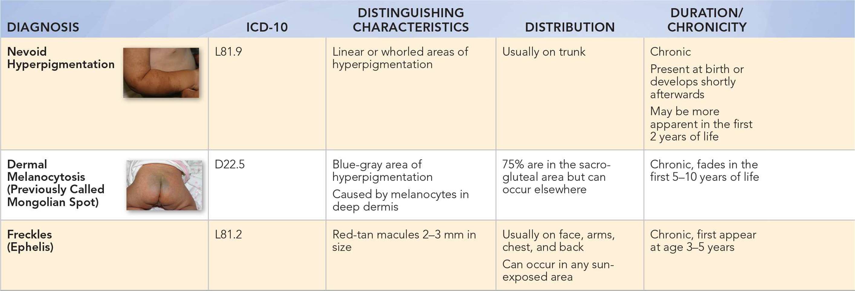
• Dermal melanocytosis (previously called Mongolian spots) fades after the first 5 to 10 years of life.
Key Points in the Physical Examination
• A combination of small 1 to 3 mm hyperpigmented macules and pustules is seen in neonatal pustular melanosis.
• A complaint of a “dirty neck” is frequently associated with the physical finding of acanthosis nigricans, which is commonly seen in association with an elevated body mass index.
• Acanthosis nigricans has a velvety quality and may have papillomatous growths within the area of hyperpigmentation.
• Weight loss can result in improvement or resolution of acanthosis nigricans.
• Dermal melanocytosis is often seen in the sacral or gluteal areas, but may also be present elsewhere on the body, including the dorsum of the hand, the upper back, and the shoulders.
• Congenital melanocytic nevi vary in size and may be associated with hair often darker and longer than in surrounding areas.
• Tinea versicolor may be hyperpigmented or hypopigmented and often has scale.
• Blue nevi get their bluish color from the location of the melanocytes deep in the dermis.
• Nevoid hypermelanosis follows Blaschko lines on the extremities and appears linear; on the trunk, it can have a whorled pattern.
• In neurofibromatosis type 1, there are more than six café au lait macules at least 0.5 cm in size in prepubertal children, and there are associated findings of axillary or inguinal freckling, Lisch nodules (iris hamartomas), and neurofibromas.
|
PHOTOGRAPHS OF SELECTED DIAGNOSES |

Figure 73-1 Postinflammatory hyperpigmentation. Hyperpigmented linear lesions following skin trauma from a razor. (Courtesy of George A. Datto, III, MD.)
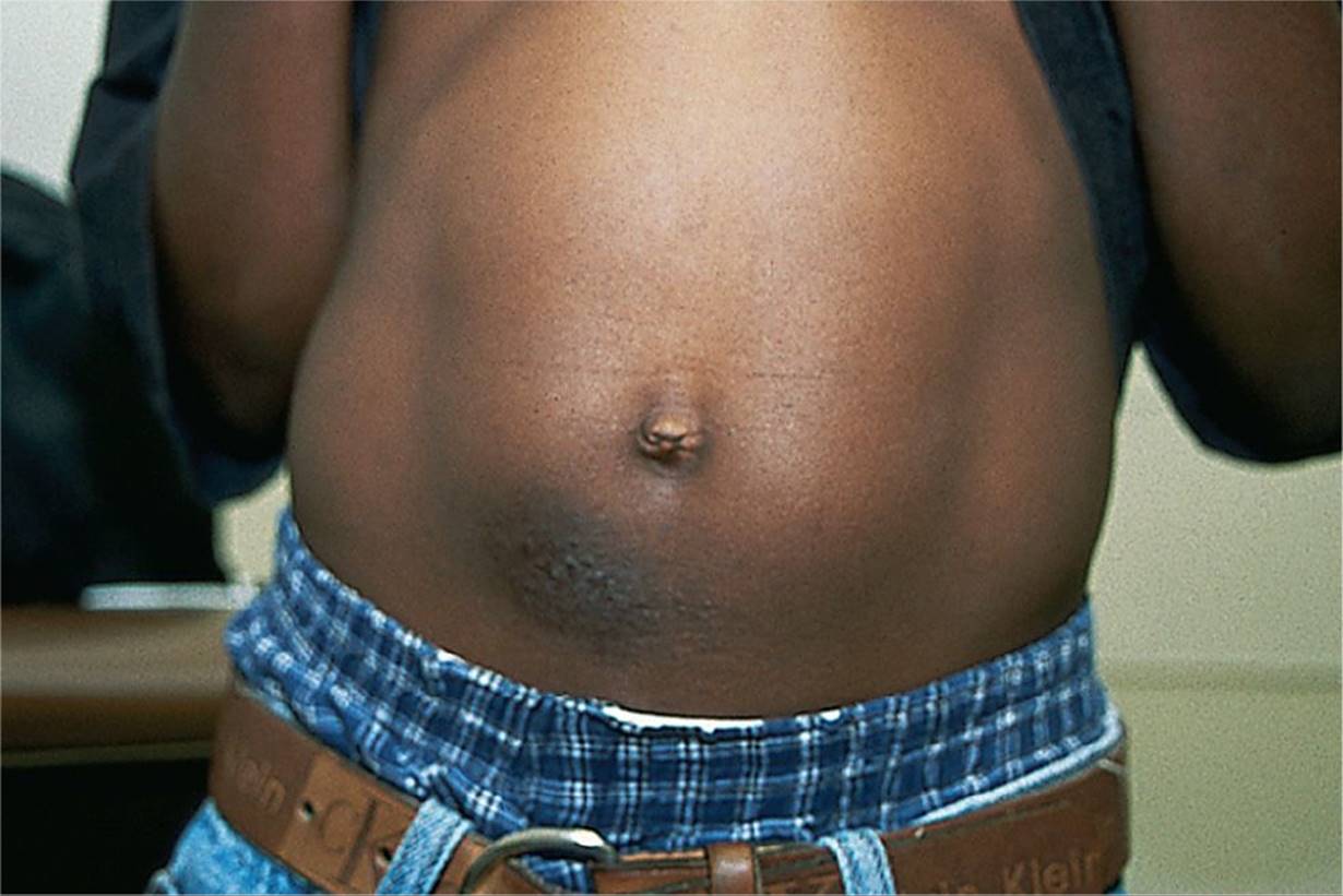
Figure 73-2 Hyperpigmentation resulting from nickel dermatitis. (Courtesy of George A. Datto, III, MD.)

Figure 73-3 Acanthosis nigricans. Thickened velvety, hyperpigmented epidermis in the neck of obese child. (Courtesy of Ilona J. Frieden, MD.)

Figure 73-4 Congenital melanocytic nevus. A medium-sized, congenital, pigmented nevus on the trunk of an infant. (Courtesy of Ilona J. Frieden, MD.)
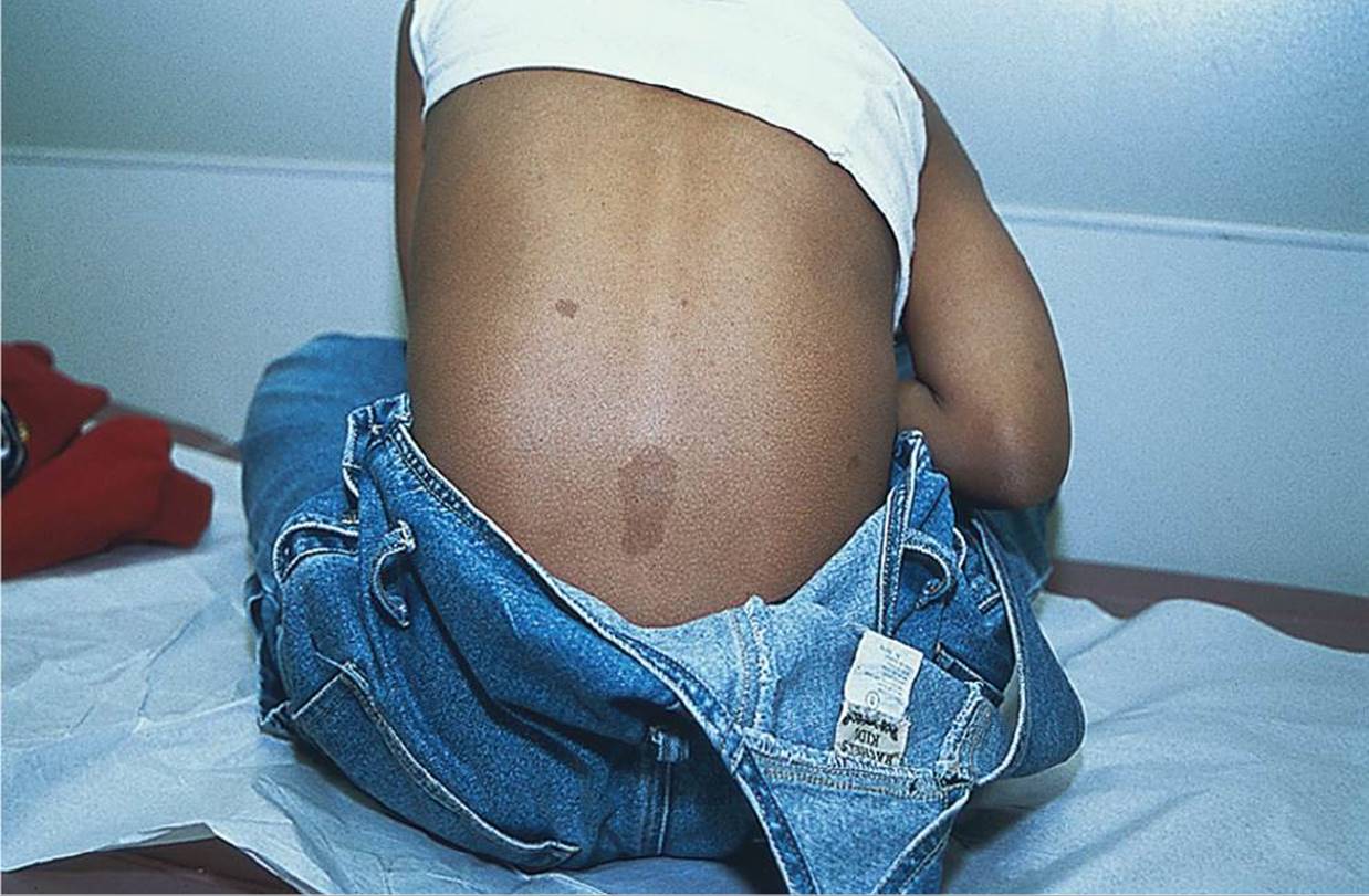
Figure 73-5 Café au lait spots. Multiple café au lait spots on the back of a child. (Courtesy of George A. Datto, III, MD.)
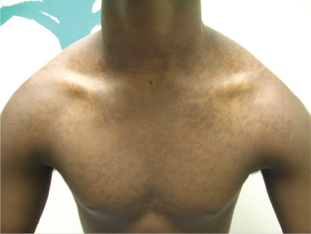
Figure 73-6 Tinea versicolor. While often hypopigmented, this rash may be hyperpigmented as in this individual. (Courtesy of Paul S. Matz, MD.)
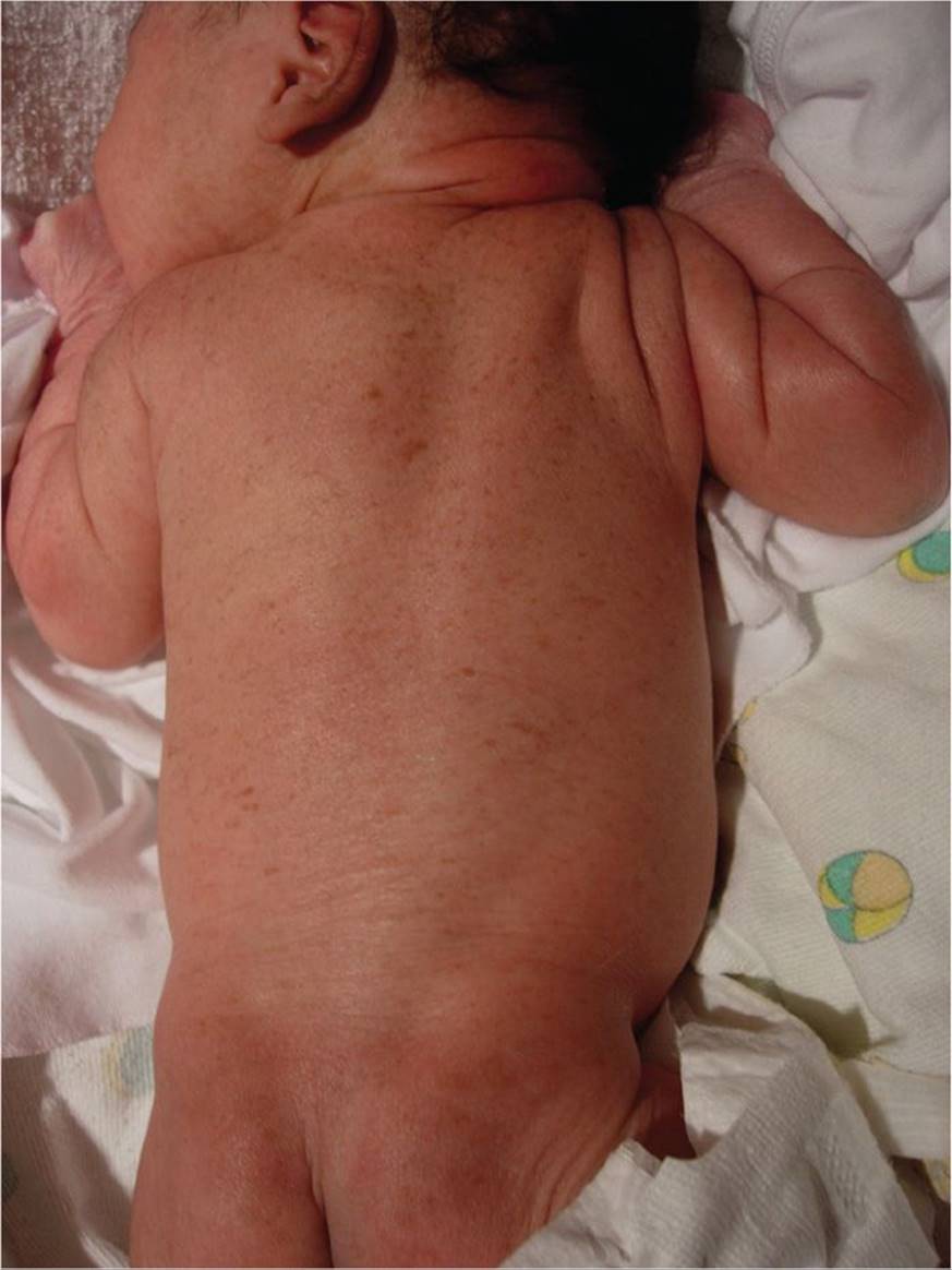
Figure 73-7 Neonatal pustular melanosis. Small hyperpigmented macules seen in a newborn. Often associated with pustules. (Courtesy of Amy Gilliam, MD.)
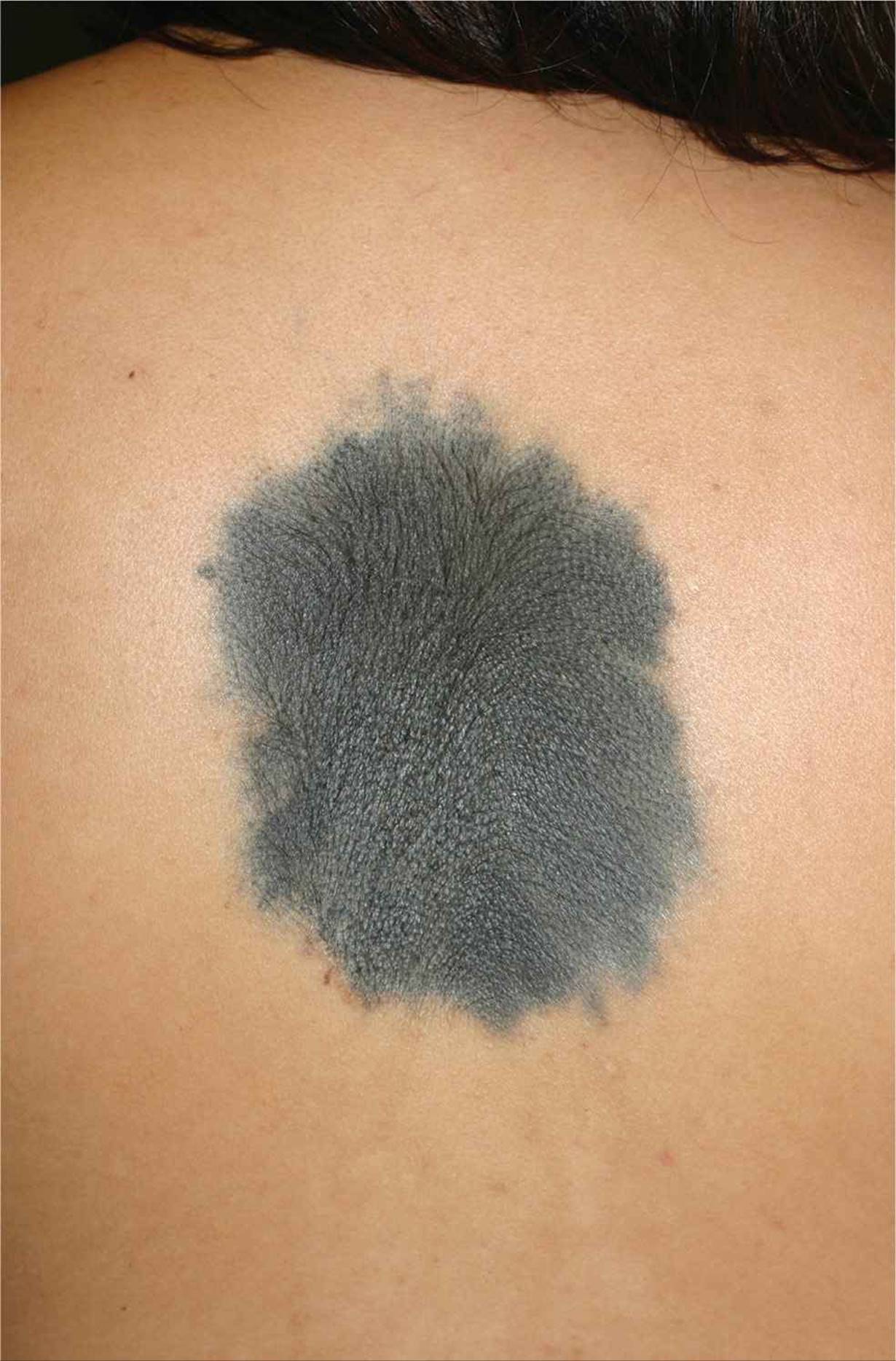
Figure 73-8 Congenital blue nevus. Bluish-discolored nevus that persists. (Courtesy of Ilona J. Frieden, MD.)

Figure 73-9 Nevoid hyperpigmentation. Previously called hypermelanosis of Ito, an irregular patterned area of hyperpigmentation. (Courtesy of Ilona J. Frieden, MD.)

Figure 73-10 Dermal melanocytosis. Slate gray-colored macule on back of infant. (Courtesy of Ilona J. Frieden, MD.)
DIFFERENTIAL DIAGNOSIS



Other Diagnoses to Consider
• Hormone-induced hyperpigmentation of the genitalia in normal newborns
• Xeroderma pigmentosa
• McCune–Albright syndrome
• Lentigines (such as Peutz–Jeghers syndrome)
• Other syndromes with associated café au lait spots, including Noonan syndrome, LEOPARD, Costello syndrome, and Cardio-Facio-Cutaneous syndrome
• Malignant melanoma
When to Consider Further Evaluation or Treatment
• Congenital melanocytic nevi have potential for malignant transformation, so they should be referred to a dermatologist for potential excision.
• Multiple café au lait spots with any associated abnormalities of the eye, seizures, developmental abnormalities, endocrine dyscrasias, or any other abnormalities should be investigated for neurofibromatosis type 1, McCune–Albright syndrome, and other associated syndromes.
• If tinea versicolor does not respond to topical treatments, consider further management with alternative topical or oral antifungal treatment.
• Children with acanthosis nigricans who are significantly overweight should be evaluated for diabetes mellitus, metabolic syndrome, or polycystic ovary syndrome. Individuals with these disorders should be evaluated by a pediatric endocrinologist.
SUGGESTED READINGS
Taïeb A, Boralevi F. Hypermelanoses of the newborn and of the infant. Dermatol Clin. 2007;25:327–336.
Weinberg S, Prose NS, Kristal L, eds. Color Atlas of Pediatric Dermatology. 4th ed. New York, NY: McGraw Hill; 2007:267–284.
Paller AS, Mancini AJ, eds. Hurwitz Clinical Pediatric Dermatology: A Textbook of Skin Disorders of Childhood and Adolescence. 4th ed. Philadelphia, PA: Elsevier Saunders; 2011:20–23.