
Approach to the Problem
Blister-associated rashes in children are divided into two groups: vesicular rashes and bullous rashes. By definition, vesicles are fluid-filled lesions that are no more than 0.5 cm in diameter and bullae are blistering lesions that are greater than 0.5 cm in diameter. They may be seen in the skin or mucous membranes and may be round or have irregular borders. Compared with adult skin, that of pediatric patients is very prone to blistering. Vesicular rashes are common in the pediatric population, and are discussed elsewhere. Bullous rashes, on the other hand, are a relatively uncommon class of pediatric skin disorders.
Bullae form secondary to fluid accumulation between cells in the epidermis or between the epidermal and dermal (subepidermal) skin layers. Bullous skin disorders can be divided into congenital disorders, such as congenital epidermolysis bullosa (EB); infectious disorders, such as bullous impetigo and staphylococcal scalded skin syndrome (SSSS), immunologic disorders, such as Stevens–Johnson syndrome (SJS), toxic epidermal necrolysis (TEN), chronic bullous dermatosis/disease of childhood, and toxin-mediated such as phototoxic or venom reactions.
Key Points in the History
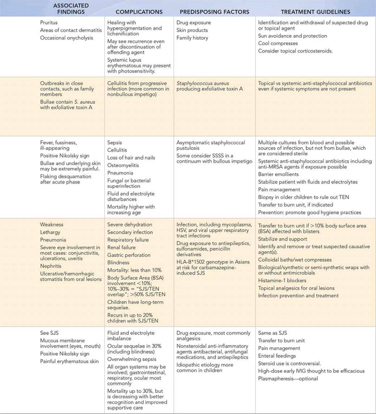
• Systemic symptoms such as fever, fussiness, ill appearance, and diffuse skin involvement are associated with SJS, TEN, and SSSS.
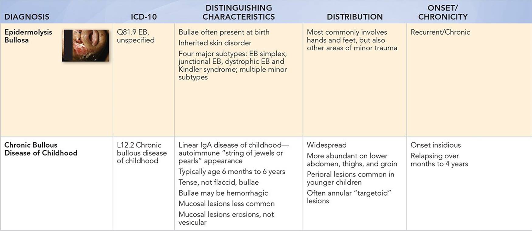
• Congenital blistering diseases, such as EB, are often present at birth or during the newborn period.
• Bullae appearing in the first few days or weeks of life may be secondary to EB, bullous impetigo of the neonate, or SSSS.
• In EB, bullae form at sites of minor skin trauma or friction.
• With the exception of TEN, the more common bullous skin disorders involve only the epidermis and may cause temporary hyperpigmentation and lichenification but do not tend to cause permanent scarring.
• Although many believe SJS and TEN are a continuum of the same disease, one convention is that infectious agents are causally related to SJS, and drugs to TEN.
• Chronic bullous disease of childhood is rare, but it is the most common acquired autoimmune bullous disease in children; it is only rarely present in the neonate.
Key Points in the Physical Examination
• Photosensitivity rashes are seen on exposed parts of the body, primarily the face and extremities.
• Bullous impetigo differs from nonbullous impetigo. Distribution is more truncal with a predilection for intertriginous areas, including the diaper area.
• Nikolsky sign—the ability to use gentle traction with a finger to separate the upper epidermis from the underlying skin in bullous lesions—is commonly seen in systemic bullous disorders, including TEN, SSSS, and EB.
• Mucous membrane involvement is seen in TEN, SJS, and in more severe forms of EB but not in SSSS.
• In bullous impetigo, photosensitivity rashes, EB, and SSSS, bullae are seen on the skin; in SJS/TEN, they are also seen on the mucous membranes of the mouth, nares, conjunctivae, and the anorectal and perineal areas.
• Conjunctivae are involved in SSSS (erythema) and SJS and TEN (mucositis).
• In chronic bullous dermatosis/disease of childhood, the lesions are often annular and appear like a “string of pearls”; lesions initially mimic bullous impetigo, herpetic diseases, or bullous erythema multiforme.
|
PHOTOGRAPHS OF SELECTED DIAGNOSES |
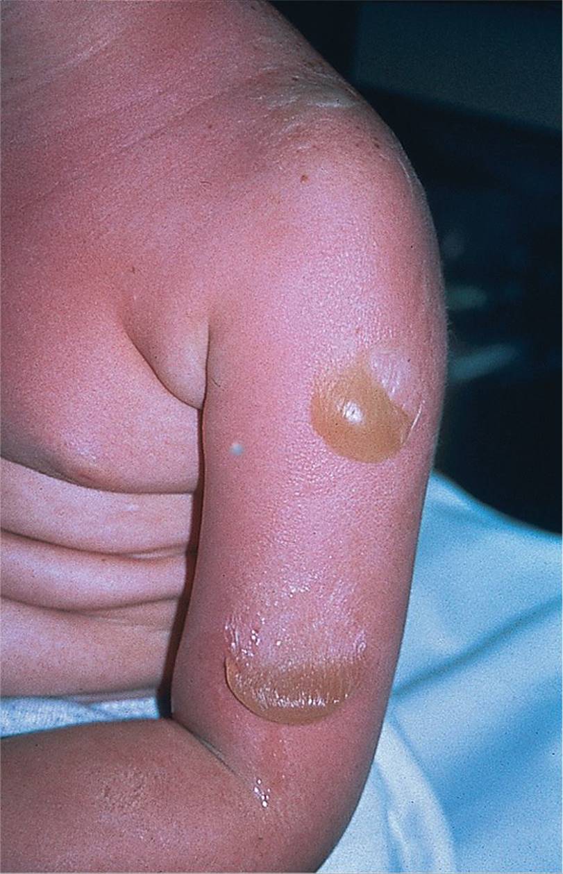
Figure 74-1 Photosensitivity. Large blisters that developed on the second day following prolonged sun exposure. (Courtesy of George A. Datto, III, MD.)
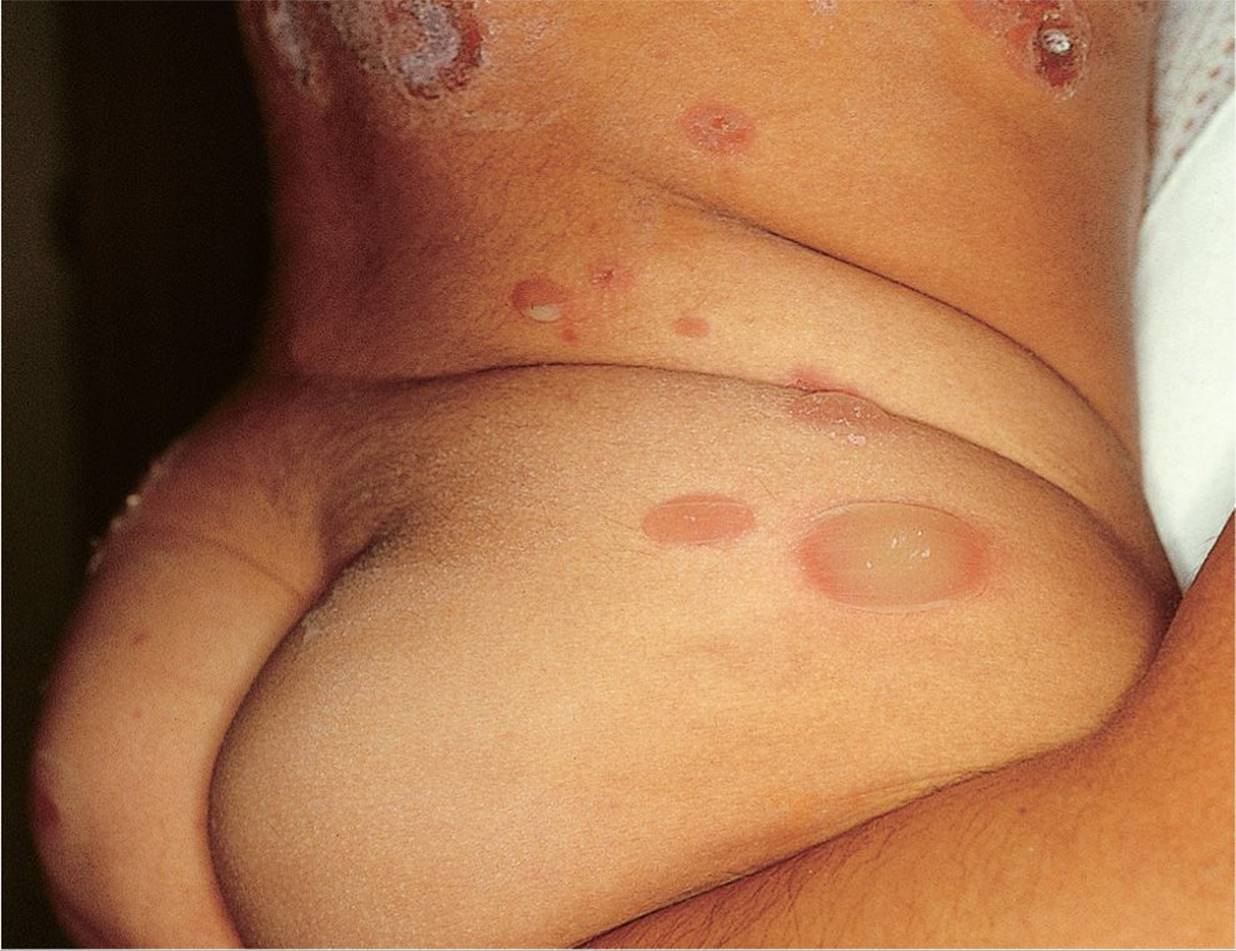
Figure 74-2 Bullous impetigo. (Used with permission from Fleisher GR, Ludwig S, Baskin MN. Atlas of Pediatric Emergency Medicine. Philadelphia, PA: Lippincott Williams & Wilkins; 2004:200.)
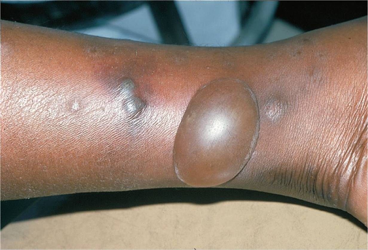
Figure 74-3 Bullous insect bite reaction. (Used with permission from Goodheart HP. Goodheart’s Photoguide to Common Skin Disorders. 2nd ed. Philadelphia, PA: Lippincott Williams & Wilkins; 2003:3.)
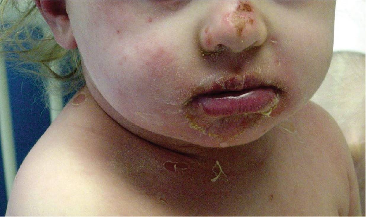
Figure 74-4 Staphylococcal scalded skin syndrome with ruptured bullae. (Courtesy of Gary Marshall, MD.)
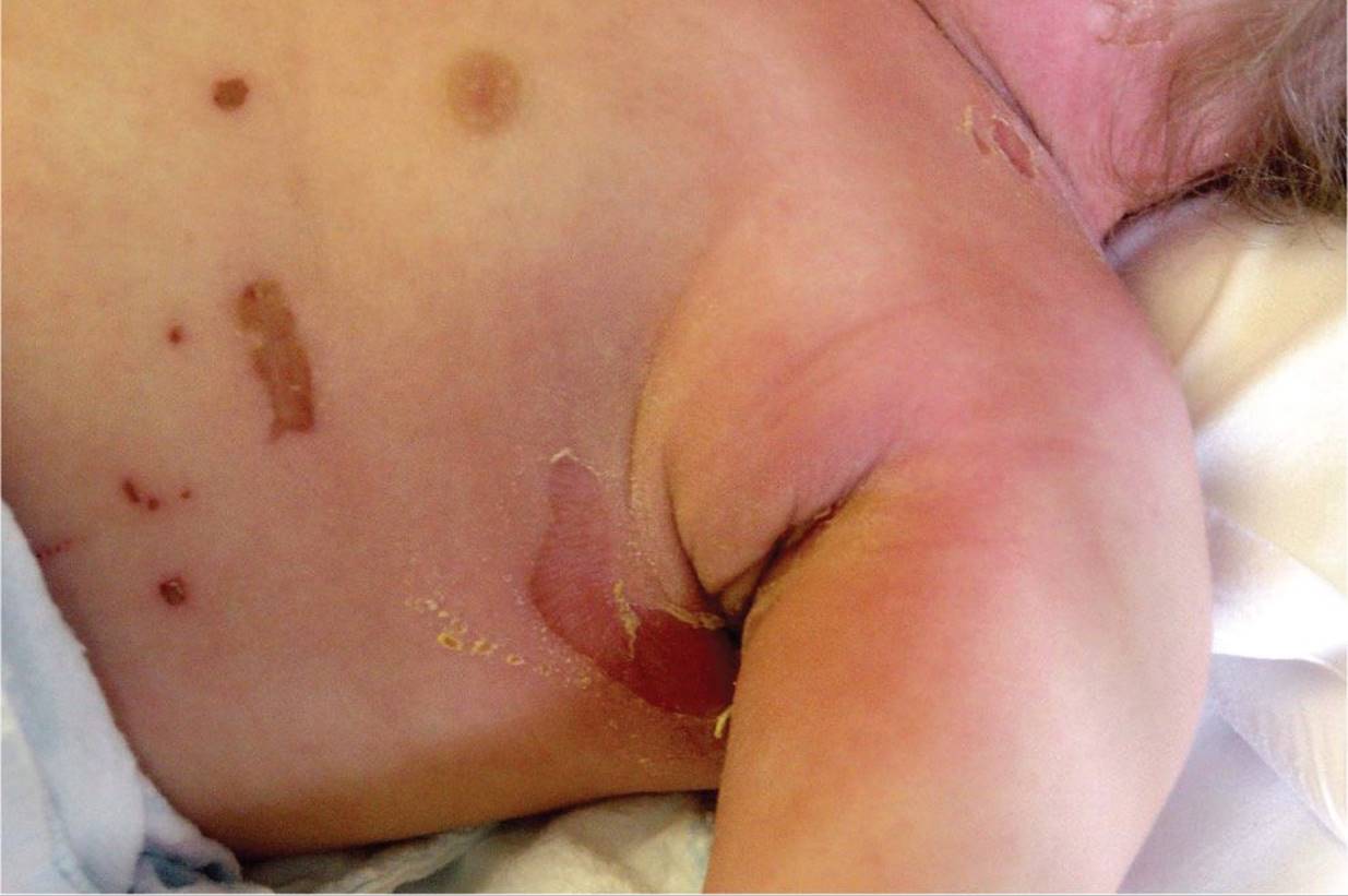
Figure 74-5 Staphylococcal scalded skin syndrome. “Scalded” skin underlying ruptured bulla in SSSS. (Courtesy of Gary Marshall, MD.)
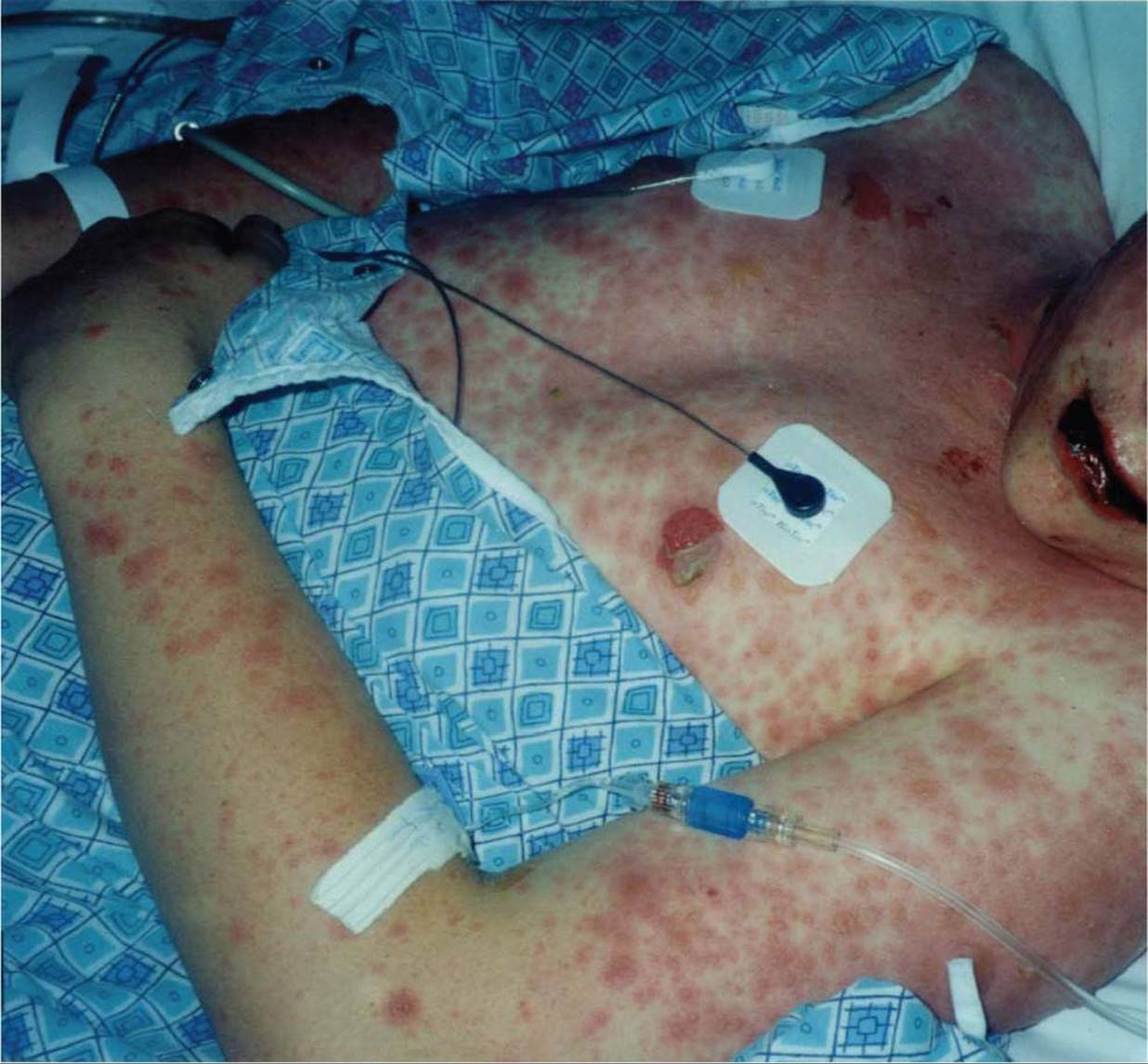
Figure 74-6 Target-like purpuric lesions of Stevens–Johnson syndrome. (Courtesy of Gary Marshall, MD.)
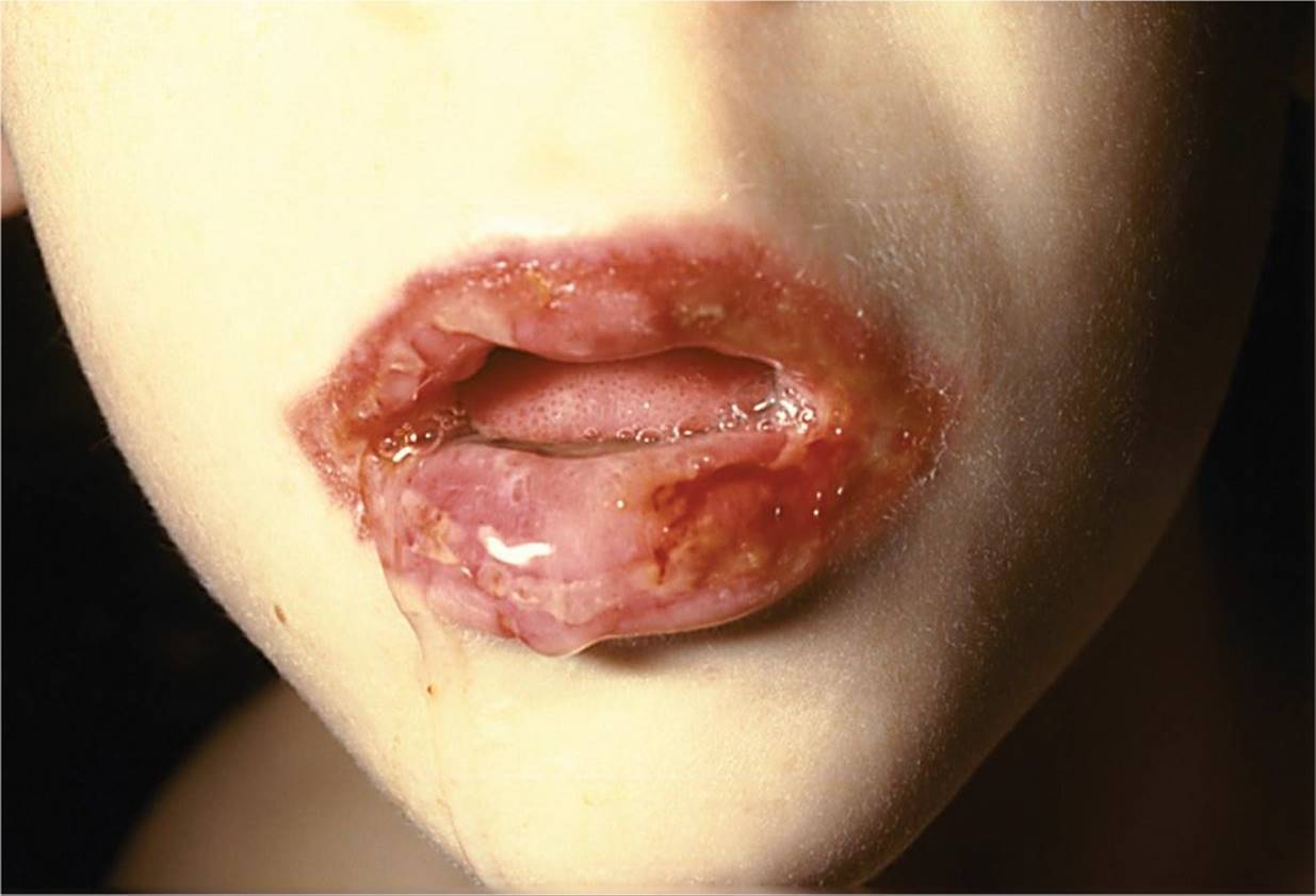
Figure 74-7 Hemorrhagic ulcerative stomatitis in SJS. (Courtesy of Joseph Lopreiato, MD.)

Figure 74-8 Epidermolysis bullosa. Note the ruptured bullous lesion. (Courtesy of Joseph Lopreiato, MD.)
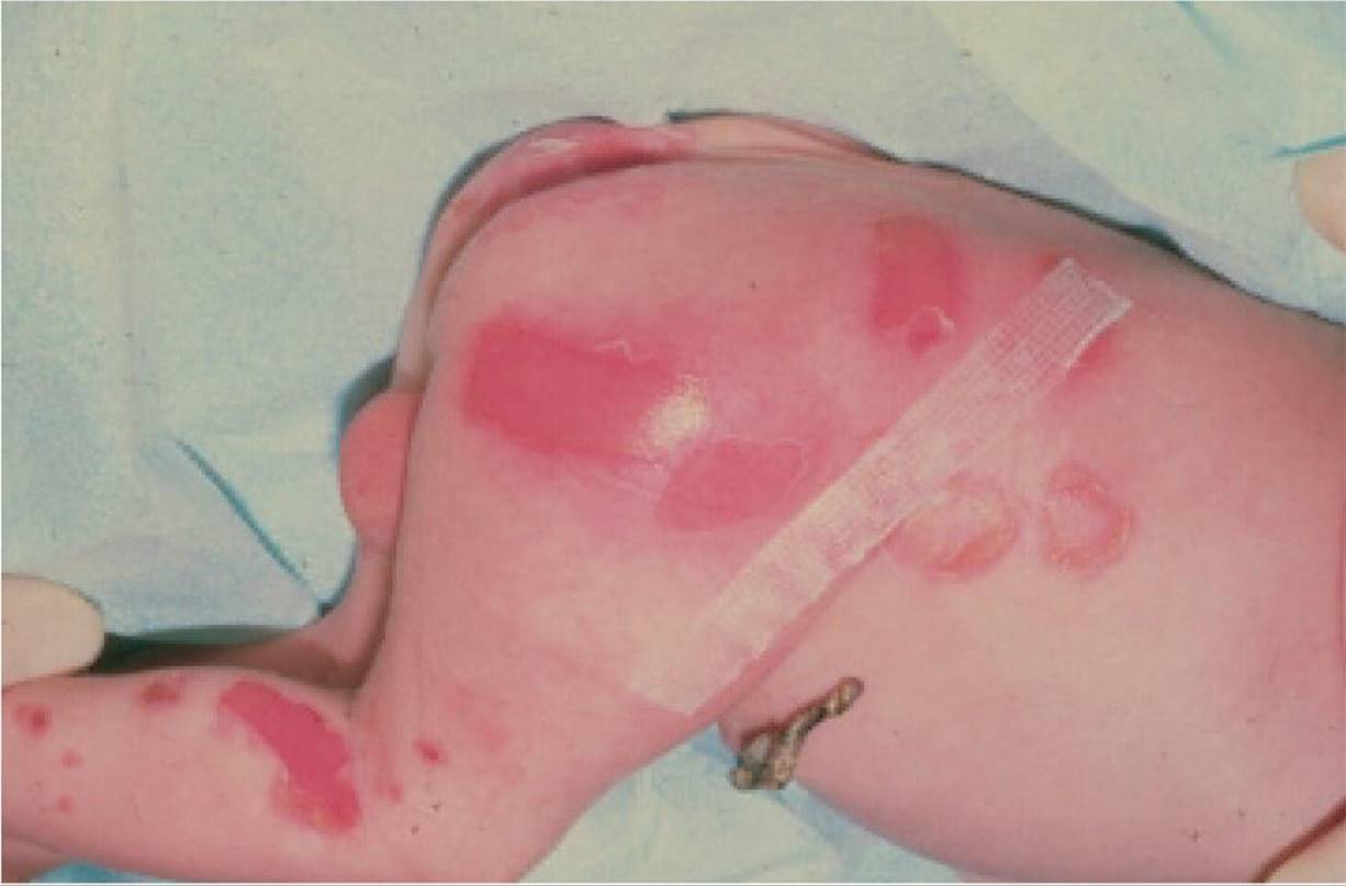
Figure 74-9 Epidermolysis bullosa congenita. (Used with permission from The Benjamin Barankin Dermatology Collection.)

Figure 74-10 Toxic epidermal necrolysis. Nikolski sign (arrow). (Used with permission from Mulholland MW, Maier RV, et al. Greenfield’s Surgery: Scientific Principles and Practice. 4th ed. Philadelphia, PA: Lippincott Williams & Wilkins; 2006.)
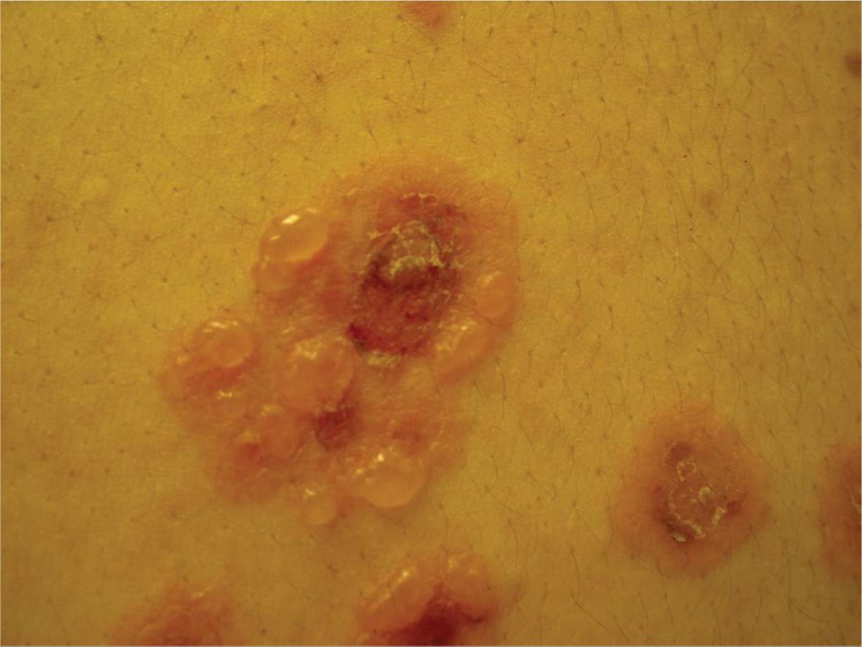
Figure 74-11 Linear IgA Disease (Chronic Bullous Disease of Childhood). Note “pearls on a string” in an annular pattern. (Used with permission from Dr. Barankin’s Dermatology Collection.)
DIFFERENTIAL DIAGNOSIS


Other Diagnoses to Consider
• Pemphigus
• Bullous pemphigoid
• Epidermolytic hyperkeratosis (bullous ichthyosis)
• Toxic shock syndrome
• Bullous scabies
• Insect and arachnid bites
• Thermal trauma
• Bullous varicella
• Juvenile dermatitis herpetiformis
• Erythema multiforme with bullae
When to Consider Further Evaluation or Treatment
• Skin biopsy strongly recommended in atypical SSSS or SJS, all TEN and persistent or refractory disease.
• Patients with suspected SSSS, SJS, and TEN need close monitoring for other organ system involvement and life-threatening complications.
• Complicated SSSS, SJS, and all TEN should be managed in a burn unit.
SUGGESTED READINGS
Cole C, Gazewood J. Diagnosis and treatment of impetigo. Am Fam Physician. 2007;75:859–864.
Drucker AM, Rosen CF. Drug-induced photosensitivity culprit drugs, management and prevention. Drug Saf. 2011;34(10):821–837.
Fine J-D. Inherited epidermolysis bullosa: recent basic and clinical advances. Curr Opin Pediatr. 2010;22:453–458.
Finkelstein Y, Soon G, Acuna P, et al. Recurrence and outcomes of Stevens-Johnson syndrome and toxic epidermal necrolysis in children. Pediatrics. 2011;128:723–728.
Gerull R, Nelle M, Schaible T. Toxic epidermal necrolysis and Stevens-Johnson syndrome: a review. Crit Care Med. 2011;39:1521–1532.
Mintz EM, Morel KD. Clinical features, diagnosis, and pathogenesis of chronic bullous disease of childhood. Dermatol Clin. 2011;29:459–462.