
Approach to the Problem
Hair loss, or alopecia, may be congenital, hereditary, or acquired. The distribution of hair loss may be described as localized, as in alopecia areata, or more diffuse, as in telogen effluvium. Though hair loss often occurs in isolation, it may be a sign of systemic illness. Hair growth cycle disruption in the anagen (active), catagen (regressive), or telogen (resting) phases may cause hair loss. Also, any damage to the follicle or shaft may result in hair loss, as in trichotillomania. Some causes of hair loss, such as tinea capitis, may lead to scalp scarring and permanent hair loss if left untreated, which can be psychologically damaging to the parents and the child.
Key Points in the History
• Tinea capitis is the primary cause of alopecia in African American children.
• Home remedies for a child’s scaling scalp, such as hair grease and oils, may mask the underlying scale of tinea capitis.
• Recent illness may cause the hair to enter the resting (telogen) phase and manifest as diffuse hair loss (telogen effluvium).
• Traction alopecia, from tight braiding, is a common cause of hair loss.
• Cutis aplasia and sebaceous nevus of Jadassohn may present at birth as well-circumscribed areas of the scalp devoid of hair.
• Hair loss in younger teens necessitates a search for autoimmune disorders, such as thyroid disorders or psychiatric problems.
• There may be a family history of hair loss or autoimmune disease, such as in systemic lupus erythematosus (SLE).
• Children with systemic symptoms, diffuse rash, and nail or teeth abnormalities may have hair loss as a manifestation of a more widespread disease, such as in acrodermatitis enteropathica.
Key Points in the Physical Examination
• A prepubescent child with a scaly scalp should warrant a scalp culture to check for tinea capitis.
• The breakage of hair shafts close to the scalp in tinea capitis causes the “black dot” sign.
• Kerions and pustules, host inflammatory responses to fungal infections, usually do not represent bacterial superinfection.
• Trichophyton species, accounting for more than 90% of tinea capitis in North America, do not fluoresce under a Wood lamp.
• Intrinsic hair shaft defects, hair pulling, or tight braiding may cause hair breakage further away from the scalp.
• Older children with hair loss need to be assessed for psychological stress, if hair pulling, or trichotillomania, is the cause of their alopecia.
• Hair pulling tends to be biased toward the side of a patient’s handedness.
• Major hair loss is often related to systemic widespread disease.
• The combination of considerable scalp erythema and hair loss should prompt an investigation into evolving psoriasis or lupus.
|
PHOTOGRAPHS OF SELECTED DIAGNOSES |

Figure 9-1 Tinea capitis. Circumscribed area of hair loss with scaliness of the scalp. (Courtesy of George A. Datto, III, MD.)
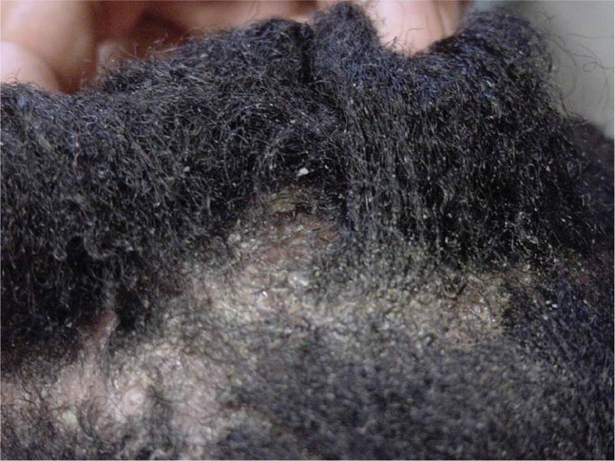
Figure 9-2 Tinea capitis. Diffuse scaling and pustules on the scalp. (Courtesy of Paul S. Matz, MD.)

Figure 9-3 “Black dot” sign. Broken hair shafts at the scalp from tinea capitis. (Courtesy of Paul S. Matz, MD.)

Figure 9-4 Kerion. Intense inflammatory response to tinea capitis. (Courtesy of Paul S. Matz, MD.)
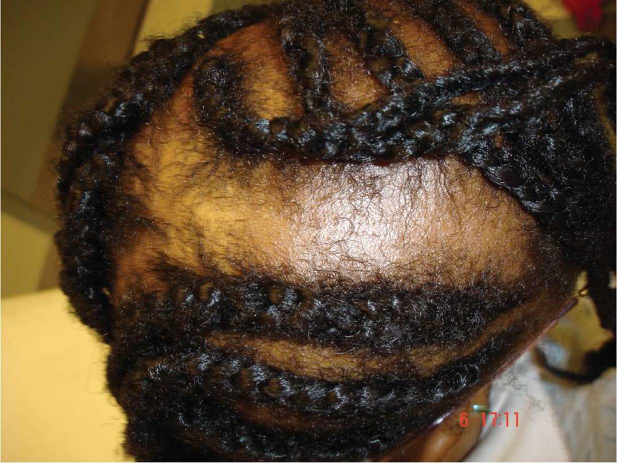
Figure 9-5 Traction alopecia. Alopecia where traction has been applied in association with hair braiding. (Courtesy of Carrie Ann Cusack, MD.)

Figure 9-6 Trichotillomania. Broken hair shafts caused by pulling of one’s hair. (Courtesy of George A. Datto, III, MD.)

Figure 9-7 Sebaceous nevus of Jadassohn. Yellowish orange verrucous plaque on the scalp. (Courtesy of the Department of Dermatology, Drexel University College of Medicine.)

Figure 9-8 Cutis aplasia. Scar on the vertex of the scalp with complete hair loss secondary to cutis aplasia. (Courtesy of Paul S. Matz, MD.)

Figure 9-9 Discoid lupus. Oval area of hair loss associated with scalp erythema, scaling, and follicular plugging. (Courtesy of George A. Datto, III, MD.)
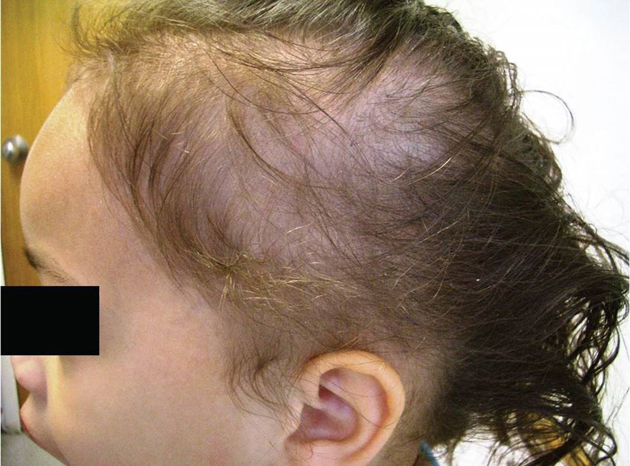
Figure 9-10 Telogen effluvium. Diffuse thinning of hair 3 months after febrile illness. (Courtesy of Paul S. Matz, MD.)
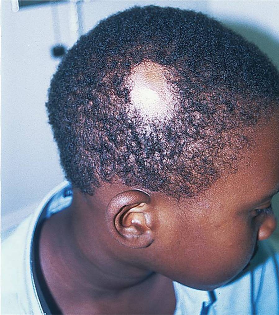
Figure 9-11 Alopecia areata. Localized circular patch of hair loss with normal scalp skin. (Courtesy of George A. Datto, III, MD.)
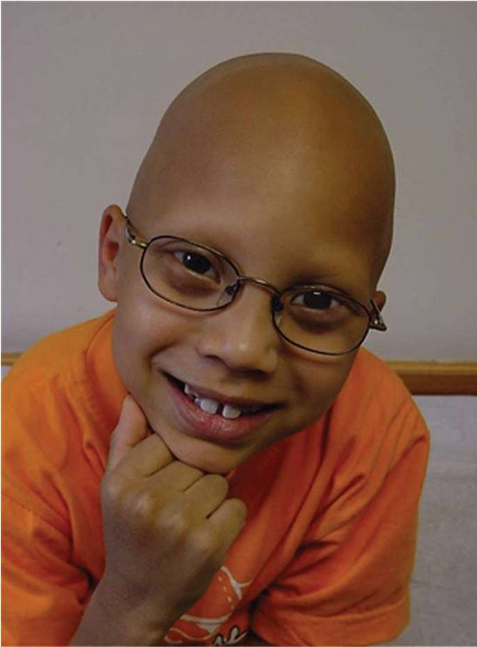
Figure 9-12 Alopecia universalis. Hair loss affecting the scalp, eyebrows, and eyelashes. (Courtesy of Paul S. Matz, MD.)
DIFFERENTIAL DIAGNOSIS


Other Diagnoses to Consider
• Monilethrix
• Pili torti
• Menkes kinky hair syndrome
• Trichorrhexis nodosa
• Loose anagen syndrome
• Ectodermal dysplasia
When to Consider Further Evaluation or Treatment
• Further evaluation and treatment should be considered for areas of intense scalp inflammation.
• Several months of treatment failure for tinea capitis with griseofulvin as first-line therapy warrant consideration of reinfection, resistance, or an alternate diagnosis, such as psoriasis.
• Consider referral to a psychologist for children with trichotillomania.
• Diffuse hair loss warrants a search for a systemic disorder, such as SLE, syphilis, thyroid disorders, or vitamin D deficiency.
SUGGESTED READINGS
Jabeen M, Mendiratta V. Hair loss and its management in children. Expert Rev Dermatol. 2011;6(6):581–590.
Shy R. Tinea corporis and tinea capitis. Pedaitr Rev. 2007;20(5):164–174.
Tay Y, Levy M, Metry D. Trichotillomania in childhood: Case series and review. Pediatrics. 2004;113(5):e494–e498.