INTRODUCTION
Cardiac arrhythmias are a frequent problem in clinical practice, occurring in up to 25% of patients treated with digitalis, 50% of anesthetized patients, and over 80% of patients with acute myocardial infarction. Arrhythmias may require treatment because rhythms that are too rapid, too slow, or asynchronous can reduce cardiac output. Some arrhythmias can precipitate more serious or even lethal rhythm disturbances¾eg, early premature ventricular depolarizations can precipitate ventricular fibrillation. In such patients, antiarrhythmic drugs may be lifesaving. On the other hand, the hazards of antiarrhythmic drugs¾and in particular the fact that they can precipitate lethal arrhythmias in some patients¾has led to a reevaluation of their relative risks and benefits. In general, treatment of asymptomatic or minimally symptomatic arrhythmias should be avoided for this reason.
Arrhythmias can be treated with the drugs discussed in this chapter and with nonpharmacologic therapies such as pacemakers, cardioversion, catheter ablation, and surgery. This chapter describes the pharmacology of drugs that suppress arrhythmias by a direct action on the cardiac cell membrane. Other modes of therapy are discussed briefly (see Box, The Nonpharmacologic Therapy of Cardiac Arrhythmias).
THE NONPHARMACOLOGIC THERAPY OF CARDIAC ARRHYTHMIAS
It was recognized over 100 years ago that reentry in simple in vitro models (eg, rings of conducting tissues) was permanently interrupted by transecting the reentry circuit. This concept is now applied in cardiac arrhythmias with defined anatomic pathways¾eg, atrioventricular reentry using accessory pathways, atrioventricular node reentry, atrial flutter, and some forms of ventricular tachycardia¾by treatment with radiofrequency catheter ablation. Recent studies have shown that paroxysmal and persistent atrial fibrillation may arise from one of the pulmonary veins. Both forms of atrial fibrillation can be cured by electrically isolating the pulmonary veins by radiofrequency catheter ablation or during concomitant cardiac surgery.
Another form of nonpharmacologic therapy is the implantable cardioverter-defibrillator (ICD), a device that can automatically detect and treat potentially fatal arrhythmias such as ventricular fibrillation. ICDs are now widely used in patients who have been resuscitated from such arrhythmias, and several trials have shown that ICD treatment reduces mortality in patients with coronary artery disease who have an ejection fraction £ 30% and in patients with class 2 or 3 heart failure and no prior history of arrhythmias. The increasing use of nonpharmacologic antiarrhythmic therapies reflects both advances in the relevant technologies and an increasing appreciation of the dangers of long-term therapy with currently available drugs.
ELECTROPHYSIOLOGY OF NORMAL CARDIAC RHYTHM
Introduction
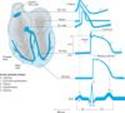
The electrical impulse that triggers a normal cardiac contraction originates at regular intervals in the sinoatrial node (Figure 14-1), usually at a frequency of 60-100 beats per minute. This impulse spreads rapidly through the atria and enters the atrioventricular node, which is normally the only conduction pathway between the atria and ventricles. Conduction through the atrioventricular node is slow, requiring about 0.15 s. (This delay provides time for atrial contraction to propel blood into the ventricles.) The impulse then propagates over the His-Purkinje system and invades all parts of the ventricles, beginning with the endocardial surface near the apex and ending with the epicardial surface at the base of the heart. Ventricular activation is complete in less than 0.1 s; therefore, contraction of all of the ventricular muscle is normally synchronous and hemodynamically effective.
Arrhythmias consist of cardiac depolarizations that deviate from the above description in one or more aspects¾ie, there is an abnormality in the site of origin of the impulse, its rate or regularity, or its conduction.
|
|
Figure 14-1. Schematic representation of the heart and normal cardiac electrical activity (intracellular recordings from areas indicated and ECG). Sinoatrial node, atrioventricular node, and Purkinje cells display pacemaker activity (phase 4 depolarization). The ECG is the body surface manifestation of the depolarization and repolarization waves of the heart. The P wave is generated by atrial depolarization, the QRS by ventricular muscle depolarization, and the T wave by ventricular repolarization. Thus, the PR interval is a measure of conduction time from atrium to ventricle, and the QRS duration indicates the time required for all of the ventricular cells to be activated (ie, the intraventricular conduction time). The QT interval reflects the duration of the ventricular action potential. |
Ionic Basis of Membrane Electrical Activity
The transmembrane potential of cardiac cells is determined by the concentrations of several ions¾chiefly sodium (Na+), potassium (K+), calcium (Ca2+), and chloride (Cl-)¾on either side of the membrane and the permeability of the membrane to each ion. These water-soluble ions are unable to freely diffuse across the lipid cell membrane in response to their electrical and concentration gradients; they require aqueous channels (specific pore-forming proteins) for such diffusion. Thus, ions move across cell membranes in response to their gradients only at specific times during the cardiac cycle when these ion channels are open. The movements of the ions produce currents that form the basis of the cardiac action potential. Individual channels are relatively ion-specific, and the flux of ions through them is thought to be controlled by "gates" (probably flexible peptide chains or energy barriers). Each type of channel has its own type of gate (sodium, calcium, and some potassium channels are each thought to have two types of gates), and each type of gate is opened and closed by specific transmembrane voltage, ionic, or metabolic conditions.
At rest, most cells are not significantly permeable to sodium, but at the start of each action potential, they become quite permeable (see below). In electrophysiologic terms, the conductance of the fast sodium channel suddenly increases in response to a depolarizing stimulus. Similarly, calcium enters and potassium leaves the cell with each action potential. Therefore, in addition to ion channels, the cell must have mechanisms to maintain stable transmembrane ionic conditions by establishing and maintaining ion gradients. The most important of these active mechanisms is the sodium pump, Na+/K+ ATPase, described in Chapter 13. This pump and other active ion carriers contribute indirectly to the transmembrane potential by maintaining the gradients necessary for diffusion through channels. In addition, some pumps and exchangers produce net current flow (eg, by exchanging three Na+ for two K+ ions) and hence are termed "electrogenic."
When the cardiac cell membrane becomes permeable to a specific ion (ie when the channels selective for that ion are open), movement of that ion across the cell membrane is determined by Ohm's law: current = voltage ¸ resistance, or current = voltage ´ conductance. Conductance is determined by the properties of the individual ion channel protein. The voltage term is the difference between the actual membrane potential and the reversal potential for that ion (the membrane potential at which no current would flow even if channels were open). For example, in the case of sodium in a cardiac cell at rest, there is a substantial concentration gradient (140 mmol/L Na+ outside; 10-15 mmol/L Na+ inside) and an electrical gradient (0 mV outside; -90 mV inside) that would drive Na+ into cells. Sodium does not enter the cell at rest because sodium channels are closed; when sodium channels open, the very large influx of Na+ ions accounts for phase 0 depolarization. The situation for K+ ions in the resting cardiac cell is quite different. Here, the concentration gradient (140 mmol/L inside; 4 mmol/L outside) would drive the ion out of the cell, but the electrical gradient would drive it in; that is, the inward gradient is in equilibrium with the outward gradient. In fact, certain potassium channels ("inward rectifier" channels) are open in the resting cell, but little current flows through them because of this balance. The equilibrium, or reversal potential, for ions is determined by the Nernst equation:
where Ce and Ci are the extracellular and intracellular concentrations, respectively, multiplied by their activity coefficients. Note that raising extracellular potassium makes EK less negative. When this occurs, the membrane depolarizes until the new EK is reached. Thus, extracellular potassium concentration and inward rectifier channel function are the major factors determining the membrane potential of the resting cardiac cell. The conditions required for application of the Nernst equation are approximated at the peak of the overshoot (using sodium concentrations) and during rest (using potassium concentrations) in most nonpacemaker cardiac cells. If the permeability is significant for both potassium and sodium, the Nernst equation is not a good predictor of membrane potential, but the Goldman-Hodgkin-Katz equation may be used:
In pacemaker cells (whether normal or ectopic), spontaneous depolarization (the pacemaker potential) occurs during diastole (phase 4, Figure 14-1). This depolarization results from a gradual increase of depolarizing current through special hyperpolarization-activated ion channels in pacemaker cells. The effect of changing extracellular potassium is more complex in a pacemaker cell than it is in a nonpacemaker cell because the effect on permeability to potassium is much more important in a pacemaker (see Box, Effects of Potassium). In a pacemaker¾especially an ectopic one¾the end result of an increase in extracellular potassium will usually be to slow or stop the pacemaker. Conversely, hypokalemia will often facilitate ectopic pacemakers.
EFFECTS OF POTASSIUM
The effects of changes in serum potassium on cardiac action potential duration, pacemaker rate, and arrhythmias can appear somewhat paradoxical if changes are predicted based solely on a consideration of changes in the potassium electrochemical gradient. In the heart, however, changes in serum potassium concentration have the additional effect of altering potassium conductance (increased extracellular potassium increases potassium conductance) independent of simple changes in electrochemical driving force, and this effect often predominates. As a result, the actual observed effects of hyperkalemia include reduced action potential duration, slowed conduction, decreased pacemaker rate, and decreased pacemaker arrhythmogenesis. Conversely, the actual observed effects of hypokalemia include prolonged action potential duration, increased pacemaker rate, and increased pacemaker arrhythmogenesis. Furthermore, pacemaker rate and arrhythmias involving ectopic pacemaker cells appear to be more sensitive to changes in serum potassium concentration, compared with cells of the sinoatrial node. These effects of serum potassium on the heart probably contribute to the observed increased sensitivity to potassium channel-blocking antiarrhythmic agents (quinidine or sotalol) during hypokalemia, eg, accentuated action potential prolongation and tendency to cause torsade de pointes.
The Active Cell Membrane
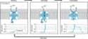
In normal atrial, Purkinje, and ventricular cells, the action potential upstroke (phase 0) is dependent on sodium current. From a functional point of view, it is convenient to describe the behavior of the sodium current in terms of three channel states (Figure 14-2). The cardiac sodium channel protein has been cloned, and it is now recognized that these channel states actually represent different protein conformations. In addition, regions of the protein that confer specific behaviors, such as voltage sensing, pore formation, and inactivation, are now being identified. The gates described below and in Figure 14-2 represent such regions.
Depolarization to the threshold voltage results in opening of the activation (m) gates of sodium channels (Figure 14-2, middle). If the inactivation (h) gates of these channels have not already closed, the channels are now open or activated, and sodium permeability is markedly increased, greatly exceeding the permeability for any other ion. Extracellular sodium therefore diffuses down its electrochemical gradient into the cell, and the membrane potential very rapidly approaches the sodium equilibrium potential, ENa (about +70 mV when Nae = 140 mmol/L and Nai = 10 mmol/L). This intense sodium current is very brief because opening of the m gates upon depolarization is promptly followed by closure of the h gates and inactivation of the sodium channels (Figure 14-2, right).
Most calcium channels become activated and inactivated in what appears to be the same way as sodium channels, but in the case of the most common type of cardiac calcium channel (the "L" type), the transitions occur more slowly and at more positive potentials. The action potential plateau (phases 1 and 2) reflects the turning off of most of the sodium current, the waxing and waning of calcium current, and the slow development of a repolarizing potassium current.
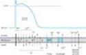
Final repolarization (phase 3) of the action potential results from completion of sodium and calcium channel inactivation and the growth of potassium permeability, so that the membrane potential once again approaches the potassium equilibrium potential. The major potassium currents involved in phase 3 repolarization include a rapidly activating potassium current (IKr) and a slowly activating potassium current (IKs). These two potassium currents are sometimes discussed together as "IK." These processes are diagrammed in Figure 14-3. It is noteworthy that a different potassium current, distinct from IKr and IKs, may control repolarization in sinoatrial nodal cells. This explains why some drugs that block either IKr or IKsmay prolong repolarization in Purkinje and ventricular cells, but have little effect on sinoatrial nodal repolarization (see Box, Molecular & Genetic Basis of Cardiac Arrhythmias).
|
|
Figure 14-2. A schematic representation of Na+ channels cycling through different conformational states during the cardiac action potential. Transitions between resting, activated, and inactivated states are dependent on membrane potential and time. The activation gate is shown as m and the inactivation gate as h. Potentials typical for each state are shown under each channel schematic as a function of time. The dashed line indicates that part of the action potential during which most Na+ channels are completely or partially inactivated and unavailable for reactivation. |
|
|
Figure 14-3. Schematic diagram of the ion permeability changes and transport processes that occur during an action potential and the diastolic period following it. The size and weight of the arrows indicate approximate magnitudes of the ion channel currents; arrows pointing down indicate inward (depolarizing) membrane currents, arrows pointing up indicate outward (repolarizing) membrane currents. Multiple subtypes of potassium and calcium currents, with different sensitivities to blocking drugs, have been identified. Chloride currents (dotted arrows) produce both inward and outward membrane currents during the cardiac action potential. |
The Effect of Resting Potential on Action Potentials
A key factor in the pathophysiology of arrhythmias and the actions of antiarrhythmic drugs is the relationship between the resting potential of a cell and the action potentials that can be evoked in it (Figure 14-4, left panel). Because the inactivation gates of sodium channels in the resting membrane close over the potential range -75 to -55 mV, fewer sodium channels are "available" for diffusion of sodium ions when an action potential is evoked from a resting potential of -60 mV than when it is evoked from a resting potential of -80 mV. Important consequences of the reduction in peak sodium permeability include reduced maximum upstroke velocity (called , for maximum rate of change of membrane voltage), reduced action potential amplitude, reduced excitability, and reduced conduction velocity.
During the plateau of the action potential, most sodium channels are inactivated. Upon repolarization, recovery from inactivation takes place (in the terminology of Figure 14-2, the h gates reopen), making the channels again available for excitation. The time between phase 0 and sufficient recovery of sodium channels in phase 3 to permit a new propagated response to an external stimulus is the refractory period. Changes in refractoriness (determined by either altered recovery from inactivation or altered action potential duration) can be important in the genesis or suppression of certain arrhythmias. Another important effect of less negative resting potential is prolongation of this recovery time, as shown in Figure 14-4(right panel). The prolongation of recovery time is reflected in an increase in the effective refractory period.
A brief, sudden, depolarizing stimulus, whether caused by a propagating action potential or by an external electrode arrangement, causes the opening of large numbers of activation gates before a significant number of inactivation gates can close. In contrast, slow reduction (depolarization) of the resting potential, whether brought about by hyperkalemia, sodium pump blockade, or ischemic cell damage, results in depressed sodium currents during the upstrokes of action potentials. Depolarization of the resting potential to levels positive to -55 mV abolishes sodium currents, since all sodium channels are inactivated. However, such severely depolarized cells have been found to support special action potentials under circumstances that increase calcium permeability or decrease potassium permeability. These "slow responses"¾slow upstroke velocity and slow conduction¾depend on a calcium inward current and constitute the normal electrical activity in the sinoatrial and atrioventricular nodes, since these tissues have a normal resting potential in the range of -50 to -70 mV. Slow responses may also be important for certain arrhythmias. Modern techniques of molecular biology and electrophysiology can identify multiple subtypes of calcium and potassium channels. One way in which such subtypes may differ is in sensitivity to drug effects, so drugs targeting specific channel subtypes may be developed in the future.
|
|
Figure 14-4. Dependence of sodium channel function on the membrane potential preceding the stimulus. Left: The fraction of sodium channels available for opening in response to a stimulus is determined by the membrane potential immediately preceding the stimulus. The decrease in the fraction available when the resting potential is depolarized in the absence of a drug (control curve) results from the voltage-dependent closure of h gates in the channels. The curve labeled Drug illustrates the effect of a typical local anesthetic antiarrhythmic drug. Most sodium channels are inactivated during the plateau of the action potential. Right: The time constant for recovery from inactivation after repolarization also depends on the resting potential. In the absence of drug, recovery occurs in less than 10 ms at normal resting potentials (-85 to -95 mV). Depolarized cells recover more slowly (note logarithmic scale). In the presence of a sodium channel-blocking drug, the time constant of recovery is increased, but the increase is far greater at depolarized potentials than at more negative ones. |
MECHANISMS OF ARRHYTHMIAS
Introduction
Many factors can precipitate or exacerbate arrhythmias: ischemia, hypoxia, acidosis or alkalosis, electrolyte abnormalities, excessive catecholamine exposure, autonomic influences, drug toxicity (eg, digitalis or antiarrhythmic drugs), overstretching of cardiac fibers, and the presence of scarred or otherwise diseased tissue. However, all arrhythmias result from (1) disturbances in impulse formation, (2) disturbances in impulse conduction, or (3) both.
Disturbances of Impulse Formation
The interval between depolarizations of a pacemaker cell is the sum of the duration of the action potential and the duration of the diastolic interval. Shortening of either duration results in an increase in pacemaker rate. The more important of the two, diastolic interval, is determined primarily by the slope of phase 4 depolarization (pacemaker potential). Vagal discharge and b-receptor-blocking drugs slow normal pacemaker rate by reducing the phase 4 slope (acetylcholine also makes the maximum diastolic potential more negative). Acceleration of pacemaker discharge is often brought about by increased phase 4 depolarization slope, which can be caused by hypokalemia, b-adrenoceptor stimulation, positive chronotropic drugs, fiber stretch, acidosis, and partial depolarization by currents of injury.
Latent pacemakers (cells that show slow phase 4 depolarization even under normal conditions, eg, some Purkinje fibers) are particularly prone to acceleration by the above mechanisms. However, all cardiac cells, including normally quiescent atrial and ventricular cells, may show repetitive pacemaker activity when depolarized under appropriate conditions, especially if hypokalemia is also present.
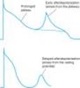
Afterdepolarizations (Figure 14-5) are depolarizations that interrupt phase 3 (early afterdepolarizations, EADs) or phase 4 (delayed afterdepolarizations, DADs). EADs are usually exacerbated at slow heart rates and are thought to contribute to the development of long QT-related arrhythmias (see Box, Molecular & Genetic Basis of Cardiac Arrhythmias). DADs on the other hand, discussed in Chapter 13, often occur when intracellular calcium is increased. They are exacerbated by fast heart rates and are thought to be responsible for some arrhythmias related to digitalis excess, to catecholamines, and to myocardial ischemia.
MOLECULAR & GENETIC BASIS OF CARDIAC ARRHYTHMIAS
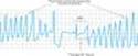
It is now possible to define the molecular basis of several congenital and acquired cardiac arrhythmias. The best example is the polymorphic ventricular tachycardia known as torsade de pointes (shown in Figure 14-7), which is associated with prolongation of the QT interval (especially at the onset of the tachycardia), syncope, and sudden death. This must represent prolongation of the action potential of at least some ventricular cells (Figure 14-1). The effect can, in theory, be attributed either to increased inward current (gain of function) or decreased outward current (loss of function) during the plateau of the action potential. In fact, recent molecular genetic studies have identified up to 300 different mutations in at least eight ion channel genes that produce the congenital long QT (LQT) syndrome (Table 14-1), and each mutation may have different clinical implications. Loss of function mutations in potassium channel genes produce decreases in outward repolarizing current and are responsible for LQT subtypes 1, 2, 5, 6, and 7. HERG and KCNE2 (MiRP1) genes encode subunits of the the rapid delayed rectifier potassium current (IKr), whereas KCNQ1 and KCNE1 (minK) encode subunits of the slow delayed rectifier potassium current (IKs). KCNJ2 encodes an inwardly rectifying potassium current (IKir). In contrast, gain of function mutations in the sodium channel gene (SCN5A) or calcium channel gene (CACNA1c) cause increases in inward plateau current and are responsible for LQT subtypes 3 and 8, respectively.
Molecular genetic studies have identified the reason why congenital and acquired cases of torsades de pointes can be so strikingly similar. The potassium channel IKr (encoded by HERG) is blocked or modified by many drugs (eg, quinidine, sotalol) or electrolyte abnormalities (hypokalemia, hypomagnesemia, hypocalcemia) that also produce torsades de pointes. Thus, the identification of the precise molecular mechanisms underlying various forms of the LQT syndromes now raises the possibility that specific therapies may be developed for individuals with defined molecular abnormalities. Indeed, preliminary reports suggest that the sodium channel blocker mexiletine can correct the clinical manifestations of congenital LQT subtype 3 syndrome. It is likely that torsade de pointes originates from triggered upstrokes arising from early afterdepolarizations (Figure 14-5). Thus, therapy is directed at correcting hypokalemia, eliminating triggered upstrokes (eg, by using b blockers or magnesium), or shortening the action potential (eg, by increasing heart rate with isoproterenol or pacing)¾or all of these.
The molecular basis of several other congenital cardiac arrhythmias associated with sudden death has also recently been identified. Three forms of short QT syndrome have been identified that are linked to gain of function mutations in three different potassium channel genes (KCNH2, KCNQ1, and KCNJ2). Catecholaminergic polymorphic ventricular tachycardia, a disease that is characterized by stress- or emotion-induced syncope, can be caused by genetic mutations in two different proteins in the sarcoplasmic reticulum that control intracellular calcium homeostasis. Mutations in two different ion channel genes (HCN4 and SCN5A) have been linked to congenital forms of sick sinus syndrome. The Brugada syndrome, which is characterized by ventricular fibrillation associated with persistent ST-segment elevation, and progressive cardiac conduction disorder (PCCD), characterized by impaired conduction in the His-Purkinje system and right or left bundle block leading to complete atrioventricular block, have both been linked to several loss-of-function mutations in the sodium channel gene, SCN5A. At least one form of familial atrial fibrillation is caused by a gain-of-function mutation in the potassium channel gene, KCNQ1.
|
|
Figure 14-5. Two forms of abnormal activity, early (top) and delayed after depolarizations (bottom). In both cases, abnormal depolarizations arise during or after a normally evoked action potential. They are therefore often referred to as "triggered" automaticity; that is, they require a normal action potential for their initiation. |
|
|
|
Figure 14-7. Electrocardiogram from a patient with the long QT syndrome during two episodes of torsade de pointes. The polymorphic ventricular tachycardia is seen at the start of this tracing and spontaneously halts at the middle of the panel. A single normal sinus beat (NSB) with an extremely prolonged QT interval follows, succeeded immediately by another episode of ventricular tachycardia of the torsade type. The usual symptoms would be dizziness or transient loss of consciousness. |
|
Disturbances of Impulse Conduction
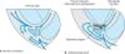
Severely depressed conduction may result in simple block, eg, atrioventricular nodal block or bundle branch block. Because parasympathetic control of atrioventricular conduction is significant, partial atrioventricular block is sometimes relieved by atropine. Another common abnormality of conduction is reentry (also known as "circus movement"), in which one impulse reenters and excites areas of the heart more than once (Figure 14-6). The path of the reentering impulse may be confined to very small areas, eg, within or near the atrioventricular node, or it may involve large portions of the atrial or ventricular walls. Some forms of reentry are strictly anatomically determined; for example, in the Wolff-Parkinson-White syndrome, the reentry circuit consists of atrial tissue, the AV node, ventricular tissue, and an accessory atrioventricular connection (a "bypass tract"). In other cases (eg, atrial or ventricular fibrillation), multiple reentry circuits, determined by the properties of the cardiac tissue, may meander through the heart in apparently random paths. Furthermore, the circulating impulse often gives off "daughter impulses" that can spread to the rest of the heart. Depending on how many round trips through the pathway the impulse makes before dying out, the arrhythmia may be manifest as one or a few extra beats or as a sustained tachycardia. In order for reentry to occur, three conditions must coexist, as indicated in Figure 14-6: (1) There must be an obstacle (anatomic or physiologic) to homogeneous conduction, thus establishing a circuit around which the reentrant wavefront can propagate; (2) there must be unidirectional block at some point in the circuit, ie conduction must die out in one direction but continue in the opposite direction (as shown in Figure 14-6, the impulse can gradually decrease as it invades progressively more depolarized tissue until it finally blocks¾a process known as decremental conduction); and (3) conduction time around the circuit must be long enough so that the retrograde impulse does not enter refractory tissue as it travels around the obstacle, ie the conduction time must exceed the effective refractory period. Importantly, reentry depends on conduction that has been depressed by some critical amount, usually as a result of injury or ischemia. If conduction velocity is too slow, bidirectional block rather than unidirectional block occurs; if the reentering impulse is too weak, conduction may fail, or the impulse may arrive so late that it collides with the next regular impulse. On the other hand, if conduction is too rapid, ie almost normal, bidirectional conduction rather than unidirectional block will occur. Even in the presence of unidirectional block, if the impulse travels around the obstacle too rapidly, it will reach tissue that is still refractory.
Slowing of conduction may be due to depression of sodium current, depression of calcium current (the latter especially in the atrioventricular node), or both. Drugs that abolish reentry usually work by further slowing depressed conduction (by blocking the sodium or calcium current) and causing bidirectional block. In theory, accelerating conduction (by increasing sodium or calcium current) would also be effective, but only under unusual circumstances does this mechanism explain the action of any available drug.
Lengthening (or shortening) of the refractory period may also make reentry less likely. The longer the refractory period in tissue near the site of block, the greater the chance that the tissue will still be refractory when reentry is attempted. (Alternatively, the shorter the refractory period in the depressed region, the less likely it is that unidirectional block will occur.) Thus, increased dispersion of refractoriness is one contributor to reentry, and drugs may suppress arrhythmias by reducing such dispersion.
|
|
Figure 14-6. Schematic diagram of a reentry circuit that might occur in small bifurcating branches of the Purkinje system where they enter the ventricular wall. A: Normally, electrical excitation branches around the circuit, is transmitted to the ventricular branches, and becomes extinguished at the other end of the circuit due to collision of impulses. B: An area of unidirectional block develops in one of the branches, preventing anterograde impulse transmission at the site of block, but the retrograde impulse may be propagated through the site of block if the impulse finds excitable tissue; that is, the refractory period is shorter than the conduction time. This impulse will then reexcite tissue it had previously passed through, and a reentry arrhythmia will be established. |
I. BASIC PHARMACOLOGY OF THE ANTIARRHYTHMIC AGENTS
Mechanisms of Action
Arrhythmias are caused by abnormal pacemaker activity or abnormal impulse propagation. Thus, the aim of therapy of the arrhythmias is to reduce ectopic pacemaker activity and modify conduction or refractoriness in reentry circuits to disable circus movement. The major mechanisms currently available for accomplishing these goals are (1) sodium channel blockade, (2) blockade of sympathetic autonomic effects in the heart, (3) prolongation of the effective refractory period, and (4) calcium channel blockade.
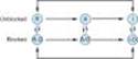
Antiarrhythmic drugs decrease the automaticity of ectopic pacemakers more than that of the sinoatrial node. They also reduce conduction and excitability and increase the refractory period to a greater extent in depolarized tissue than in normally polarized tissue. This is accomplished chiefly by selectively blocking the sodium or calcium channels of depolarized cells (Figure 14-8). Therapeutically useful channel-blocking drugs bind readily to activated channels (ie, during phase 0) or inactivated channels (ie, during phase 2) but bind poorly or not at all to rested channels. Therefore, these drugs block electrical activity when there is a fast tachycardia (many channel activations and inactivations per unit time) or when there is significant loss of resting potential (many inactivated channels during rest). This type of drug action is often described as use-dependent or state-dependent; that is, channels that are being used frequently, or in an inactivated state, are more susceptible to block. Channels in normal cells that become blocked by a drug during normal activation-inactivation cycles will rapidly lose the drug from the receptors during the resting portion of the cycle (Figure 14-8). Channels in myocardium that is chronically depolarized (ie, has a resting potential more positive than -75 mV) will recover from block very slowly if at all (see also right panel, Figure 14-4).
In cells with abnormal automaticity, most of these drugs reduce the phase 4 slope by blocking either sodium or calcium channels, thereby reducing the ratio of sodium (or calcium) permeability to potassium permeability. As a result, the membrane potential during phase 4 stabilizes closer to the potassium equilibrium potential. In addition, some agents may increase the threshold (make it more positive). Beta-adrenoceptor-blocking drugs indirectly reduce the phase 4 slope by blocking the positive chronotropic action of norepinephrine in the heart.
In reentry arrhythmias, which depend on critically depressed conduction, most antiarrhythmic agents slow conduction further by one or both of two mechanisms: (1) steady-state reduction in the number of available unblocked channels, which reduces the excitatory currents to a level below that required for propagation (Figure 14-4, left); and (2) prolongation of recovery time of the channels still able to reach the rested and available state, which increases the effective refractory period (Figure 14-4, right). As a result, early extrasystoles are unable to propagate at all; later impulses propagate more slowly and are subject to bidirectional conduction block.
By these mechanisms, antiarrhythmic drugs can suppress ectopic automaticity and abnormal conduction occurring in depolarized cells¾rendering them electrically silent¾while minimally affecting the electrical activity in normally polarized parts of the heart. However, as dosage is increased, these agents also depress conduction in normal tissue, eventually resulting in drug-induced arrhythmias. Furthermore, a drug concentration that is therapeutic (antiarrhythmic) under the initial circumstances of treatment may become "proarrhythmic" (arrhythmogenic) during fast heart rates (more development of block), acidosis (slower recovery from block for most drugs), hyperkalemia, or ischemia.
|
|
Figure 14-8. Diagram of a mechanism for the selective depressant action of antiarrhythmic drugs on sodium channels. The upper portion of the figure shows the population of channels moving through a cycle of activity during an action potential in the absence of drugs: R (rested) ® A (activated) ® I (inactivated). Recovery takes place via the I ® R pathway. Antiarrhythmic drugs (D) that act by blocking sodium channels can bind to their receptors in the channels, as shown by the vertical arrows, to form drug-channel complexes, indicated as R-D, A-D, and I-D. Binding of the drugs to the receptor varies with the state of the channel. The data available for a variety of sodium channel blockers indicate that the binding of the drugs to the active and inactivated channel receptor is much greater than the binding to the rested channel. Furthermore, recovery from the I-D state to the R-D state is much slower than from I to R. As a result, rapid activity (more activations and inactivations) and depolarization of the resting potential (more channels in the I state) will favor blockade of the channels and selectively suppress arrhythmic cells. |
II. SPECIFIC ANTIARRHYTHMIC AGENTS
INTRODUCTION
The most widely used scheme for the classification of antiarrhythmic drug actions recognizes four classes:
1. Class 1 action is sodium channel blockade. Subclasses of this action reflect effects on the action potential duration (APD) and the kinetics of sodium channel blockade. Drugs with class 1A action prolong the APD and dissociate from the channel with intermediate kinetics; drugs with class 1B action shorten the APD in some tissues of the heart and dissociate from the channel with rapid kinetics; and drugs with class 1C action have minimal effects on the APD and dissociate from the channel with slow kinetics.
2. Class 2 action is sympatholytic. Drugs with this action reduce b-adrenergic activity in the heart.
3. Class 3 action is manifest by prolongation of the APD. Most drugs with this action block the rapid component of the delayed rectifier potassium current, IKr.
4. Class 4 action is blockade of the cardiac calcium current. This action slows conduction in regions where the action potential upstroke is calcium dependent, eg, the sinoatrial and atrioventricular nodes.
A given drug may have multiple classes of action as indicated by its membrane and electrocardiographic
(ECG) effects (Tables 14-2 and 14-3). For example, amiodarone shares all four classes of action. Drugs are usually discussed according to the predominant class of action. Certain antiarrhythmic agents, eg, adenosine and magnesium, do not fit readily into this scheme and are described separately.
SODIUM CHANNEL-BLOCKING DRUGS (CLASS 1)
PROCAINAMIDE (SUBGROUP 1A)
Cardiac Effects
By blocking sodium channels, procainamide slows the upstroke of the action potential, slows conduction, and prolongs the QRS duration of the ECG. The drug also prolongs the action potential duration by nonspecific blockade of potassium channels. The drug may be somewhat less effective than quinidine (see below) in suppressing abnormal ectopic pacemaker activity but more effective in blocking sodium channels in depolarized cells.
Procainamide has directly depressant actions on sinoatrial and atrioventricular nodes that are only slightly counterbalanced by drug-induced vagal block.
Extracardiac Effects
Procainamide has ganglion-blocking properties. This action reduces peripheral vascular resistance and can cause hypotension, particularly with intravenous use. However, in therapeutic concentrations, its peripheral vascular effects are less prominent than those of quinidine. Hypotension is usually associated with excessively rapid procainamide infusion or the presence of severe underlying left ventricular dysfunction.
Toxicity
A. CARDIAC
Procainamide's cardiotoxic effects include excessive action potential prolongation, QT interval prolongation, and induction of torsade de pointes arrhythmia and syncope. Excessive slowing of conduction can also occur. New arrhythmias can be precipitated.
B. EXTRACARDIAC
The most troublesome adverse effect of long-term procainamide therapy is a syndrome resembling lupus erythematosus and usually consisting of arthralgia and arthritis. In some patients, pleuritis, pericarditis, or parenchymal pulmonary disease also occurs. Renal lupus is rarely induced by procainamide. During long-term therapy, serologic abnormalities (eg, increased antinuclear antibody titer) occur in nearly all patients, and in the absence of symptoms these are not an indication to stop drug therapy. Approximately one third of patients receiving long-term procainamide therapy develop these reversible lupus-related symptoms.
Other adverse effects include nausea and diarrhea (about 10% of cases), rash, fever, hepatitis (< 5%), and agranulocytosis (approximately 0.2%).
Pharmacokinetics & Dosage
Procainamide can be administered safely by the intravenous and intramuscular routes and is well absorbed orally. A metabolite (N-acetylprocainamide, NAPA) has class 3 activity. Excessive accumulation of NAPA has been implicated in torsade de pointes during procainamide therapy, especially in patients with renal failure. Some individuals rapidly acetylate procainamide and develop high levels of NAPA. The lupus syndrome appears to be less common in these patients.
Procainamide is eliminated by hepatic metabolism to NAPA and by renal elimination. Its half-life is only 3-4 hours, which necessitates frequent dosing or use of a slow-release formulation (the usual practice). NAPA is eliminated by the kidneys. Thus, procainamide dosage must be reduced in patients with renal failure. The reduced volume of distribution and renal clearance associated with heart failure also require reduction in dosage. The half-life of NAPA is considerably longer than that of procainamide, and it therefore accumulates more slowly. Thus, it is important to measure plasma levels of both procainamide and NAPA, especially in patients with circulatory or renal impairment.
If a rapid procainamide effect is needed, an intravenous loading dose of up to 12 mg/kg can be given at a rate of 0.3 mg/kg/min or less rapidly. This dose is followed by a maintenance dosage of 2-5 mg/min, with careful monitoring of plasma levels. The risk of gastrointestinal or cardiac toxicity rises at plasma concentrations greater than 8 mcg/mL or NAPA concentrations greater than 20 mcg/mL.
In order to control ventricular arrhythmias, a total procainamide dosage of 2-5 g/d is usually required. In an occasional patient who accumulates high levels of NAPA, less frequent dosing may be possible. This is also possible in renal disease, where procainamide elimination is slowed.
Therapeutic Use
Procainamide is effective against most atrial and ventricular arrhythmias. However, many clinicians attempt to avoid long-term therapy because of the requirement for frequent dosing and the common occurrence of lupus-related effects. Procainamide is the drug of second choice (after lidocaine) in most coronary care units for the treatment of sustained ventricular arrhythmias associated with acute myocardial infarction.
QUINIDINE (SUBGROUP 1A)
Cardiac Effects
Quinidine has actions similar to those of procainamide: it slows the upstroke of the action potential and conduction, and prolongs the QRS duration of the ECG, by blockade of sodium channels. The drug also prolongs the action potential duration by nonspecific blockade of potassium channels. It has more pronounced cardiac antimuscarinic effects than procainamide. Its toxic cardiac effects include excessive QT interval prolongation and induction of torsade de pointes arrhythmia. Toxic concentrations of quinidine also produce excessive sodium channel blockade with slowed conduction throughout the heart.
Extracardiac Effects
Gastrointestinal side effects of diarrhea, nausea, and vomiting are observed in one third to one half of patients. A syndrome of headache, dizziness, and tinnitus (cinchonism) is observed at toxic drug concentrations. Idiosyncratic or immunologic reactions, including thrombocytopenia, hepatitis, angioneurotic edema, and fever, are observed rarely.
Pharmacokinetics
Quinidine is absorbed readily following oral administration, bound to albumin and a1-acid glycoprotein, and eliminated primarily by hepatic metabolism. The elimination half-life is 6-8 hours. Quinidine is usually administered in a slow release formulation, eg, that of the gluconate salt.
Therapeutic Use
Quinidine is used only occasionally to maintain normal sinus rhythm in patients with atrial flutter/fibrillation. Because of its cardiac and extracardiac side effects, its use is now largely restricted to patients with normal (but arrhythmic) hearts. In randomized, controlled clinical trials, quinidine-treated patients are twice as likely to remain in normal sinus rhythm compared with controls, but the risk of death is increased two- to threefold. Quinidine is used rarely in patients with ventricular tachycardia. Quinidine is the optical isomer of quinine and is sometimes used intravenously for the treatment of acute, severe malaria (see Chapter 53).
DISOPYRAMIDE (SUBGROUP 1A)
Cardiac Effects
The effects of disopyramide are very similar to those of procainamide and quinidine. Its cardiac antimuscarinic effects are even more marked than those of quinidine. Therefore, a drug that slows atrioventricular conduction should be administered with disopyramide when treating atrial flutter or fibrillation.
Toxicity
A. CARDIAC
Toxic concentrations of disopyramide can precipitate all of the electrophysiologic disturbances described under quinidine. As a result of its negative inotropic effect, disopyramide may precipitate heart failure de novo or in patients with preexisting depression of left ventricular function. Because of this effect, disopyramide is not used as a first-line antiarrhythmic agent in the USA. It should not be used in patients with heart failure.
B. EXTRACARDIAC
Disopyramide's atropine-like activity accounts for most of its symptomatic adverse effects: urinary retention (most often, but not exclusively, in male patients with prostatic hyperplasia), dry mouth, blurred vision, constipation, and worsening of preexisting glaucoma. These effects may require discontinuation of the drug.
Pharmacokinetics & Dosage
In the USA, disopyramide is only available for oral use. The usual oral dosage of disopyramide is 150 mg three times a day, but as much as 1 g/d has been used. In patients with renal impairment, dosage must be reduced. Because of the danger of precipitating heart failure, the use of loading doses is not recommended.
Therapeutic Use
Although disopyramide has been shown to be effective in a variety of supraventricular arrhythmias, in the USA it is approved only for the treatment of ventricular arrhythmias.
LIDOCAINE (SUBGROUP 1B)
Introduction
Lidocaine has a low incidence of toxicity and a high degree of effectiveness in arrhythmias associated with acute myocardial infarction. It is used only by the intravenous route.
Cardiac Effects
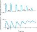
Lidocaine blocks activated and inactivated sodium channels with rapid kinetics (Figure 14-9); the inactivated state block ensures greater effects on cells with long action potentials such as Purkinje and ventricular cells, compared with atrial cells. The rapid kinetics at normal resting potentials result in recovery from block between action potentials and no effect on conduction. The increased inactivation and slower unbinding kinetics result in the selective depression of conduction in depolarized cells.
|
|
Figure 14-9. Computer simulation of the effect of resting membrane potential on the blocking and unblocking of sodium channels by lidocaine. Upper tracing: Action potentials in a ventricular muscle cell. Lower tracing: Percentage of channels blocked by the drug. As the membrane depolarizes through -80, -75, and -70 mV, an 800 ms time segment is shown. Extra passage of time is indicated by breaks in the traces. Left side: At the normal resting potential of -85 mV, the drug combines with open (activated) and inactivated channels during each action potential, but block is rapidly reversed during diastole because the affinity of the drug for its receptor is so low when the channel recovers to the resting state at -85 mV. Middle: Metabolic injury has occurred, eg, ischemia due to coronary occlusion, that causes gradual depolarization over time. With subsequent action potentials arising from more depolarized potentials, the fraction of channels blocked increases because more channels remain in the inactivated state at less negative potentials (Figure 14-4, left), and the time constant for unblocking during diastole rapidly increases at less negative resting potentials (Figure 14-4, right). Right: Because of marked drug binding, conduction block and loss of excitability in this tissue result; that is, the "sick" (depolarized) tissue is selectively suppressed. |
Toxicity
A. CARDIAC
Lidocaine is one of the least cardiotoxic of the currently used sodium channel blockers. Proarrhythmic effects, including sinoatrial node arrest, worsening of impaired conduction, and ventricular arrhythmias, are uncommon with lidocaine use. In large doses, especially in patients with preexisting heart failure, lidocaine may cause hypotension¾partly by depressing myocardial contractility.
B. EXTRACARDIAC
Lidocaine's most common adverse effects¾like those of other local anesthetics¾are neurologic: paresthesias, tremor, nausea of central origin, lightheadedness, hearing disturbances, slurred speech, and convulsions. These occur most commonly in elderly or otherwise vulnerable patients or when a bolus of the drug is given too rapidly. The effects are dose-related and usually short-lived; seizures respond to intravenous diazepam. In general, if plasma levels above 9 mcg/mL are avoided, lidocaine is well tolerated.
Pharmacokinetics & Dosage
Because of its extensive first-pass hepatic metabolism, only 3% of orally administered lidocaine appears in the plasma. Thus, lidocaine must be given parenterally. Lidocaine has a half-life of 1-2 hours. In adults, a loading dose of 150-200 mg administered over about 15 minutes (as a single infusion or as a series of slow boluses) should be followed by a maintenance infusion of 2-4 mg/min to achieve a therapeutic plasma level of 2-6 mcg/mL. Determination of lidocaine plasma levels is of great value in adjusting the infusion rate. Occasional patients with myocardial infarction or other acute illness require (and tolerate) higher concentrations. This may be due to increased plasma a1-acid glycoprotein, an acute phase reactant protein that binds lidocaine, making less free drug available to exert its pharmacologic effects.
In patients with heart failure, lidocaine's volume of distribution and total body clearance may both be decreased. Thus, both loading and maintenance doses should be decreased. Since these effects counterbalance each other, the half-life may not be increased as much as predicted from clearance changes alone. In patients with liver disease, plasma clearance is markedly reduced and the volume of distribution is often increased; the elimination half-life in such cases may be increased threefold or more. In liver disease, the maintenance dose should be decreased, but usual loading doses can be given. Elimination half-life determines the time to steady state. Thus, while steady-state concentrations may be achieved in 8-10 hours in normal patients and patients with heart failure, 24-36 hours may be required in those with liver disease. Drugs that decrease liver blood flow (eg, propranolol, cimetidine) reduce lidocaine clearance and so increase the risk of toxicity unless infusion rates are decreased. With infusions lasting more than 24 hours, clearance falls and plasma concentrations rise. Renal disease has no major effect on lidocaine disposition.
Therapeutic Use
Lidocaine is the agent of choice for termination of ventricular tachycardia and prevention of ventricular fibrillation after cardioversion in the setting of acute ischemia. However, routine prophylactic use of lidocaine in this setting may actually increase total mortality, possibly by increasing the incidence of asystole, and is not the standard of care. Most physicians administer IV lidocaine only to patients with arrhythmias.
MEXILETINE (SUBGROUP 1B)
Mexiletine is an orally active congener of lidocaine. Its electrophysiologic and antiarrhythmic actions are similar to those of lidocaine. (The anticonvulsant phenytoin [see Chapter 24] also exerts similar electrophysiologic effects and has been used as an antiarrhythmic.) Mexiletine is used in the treatment of ventricular arrhythmias. The elimination half-life is 8-20 hours and permits administration two or three times a day. The usual daily dosage of mexiletine is 600-1200 mg/d. Dose-related adverse effects are seen frequently at therapeutic dosage. These are predominantly neurologic, including tremor, blurred vision, and lethargy. Nausea is also a common effect.
Mexiletine has also shown significant efficacy in relieving chronic pain, especially pain due to diabetic neuropathy and nerve injury. The usual dosage is 450-750 mg/d orally. This application is unlabeled.
FLECAINIDE (SUBGROUP 1C)
Flecainide is a potent blocker of sodium and potassium channels with slow unblocking kinetics. (Note that although it does block certain potassium channels, it does not prolong the action potential or the QT interval.) It is currently used for patients with otherwise normal hearts who have supraventricular arrhythmias. It has no antimuscarinic effects.
Flecainide is very effective in suppressing premature ventricular contractions. However, it may cause severe exacerbation of arrhythmia even when normal doses are administered to patients with preexisting ventricular tachyarrhythmias and those with a previous myocardial infarction and ventricular ectopy. This was dramatically demonstrated in the Cardiac Arrhythmia Suppression Trial (CAST), which was terminated prematurely because of a two and one-half-fold increase in mortality in the patients receiving flecainide and similar Group 1c drugs. Flecainide is well absorbed and has a half-life of approximately 20 hours. Elimination is both by hepatic metabolism and by the kidney. The usual dosage of flecainide is 100-200 mg twice a day.
PROPAFENONE (SUBGROUP 1C)
Propafenone has some structural similarities to propranolol and possesses weak b-blocking activity. Its spectrum of action is very similar to that of quinidine. Its sodium channel blocking kinetics are similar to that of flecainide. Propafenone is metabolized in the liver, with an average half-life of 5-7 hours. The usual daily dosage of propafenone is 450-900 mg in three doses. The drug is used primarily for supraventricular arrhythmias. The most common adverse effects are a metallic taste and constipation; arrhythmia exacerbation can occur.
MORICIZINE (SUBGROUP 1C)
Moricizine is an antiarrhythmic phenothiazine derivative that is used for treatment of ventricular arrhythmias. It is a relatively potent sodium channel blocker that does not prolong action potential duration.
Moricizine has multiple metabolites, some of which are probably active and have long half-lives. Its most common adverse effects are dizziness and nausea. Like other potent sodium channel blockers, it can exacerbate arrhythmias. The usual dosage of moricizine is 200-300 mg by mouth three times a day.
BETA-ADRENOCEPTOR-BLOCKING DRUGS (CLASS 2)
Cardiac Effects
Propranolol and similar drugs have antiarrhythmic properties by virtue of their b-receptor-blocking action and direct membrane effects. As described in Chapter 10, some of these drugs have selectivity for cardiac b1 receptors, some have intrinsic sympathomimetic activity, some have marked direct membrane effects, and some prolong the cardiac action potential. The relative contributions of the b-blocking and direct membrane effects to the antiarrhythmic effects of these drugs are not fully known. Although b blockers are fairly well tolerated, their efficacy for suppression of ventricular ectopic depolarizations is lower than that of sodium channel blockers. However, there is good evidence that these agents can prevent recurrent infarction and sudden death in patients recovering from acute myocardial infarction (see Chapter 10).
Esmolol is a short-acting b blocker used primarily as an antiarrhythmic drug for intraoperative and other acute arrhythmias. See Chapter 10 for more information. Sotalol is a nonselective b-blocking drug that prolongs the action potential (class 3 action).
DRUGS THAT PROLONG EFFECTIVE REFRACTORY PERIOD BY PROLONGING ACTION POTENTIAL (CLASS 3)
INTRODUCTION
These drugs prolong action potentials, usually by blocking potassium channels in cardiac muscle or by enhancing inward current, eg, through sodium channels. Action potential prolongation by most of these drugs often exhibits the undesirable property of "reverse use-dependence": action potential prolongation is least marked at fast rates (where it is desirable) and most marked at slow rates, where it can contribute to the risk of torsade de pointes.
AMIODARONE
Introduction
In the USA, amiodarone is approved for oral and intravenous use to treat serious ventricular arrhythmias. However, the drug is also highly effective for the treatment of supraventricular arrhythmias such as atrial fibrillation. Amiodarone has a broad spectrum of cardiac actions, unusual pharmacokinetics, and important extracardiac adverse effects. Dronedarone, an analog that lacks iodine atoms, is under investigation.
Cardiac Effects
Amiodarone markedly prolongs the action potential duration (and the QT interval on the ECG) by blockade of IKr. During chronic administration, IKs is also blocked. The action potential duration is prolonged uniformly over a wide range of heart rates; that is, the drug does not have reverse use-dependent action. In spite of its present classification as a class 3 agent, amiodarone also significantly blocks inactivated sodium channels. Its action potential prolonging action reinforces this effect. Amiodarone also has weak adrenergic and calcium channel blocking actions. Consequences of these actions include slowing of the heart rate and atrioventricular node conduction. The broad spectrum of actions may account for its relatively high efficacy and low incidence of torsade de pointes despite significant QT interval prolongation.
Extracardiac Effects
Amiodarone causes peripheral vasodilation. This action is prominent following intravenous administration and may be related to the action of the vehicle.
Toxicity
A. CARDIAC
Amiodarone may produce symptomatic bradycardia and heart block in patients with preexisting sinus or atrioventricular node disease.
B. EXTRACARDIAC
Amiodarone accumulates in many tissues, including the heart (10-50 times greater than plasma), lung, liver, and skin, and is concentrated in tears. Dose-related pulmonary toxicity is the most important adverse effect. Even on a low dose of £ 200 mg/d, fatal pulmonary fibrosis may be observed in 1% of patients. Abnormal liver function tests and hepatitis may develop during amiodarone treatment. The skin deposits result in a photodermatitis and a gray-blue skin discoloration in sun-exposed areas, eg, the malar regions. After a few weeks of treatment, asymptomatic corneal microdeposits are present in virtually all patients treated with amiodarone. Halos develop in the peripheral visual fields of some patients. Drug discontinuation is usually not required. Rarely, an optic neuritis may progress to blindness.
Amiodarone blocks the peripheral conversion of thyroxine (T4) to triiodothyronine (T3). It is also a potential source of large amounts of inorganic iodine. Amiodarone may result in hypothyroidism or hyperthyroidism. Thyroid function should be evaluated prior to initiation of treatment and monitored periodically. Because effects have been described in virtually every organ system, amiodarone treatment should be reevaluated whenever new symptoms develop in a patient, including arrhythmia aggravation.
Pharmacokinetics
Amiodarone is variably absorbed with a bioavailability of 35-65%. It undergoes hepatic metabolism, and the major metabolite, desethylamiodarone, is bioactive. The elimination half-life is complex, with a rapid component of 3-10 days (50% of the drug) and a slower component of several weeks. Following discontinuation of the drug, effects are maintained for 1-3 months. Measurable tissue levels may be observed up to 1 year after discontinuation. A total loading dose of 10 g is usually achieved with 0.8-1.2 g daily doses. The maintenance dose is 200-400 mg daily. Pharmacologic effects may be achieved rapidly by intravenous loading. QT-prolonging effect is modest with this route of administration, whereas bradycardia and atrioventricular block may be significant.
Amiodarone has many important drug interactions and all medications should be reviewed during drug initiation or dose adjustments. Amiodarone is a substrate for the liver cytochrome metabolizing enzyme CYP3A4 and its levels are increased by drugs that inhibit this enzyme, eg, the histamine H2 blocker cimetidine. Drugs that induce CYP3A4, eg, rifampin, decrease amiodarone concentration when coadministered. Amiodarone inhibits the other liver cytochrome metabolizing enzymes and may result in high levels of drugs that are substrates for these enzymes, eg, digoxin and warfarin.
Therapeutic Use
Low doses (100-200 mg/d) of amiodarone are effective in maintaining normal sinus rhythm in patients with atrial fibrillation. The drug is effective in the prevention of recurrent ventricular tachycardia. Its use is not associated with an increase in mortality in patients with coronary artery disease or heart failure. In many centers, the implanted cardioverter-defibrillator (ICD) has succeeded drug therapy as the primary treatment modality for ventricular tachycardia, but amiodarone may be used for ventricular tachycardia as adjuvant therapy to decrease the frequency of uncomfortable ICD discharges. The drug increases the pacing and defibrillation threshold and these devices require retesting after a maintenance dose has been achieved.
BRETYLIUM
Introduction
Bretylium was first introduced as an antihypertensive agent. It interferes with the neuronal release of catecholamines but also has direct antiarrhythmic properties.
Cardiac & Extracardiac Effects
Bretylium lengthens the ventricular (but not the atrial) action potential duration and effective refractory period. This effect is most pronounced in ischemic cells, which have shortened action potential durations. Thus, bretylium may reverse the shortening of action potential duration caused by ischemia.
Since bretylium causes an initial release of catecholamines, it has some positive inotropic actions when first administered. This action may also precipitate ventricular arrhythmias and must be watched for at the onset of therapy with the drug.
The drug's sympathoplegic actions may result in postural hypotension. This effect can be almost totally prevented by concomitant administration of a tricyclic antidepressant agent such as protriptyline. Nausea and vomiting may occur after the intravenous administration of a bolus of bretylium.
Pharmacokinetics & Dosage
Bretylium is available only for intravenous use in the USA. In adults, an intravenous bolus of bretylium tosylate, 5 mg/kg, is administered over a 10-minute period. This dosage may be repeated after 30 minutes. Maintenance therapy is achieved by a similar bolus every 4-6 hours or by a constant infusion of 0.5-2 mg/min.
Therapeutic Use
Bretylium is rarely used and then only in an emergency setting, often during attempted resuscitation from ventricular fibrillation when lidocaine and cardioversion have failed. In most centers amiodarone is preferred for this indication.
SOTALOL
Sotalol has both b-adrenergic receptor-blocking (class 2) and action potential prolonging (class 3) actions. The drug is formulated as a racemic mixture of d- and l-sotalol. All the b-adrenergic blocking activity resides in the l-isomer; the d- and l-isomers share action potential prolonging actions. Beta-adrenergic blocking action is not cardioselective and is maximal at doses below those required for action potential prolongation.
Sotalol is well absorbed orally with bioavailability of approximately 100%. It is not metabolized in the liver and it is not bound to plasma proteins. Excretion is predominantly by the kidneys in the unchanged form with a half-life of approximately 12 hours. Because of its relatively simple pharmacokinetics, it exhibits few direct drug interactions. Its most significant cardiac adverse effect is an extension of its pharmacologic action: a dose-related incidence of torsade de pointes that approaches 6% at the highest recommended daily dose. Patients with overt heart failure may experience further depression of left ventricular function during treatment with sotalol.
Sotalol is approved for the treatment of life-threatening ventricular arrhythmias and the maintenance of sinus rhythm in patients with atrial fibrillation. It is also approved for treatment of supraventricular and ventricular arrhythmias in the pediatric age group. Sotalol decreases the threshold for cardiac defibrillation.
DOFETILIDE
Dofetilide has class 3 action potential prolonging action. This action is effected by a dose-dependent blockade of the rapid component of the delayed rectifier potassium current, IKr. Dofetilide block of IKr increases in hypokalemia. Dofetilide produces no relevant blockade of the other potassium channels or the sodium channel. Because of the slow rate of recovery from blockade, the extent of blockade shows little dependence on stimulation frequency. However, dofetilide does show less action potential prolongation at rapid rates because of the increased importance of other potassium channels such as IKs at higher frequencies.
Dofetilide is 100% bioavailable. Verapamil increases peak plasma dofetilide concentration by increasing intestinal blood flow. Eighty percent of an oral dose is eliminated by the kidneys unchanged; the remainder is eliminated in the urine as inactive metabolites. Inhibitors of the renal cation secretion mechanism, eg, cimetidine, prolong the half-life of dofetilide. Since the QT-prolonging effects and risks of ventricular proarrhythmia are directly related to plasma concentration, dofetilide dosage must be based on the estimated creatinine clearance. Treatment with dofetilide should be initiated in hospital after baseline measurement of the rate-corrected QT interval (QTC) and serum electrolytes. A baseline QTC of > 450 ms (500 ms in the presence of an intraventricular conduction delay), bradycardia of < 50 beats/min, and hypokalemia are relative contraindications to its use.
Dofetilide is approved for the maintenance of normal sinus rhythm in patients with atrial fibrillation. It is also effective in restoring normal sinus rhythm in patients with atrial fibrillation.
IBUTILIDE
Ibutilide slows cardiac repolarization by blockade of the rapid component of the delayed rectifier potassium current. Activation of slow inward sodium current has also been suggested as an additional mechanism of action. After intravenous administration, ibutilide is rapidly cleared from the plasma by hepatic metabolism. The metabolites are excreted by the kidney. The elimination half-life averages 6 hours.
Intravenous ibutilide is used for the acute conversion of atrial flutter and atrial fibrillation to normal sinus rhythm. The drug is more effective in atrial flutter than fibrillation, with a mean time to termination of 20 minutes. The most important adverse effect is excessive QT interval prolongation and torsade de pointes. Patients require continuous ECG monitoring for 4 hours following ibutilide infusion or until QTC returns to baseline.
CALCIUM CHANNEL-BLOCKING DRUGS (CLASS 4)
INTRODUCTION
These drugs, of which verapamil is the prototype, were first introduced as antianginal agents and are discussed in greater detail in Chapter 12. Verapamil and diltiazem also have antiarrhythmic effects.
VERAPAMIL
Cardiac Effects
Verapamil blocks both activated and inactivated L-type calcium channels. Thus, its effect is more marked in tissues that fire frequently, those that are less completely polarized at rest, and those in which activation depends exclusively on the calcium current, such as the sinoatrial and atrioventricular nodes. Atrioventricular nodal conduction time and effective refractory period are invariably prolonged by therapeutic concentrations. Verapamil usually slows the sinoatrial node by its direct action, but its hypotensive action may occasionally result in a small reflex increase of sinoatrial nodal rate.
Verapamil can suppress both early and delayed afterdepolarizations and may antagonize slow responses arising in severely depolarized tissue.
Extracardiac Effects
Verapamil causes peripheral vasodilation, which may be beneficial in hypertension and peripheral vasospastic disorders. Its effects upon smooth muscle produce a number of extracardiac effects (see Chapter 12).
Toxicity
A. CARDIAC
Verapamil's cardiotoxic effects are dose-related and usually avoidable. A common error has been to administer intravenous verapamil to a patient with ventricular tachycardia misdiagnosed as supraventricular tachycardia. In this setting, hypotension and ventricular fibrillation can occur. Verapamil's negative inotropic effects may limit its clinical usefulness in diseased hearts (see Chapter 12). Verapamil can induce atrioventricular block when used in large doses or in patients with atrioventricular nodal disease. This block can be treated with atropine and b-receptor stimulants. In patients with sinus node disease, verapamil can precipitate sinus arrest.
B. EXTRACARDIAC
Adverse effects include constipation, lassitude, nervousness, and peripheral edema.
Pharmacokinetics & Dosage
The half-life of verapamil is approximately 7 hours. It is extensively metabolized by the liver; after oral administration, its bioavailability is only about 20%. Therefore, verapamil must be administered with caution in patients with hepatic dysfunction.
In adult patients without heart failure or sinoatrial or atrioventricular nodal disease, parenteral verapamil can be used to terminate supraventricular tachycardia, although adenosine is the agent of first choice. Verapamil dosage is an initial bolus of 5 mg administered over 2-5 minutes, followed a few minutes later by a second 5 mg bolus if needed. Thereafter, doses of 5-10 mg can be administered every 4-6 hours, or a constant infusion of 0.4 mcg/kg/min may be used.
Effective oral dosages are higher than intravenous dosage because of first-pass metabolism and range from 120 to 640 mg daily, divided into three or four doses.
Therapeutic Use
Supraventricular tachycardia is the major arrhythmia indication for verapamil. Adenosine or verapamil are preferred over older treatments (propranolol, digoxin, edrophonium, vasoconstrictor agents, and cardioversion) for termination. Verapamil can also reduce the ventricular rate in atrial fibrillation and flutter. It only rarely converts atrial flutter and fibrillation to sinus rhythm. Verapamil is occasionally useful in ventricular arrhythmias. However, the use of intravenous verapamil in a patient with sustained ventricular tachycardia can cause hemodynamic collapse.
DILTIAZEM
Diltiazem appears to be similar in efficacy to verapamil in the management of supraventricular arrhythmias, including rate control in atrial fibrillation. An intravenous form of diltiazem is available for the latter indication and causes hypotension or bradyarrhythmias relatively infrequently.
MISCELLANEOUS ANTIARRHYTHMIC AGENTS
Certain agents used for the treatment of arrhythmias do not fit the conventional class 1-4 organization. These include digitalis (discussed in Chapter 13), adenosine, magnesium, and potassium.
ADENOSINE
Mechanism & Clinical Use
Adenosine is a nucleoside that occurs naturally throughout the body. Its half-life in the blood is less than 10 seconds. Its mechanism of action involves activation of an inward rectifier K+current and inhibition of calcium current. The results of these actions are marked hyperpolarization and suppression of calcium-dependent action potentials. When given as a bolus dose, adenosine directly inhibits atrioventricular nodal conduction and increases the atrioventricular nodal refractory period but has lesser effects on the sinoatrial node. Adenosine is currently the drug of choice for prompt conversion of paroxysmal supraventricular tachycardia to sinus rhythm because of its high efficacy (90-95%) and very short duration of action. It is usually given in a bolus dose of 6 mg followed, if necessary, by a dose of 12 mg. An uncommon variant of ventricular tachycardia is adenosine sensitive. The drug is less effective in the presence of adenosine receptor blockers such as theophylline or caffeine, and its effects are potentiated by adenosine uptake inhibitors such as dipyridamole.
Toxicity
Adenosine causes flushing in about 20% of patients and shortness of breath or chest burning (perhaps related to bronchospasm) in over 10%. Induction of high-grade atrioventricular block may occur but is very short-lived. Atrial fibrillation may occur. Less common toxicities include headache, hypotension, nausea, and paresthesias.
MAGNESIUM
Originally used for patients with digitalis-induced arrhythmias who were hypomagnesemic, magnesium infusion has been found to have antiarrhythmic effects in some patients with normal serum magnesium levels. The mechanisms of these effects are not known, but magnesium is recognized to influence Na+/K+ ATPase, sodium channels, certain potassium channels, and calcium channels. Magnesium therapy appears to be indicated in patients with digitalis-induced arrhythmias if hypomagnesemia is present; it is also indicated in some patients with torsade de pointes even if serum magnesium is normal. The usual dosage is 1 g (as sulfate) given intravenously over 20 minutes and repeated once if necessary. A full understanding of the action and indications of magnesium as an antiarrhythmic drug awaits further investigation.
POTASSIUM
The significance of the potassium ion concentrations inside and outside the cardiac cell membrane has been discussed earlier in this chapter. The effects of increasing serum K+ can be summarized as (1) a resting potential depolarizing action and (2) a membrane potential stabilizing action, the latter caused by increased potassium permeability. Hypokalemia results in an increased risk of early and delayed afterdepolarizations, and ectopic pacemaker activity, especially in the presence of digitalis; hyperkalemia depresses ectopic pacemakers (severe hyperkalemia is required to suppress the sinoatrial node) and slows conduction. Because both insufficient and excess potassium are potentially arrhythmogenic, potassium therapy is directed toward normalizing potassium gradients and pools in the body.
III. PRINCIPLES IN THE CLINICAL USE OF ANTIARRHYTHMIC AGENTS
Introduction
The margin between efficacy and toxicity is particularly narrow for antiarrhythmic drugs. Risks and benefits must be carefully considered (see Box, Antiarrhythmic Drug-Use Principles Applied to Atrial Fibrillation).
ANTIARRHYTHMIC DRUG-USE PRINCIPLES APPLIED TO ATRIAL FIBRILLATION
Atrial fibrillation is the most common sustained arrhythmia observed clinically. Its prevalence increases from ~ 0.5% in individuals younger than 65 years of age to 10% in individuals older than 80. Diagnosis is usually straightforward by means of an ECG. The ECG may also enable the identification of a prior myocardial infarction, left ventricular hypertrophy, and ventricular pre-excitation. Hyperthyroidism is an important treatable cause of atrial fibrillation, and a thyroid panel should be obtained at the time of diagnosis to exclude this possibility. With the clinical history and physical examination as a guide, the presence and extent of the underlying heart disease should be evaluated, preferably using noninvasive techniques such as echocardiography.
Treatment of atrial fibrillation is initiated to relieve patient symptoms and prevent the complications of thromboembolism and tachycardia-induced heart failure, the result of prolonged uncontrolled heart rates. The initial treatment objective is control of the ventricular response. This is usually achieved by use of a calcium channel blocking drug alone or in combination with a b-adrenergic blocker. Digoxin may be of value in the presence of heart failure. A second objective is a restoration and maintenance of normal sinus rhythm. Several studies show that rate control (maintenance of ventricular rate in the range of 60-80 beats/min) has a better benefit-to-risk outcome than rhythm control (conversion to normal sinus rhythm) in the long-term health of patients with atrial fibrillation. If rhythm control is deemed desirable, sinus rhythm is usually restored by DC cardioversion in the USA; in some countries, a class 1 antiarrhythmic drug is used initially. For patients with paroxysmal atrial fibrillation, normal sinus rhythm may be restored with a single large oral dose of propafenone or flecainide, provided that safety is initially documented in a monitored setting. Intravenous ibutilide can restore sinus rhythm promptly. For restoration of sinus rhythm in an emergency, eg, atrial fibrillation associated with hypotension or angina, DC cardioversion is the preferred modality. A class 1 or class 3 antiarrhythmic drug is used to maintain normal sinus rhythm.
Pretreatment Evaluation
Several important determinations must be made prior to initiation of any antiarrhythmic therapy:
(1) Eliminate the cause if possible. Precipitating factors must be recognized and eliminated if possible. These include not only abnormalities of internal homeostasis, such as hypoxia or electrolyte abnormalities (especially hypokalemia or hypomagnesemia), but also drug therapy and underlying disease states such as hyperthyroidism or underlying cardiac disease. It is important to separate this abnormal substrate from triggering factors, such as myocardial ischemia or acute cardiac dilation, which may be treatable and reversible.
(2) Make a firm diagnosis. A firm arrhythmia diagnosis should be established. For example, the misuse of verapamil in patients with ventricular tachycardia mistakenly diagnosed as supraventricular tachycardia can lead to catastrophic hypotension and cardiac arrest. As increasingly sophisticated methods to characterize underlying arrhythmia mechanisms become available and are validated, it may be possible to direct certain drugs toward specific arrhythmia mechanisms.
(3) Determine the baseline condition. A reliable baseline should be established against which to judge the efficacy of any subsequent antiarrhythmic intervention. Several methods are now available for such baseline quantitation. These include prolonged ambulatory monitoring, electrophysiologic studies that reproduce a target arrhythmia, reproduction of a target arrhythmia by treadmill exercise, or the use of transtelephonic monitoring for recording of sporadic but symptomatic arrhythmias.
(4) Question the need for therapy. The mere identification of an abnormality of cardiac rhythm does not necessarily require that the arrhythmia be treated. An excellent justification for conservative treatment was provided by the Cardiac Arrhythmia Suppression Trial (CAST) referred to earlier.
Benefits & Risks
The benefits of antiarrhythmic therapy are actually relatively difficult to establish. Two types of benefits can be envisioned: reduction of arrhythmia-related symptoms, such as palpitations, syncope, or cardiac arrest; or reduction in long-term mortality in asymptomatic patients. Among drugs discussed here, only b blockers have been definitely associated with reduction of mortality in relatively asymptomatic patients, and the mechanism underlying this effect is not established (see Chapter 10).
Antiarrhythmic therapy carries with it a number of risks. In some cases, the risk of an adverse reaction is clearly related to high dosages or plasma concentrations. Examples include lidocaine-induced tremor or quinidine-induced cinchonism. In other cases, adverse reactions are unrelated to high plasma concentrations (eg, procainamide-induced agranulocytosis). For many serious adverse reactions to antiarrhythmic drugs, the combination of drug therapy and the underlying heart disease appears important.
Several specific syndromes of arrhythmia provocation by antiarrhythmic drugs have also been identified, each with its underlying pathophysiologic mechanism and risk factors. Drugs such as quinidine, sotalol, ibutilide, and dofetilide, which act¾at least in part¾by slowing repolarization and prolonging cardiac action potentials, can result in marked QT prolongation and torsade de pointes. Treatment of torsade de pointes requires recognition of the arrhythmia, withdrawal of any offending agent, correction of hypokalemia, and treatment with maneuvers to increase heart rate (pacing or isoproterenol); intravenous magnesium also appears effective, even in patients with normal magnesium levels.
Drugs that markedly slow conduction, such as flecainide, or high concentrations of quinidine, can result in an increased frequency of reentry arrhythmias, notably ventricular tachycardia in patients with prior myocardial infarction in whom a potential reentry circuit may be present. Treatment here consists of recognition, withdrawal of the offending agent, and intravenous sodium.
Conduct of Antiarrhythmic Therapy
The urgency of the clinical situation determines the route and rate of drug initiation. When immediate drug action is required, the intravenous route is preferred. Therapeutic drug levels can be achieved by administration of multiple intravenous boluses. Drug therapy can be considered effective when the target arrhythmia is suppressed (according to the measure used to quantify at baseline) and toxicities are absent. Conversely, drug therapy should not be considered ineffective unless toxicities occur at a time when arrhythmias are not suppressed.
Monitoring plasma drug concentrations can be a useful adjunct to managing antiarrhythmic therapy. Plasma drug concentrations are also important in establishing compliance during long-term therapy as well as in detecting drug interactions that may result in very high concentrations at low drug dosages or very low concentrations at high dosages.
PREPARATIONS AVAILABLE
SODIUM CHANNEL BLOCKERS
Disopyramide (generic, Norpace)
Oral: 100, 150 mg capsules
Oral controlled-release (generic, Norpace CR): 100, 150 capsules
Flecainide (Tambocor)
Oral: 50, 100, 150 mg tablets
Lidocaine (generic, Xylocaine)
Parenteral: 100 mg/mL for IM injection; 10, 20 mg/mL for IV injection; 40, 100, 200 mg/mL for IV admixtures; 2, 4, 8 mg/mL premixed IV (5% D/W) solution
Mexiletine (Mexitil)
Oral: 150, 200, 250 mg capsules
Moricizine (Ethmozine)
Oral: 200, 250, 300 mg tablets
Procainamide (generic, Pronestyl, others)
Oral: 250, 375, 500 mg tablets and capsules
Oral sustained-release (generic, Procan-SR): 250, 500, 750, 1000 mg tablets
Parenteral: 100, 500 mg/mL for injection
Propafenone (Rythmol)
Oral: 150, 225, 300 mg tablets
Quinidine sulfate [83% quinidine base] (generic)
Oral: 200, 300 mg tablets
Oral sustained-release (Quinidex Extentabs): 300 mg tablets
Quinidine gluconate [62% quinidine base] (generic)
Oral sustained-release: 324 mg tablets
Parenteral: 80 mg/mL for injection
Quinidine polygalacturonate [60% quinidine base] (Cardioquin)
Oral: 275 mg tablets
b-BLOCKERS LABELED FOR USE AS ANTIARRHYTHMICS
Acebutolol (generic, Sectral)
Oral: 200, 400 mg capsules
Esmolol (Brevibloc)
Parenteral: 10 mg/mL, 250 mg/mL for IV injection
Propranolol (generic, Inderal)
Oral: 10, 20, 40, 60, 80, 90 mg tablets
Oral sustained-release: 60, 80, 120, 160 mg capsules
Oral solution: 4, 8 mg/mL
Parenteral: 1 mg/mL for injection
ACTION POTENTIAL-PROLONGING AGENTS
Amiodarone (Cordarone)
Oral: 200, 400 mg tablets
Parenteral: 150 mg/3 mL for IV infusion
Bretylium (generic)
Parenteral: 2, 4, 50 mg/mL for injection
Dofetilide (Tikosyn)
Oral: 125, 250, 500 mcg capsules
Ibutilide (Corvert)
Parenteral: 0.1 g/mL solution for IV infusion
Sotalol (generic, Betapace)
Oral: 80, 120, 160, 240 mg capsules
CALCIUM CHANNEL BLOCKERS
Diltiazem (generic, Cardizem, Dilacor)
Oral: 30, 60, 90, 120 mg tablets; 60, 90, 120, 180, 240, 300, 340, 420 mg extended- or sustained-release capsules (not labeled for use in arrhythmias)
Parenteral: 5 mg/mL for IV injection
Verapamil (generic, Calan, Isoptin)
Oral: 40, 80, 120 mg tablets
Oral sustained-release (Calan SR, Isoptin SR): 100, 120, 180, 240 mg capsules
Parenteral: 5 mg/2 mL for injection
MISCELLANEOUS
Adenosine (Adenocard)
Parenteral: 3 mg/mL for injection
Magnesium sulfate
Parenteral: 125, 500 mg/mL for IV infusion
REFERENCES
Antzelevitch C, Shimizu W: Cellular mechanisms underlying the long QT syndrome. Curr Opin Cardiol 2002;17:43.
Chen YH et al: KCNQ1 gain-of-function mutation in familial atrial fibrillation. Science 2003;299:251.
Cho H-S, Takano M, Noma A: The electrophysiological properties of spontaneously beating pacemaker cells isolated from mouse sinoatrial node. J Physiol 2003;550:169.
Duan D et al: Functional role of anion channels in cardiac diseases. Acta Pharmacol Sin 2005;26:265.
Dumaine R, Antzelevitch C: Molecular mechanisms underlying the long QT syndrome. Curr Opin Cardiol 2002;17:36.
Echt DS et al for the CAST Investigators: Mortality and morbidity in patients receiving encainide, flecainide, or placebo. The Cardiac Arrhythmia Suppression Trial. N Engl J Med 1991; 324:781.
Fuster V et al: ACC/AHA/ESC Guidelines for the management of patients with atrial fibrillation. Circulation 2001;104:2118.
Gollob MH, Seger JJ: Current status of the implantable cardioverter-defibrillator. Chest 2001;119:1210.
Grant AO: Molecular biology of sodium channels and their role in cardiac arrhythmias. Am J Med 2001;110:296.
Grant AO: Recent advances in the treatment of arrhythmias. Circ J 2003;67:651.
Hohnloser SH, Woosley RL: Sotalol. N Engl J Med 1994;331:31.
IRCCS Fondazione Salvatore Maugeri: Gene connection for the heart 2005. Available at: http://pc4.fsm.it:81/cardmoc.
Keating MT, Sanguinetti MC: Molecular and cellular mechanisms of cardiac arrhythmias. Cell 2001;104:569.
Khan IA: Clinical and therapeutic aspects of congenital and acquired long QT syndrome. Am J Med 2002;112:58.
Mohler PJ, Gramolini AO, Bennett V: Ankyrins. J Cell Biol 2002;115:1565.
Morady F: Catheter ablation of supraventricular arrhythmias: state of the art. J Cardiovasc Electrophysiol 2004;15:124.
Nattel S: New ideas about atrial fibrillation 50 years on. Nature 2002;415:219.
Priori SG, Napolitano C: Genetics of cardiac arrhythmias and sudden cardiac death. NY Acad Sci 2004;1015:96.
Splawski I, Tomothy KW, Decher N et al: Severe arrhythmia disorder caused by cardiac L-type calcium channel mutations. Proc Natl Acad Sci USA 2005;102:8089.
Splawski I et al: Variant of SCN5A sodium channel implicated in risk of cardiac arrhythmia. Science 2002;297:1333.
Srivatsa U, Wadhani N, Singh AB: Mechanisms of antiarrhythmic drug actions and their clinical relevance for controlling disorders of cardiac rhythm. Curr Cardiol Rep 2002;4:401.
Subbiah RN, Campbell TJ, Vandenberg JI: Inherited cardiac arrhythmia syndromes: What have they taught us about arrhythmias and anti-arrhythmic therapy? Clin Exp Pharmacol Physiol 2004;31:906.
Tristani-Firouzi M et al: Molecular biology of K(+) channels and their role in cardiac arrhythmias. Am J Med 2001;110:50.
van Gelder I et al: A comparison of rate control and rhythm control in patients with recurrent persistent atrial fibrillation, N Engl J Med 2002;347:1834.
Wyse DG et al: A comparison of rate control and rhythm control in patients with atrial fibrillation. N Engl J Med 2002; 347:1825.
Wehrens XHT, Lehnart SE, Marks AR: Ryanodine receptor-targeted anti-arrhythmic therapy. NY Acad Sci 2005;1047:366.