INTRODUCTION
Peptides are used by most tissues for cell-to-cell communication. As noted in Chapters 6 and 21, they play important roles in the autonomic and central nervous systems. Several peptides exert important direct effects on vascular and other smooth muscles. These peptides include vasoconstrictors (angiotensin II, vasopressin, endothelins, neuropeptide Y, and urotensin) and vasodilators (bradykinin and related kinins, natriuretic peptides, vasoactive intestinal peptide, substance P, neurotensin, calcitonin gene-related peptide, and adrenomedullin). This chapter focuses on the smooth muscle actions of the peptides.
ANGIOTENSIN
BIOSYNTHESIS OF ANGIOTENSIN
Introduction
The pathway for the formation and metabolism of angiotensin II is summarized in Figure 17-1. The principal steps include enzymatic cleavage of angiotensin I from angiotensinogen by renin, conversion of angiotensin I to angiotensin II by converting enzyme, and degradation of angiotensin II by several peptidases.
|
|
Figure 17-1. Chemistry of the renin-angiotensin system. The amino acid sequence of the amino terminal of human angiotensinogen is shown. R denotes the remainder of the protein molecule. See text for additional steps in the formation and metabolism of angiotensin peptides. |
Renin & Factors Controlling Renin Secretion
Renin is an aspartyl protease that specifically catalyzes the hydrolytic release of the decapeptide angiotensin I from angiotensinogen. It is synthesized as a preprohormone that is processed to prorenin, which is inactive, and then to active renin, a glycoprotein consisting of 340 amino acids.
Renin in the circulation originates in the kidneys. Enzymes with renin-like activity are present in several extrarenal tissues, including blood vessels, uterus, salivary glands, and adrenal cortex, but no physiologic role for these enzymes has been established. Within the kidney, renin is synthesized and stored in the juxtaglomerular apparatus of the nephron. Specialized granular cells called juxtaglomerular cells are the site of synthesis, storage, and release of renin. The macula densa is a specialized segment of the nephron that is closely associated with the vascular components of the juxtaglomerular apparatus. The vascular and tubular components of the juxtaglomerular apparatus, including the juxtaglomerular cells, are innervated by noradrenergic neurons.
The rate at which renin is secreted by the kidney is the primary determinant of activity of the renin-angiotensin system. Active renin is released immediately upon stimulation of the juxtaglomerular apparatus. Prorenin is released constitutively and, for unknown reasons, circulates at levels that can be considerably higher than those of active renin. Active renin secretion is controlled by a variety of factors, including a renal vascular receptor, the macula densa, the sympathetic nervous system, and angiotensin II.
A. RENAL VASCULAR RECEPTOR
The renal vascular receptor functions as a stretch receptor, with decreased stretch leading to increased renin release and vice versa. The receptor is apparently located in the afferent arteriole, possibly in the juxtaglomerular cells. Stretch-induced changes in renin release are mediated by changes in Ca2+ concentration in the juxtaglomerular cells.
B. MACULA DENSA
The macula densa contains a different type of receptor, sensitive to changes in the rate of delivery of sodium or chloride to the distal tubule. Decreases in distal delivery result in stimulation of renin secretion and vice versa. Potential candidates for signal transmission between the macula densa and the juxtaglomerular cells include adenosine, prostaglandins, and nitric oxide.
C. SYMPATHETIC NERVOUS SYSTEM
Maneuvers that increase renal nerve activity cause stimulation of renin secretion, whereas renal denervation results in suppression of renin secretion. Norepinephrine stimulates renin secretion by a direct action on the juxtaglomerular cells. In humans, this effect is mediated by b1 adrenoceptors.
Circulating epinephrine and norepinephrine may act via the same mechanisms as the norepinephrine released locally from the renal sympathetic nerves, but there is evidence that a major component of the renin secretory response to circulating catecholamines is mediated by extrarenal b receptors.
D. ANGIOTENSIN
Angiotensin II inhibits renin secretion. The inhibition, which results from a direct action of the peptide on the juxtaglomerular cells, forms the basis of a short-loop negative feedback mechanism controlling renin secretion. Interruption of this feedback with inhibitors of the renin-angiotensin system (see below) results in stimulation of renin secretion.
E. PHARMACOLOGIC ALTERATION OF RENIN RELEASE
The release of renin is altered by a wide variety of pharmacologic agents. Renin release is stimulated by vasodilators (hydralazine, minoxidil, nitroprusside), b-adrenoceptor agonists (isoproterenol), a-adrenoceptor antagonists, phosphodiesterase inhibitors (theophylline, milrinone, rolipram), and most diuretics and anesthetics. This stimulation can be accounted for by the control mechanisms just described. Drugs that inhibit renin release are discussed below in the section on inhibition of the renin-angiotensin system.
Angiotensinogen
Angiotensinogen is the circulating protein substrate from which renin cleaves angiotensin I. It is synthesized in the liver. Human angiotensinogen is a glycoprotein with a molecular weight of approximately 57,000. The 14 amino acids at the amino terminal of the molecule are shown in Figure 17-1. In humans, the concentration of angiotensinogen in the circulation is less than the Km of the renin-angiotensinogen reaction and is therefore an important determinant of the rate of formation of angiotensin.
The production of angiotensinogen is increased by corticosteroids, estrogens, thyroid hormones, and angiotensin II. It is also elevated during pregnancy and in women taking estrogen-containing oral contraceptives. The increased plasma angiotensinogen concentration is thought to contribute to the hypertension that may occur in these situations.
Angiotensin I
Although angiotensin I contains the peptide sequences necessary for all of the actions of the renin-angiotensin system, it has little or no biologic activity. Instead, it must be converted to angiotensin II by converting enzyme (Figure 17-1). Angiotensin I may also be acted on by plasma or tissue aminopeptidases to form [des-Asp1]angiotensin I; this in turn is converted to [des-Asp1]angiotensin II (commonly known as angiotensin III) by converting enzyme.
Converting Enzyme (ACE, Peptidyl Dipeptidase, Kininase II)
Converting enzyme is a dipeptidyl carboxypeptidase that catalyzes the cleavage of dipeptides from the carboxyl terminal of certain peptides. Its most important substrates are angiotensin I, which it converts to angiotensin II, and bradykinin, which it inactivates (see below). It also cleaves enkephalins and substance P, but the physiologic significance of these effects has not been established. The action of converting enzyme is prevented by a penultimate prolyl residue, and angiotensin II is therefore not hydrolyzed by converting enzyme. Converting enzyme is distributed widely in the body. In most tissues, converting enzyme is located on the luminal surface of vascular endothelial cells and is thus in close contact with the circulation.
A homolog of converting enzyme known as ACE2 was recently discovered. ACE2 is highly expressed in vascular endothelial cells of the kidneys, heart, and testes. Unlike converting enzyme, ACE2 has only one active site and functions as a carboxypeptidase rather than a dipeptidyl carboxypeptidase. It removes a single amino acid from the C-terminal of angiotensin I and II forming angiotensin (1-9), which has no known function, and angiotensin (1-7), which has vasodilator activity and may serve to counteract the vasoconstrictor activity of angiotensin II. ACE2 also differs from converting enzyme in that it does not hydrolyze bradykinin and is not inhibited by converting enzyme inhibitors (see below). Thus, the enzyme might better be regarded as an angiotensinase rather than a converting enzyme. (An interesting recent finding is that ACE2 is a functional receptor for coronaviruses including the virus that causes severe acute respiratory syndrome.)
Angiotensinase
Angiotensin II, which has a plasma half-life of 15-60 seconds, is removed rapidly from the circulation by a variety of peptidases collectively referred to as angiotensinase. It is metabolized during passage through most vascular beds (a notable exception being the lung). Most metabolites of angiotensin II are biologically inactive, but the initial product of aminopeptidase action¾[des-Asp1]angiotensin II¾retains considerable biologic activity.
ACTIONS OF ANGIOTENSIN II
Introduction
Angiotensin II exerts important actions at vascular smooth muscle, adrenal cortex, kidney, heart, and brain. Through these actions, the renin-angiotensin system plays a key role in the regulation of fluid and electrolyte balance and arterial blood pressure. Excessive activity of the renin-angiotensin system can result in hypertension and disorders of fluid and electrolyte homeostasis.
Blood Pressure
Angiotensin II is a very potent pressor agent¾on a molar basis, approximately 40 times more potent than norepinephrine. The pressor response to intravenous angiotensin II is rapid in onset (10-15 seconds) and sustained during long-term infusions. A large component of the pressor response is due to direct contraction of vascular¾especially arteriolar¾smooth muscle. In addition, however, angiotensin II can also increase blood pressure through actions on the brain and autonomic nervous system. The pressor response to angiotensin is usually accompanied by little or no reflex bradycardia because the peptide acts on the brain to reset the baroreceptor reflex control of heart rate to a higher pressure.
Angiotensin II also interacts with the autonomic nervous system. It stimulates autonomic ganglia, increases the release of epinephrine and norepinephrine from the adrenal medulla, and¾what is most important¾facilitates sympathetic transmission by an action at adrenergic nerve terminals. The latter effect involves both increased release and reduced reuptake of norepinephrine. Angiotensin II also has a less important direct positive inotropic action on the heart.
Adrenal Cortex
Angiotensin II acts directly on the zona glomerulosa of the adrenal cortex to stimulate aldosterone biosynthesis. At higher concentrations, angiotensin II also stimulates glucocorticoid biosynthesis.
Kidney
Angiotensin II acts on the kidney to cause renal vasoconstriction, increase proximal tubular sodium reabsorption, and inhibit the secretion of renin.
Central Nervous System
In addition to its central effects on blood pressure, angiotensin II acts on the central nervous system to stimulate drinking (dipsogenic effect) and increase the secretion of vasopressin and adrenocorticotropic hormone (ACTH). The physiologic significance of the effects of angiotensin II on drinking and pituitary hormone secretion is not known.
Cell Growth
Angiotensin II is mitogenic for vascular and cardiac muscle cells and may contribute to the development of cardiovascular hypertrophy. It also exerts a variety of important effects on the vascular endothelium. Considerable evidence now indicates that ACE inhibitors and angiotensin II receptor antagonists (see below) slow or prevent morphologic changes (remodeling) following myocardial infarction that would otherwise lead to heart failure.
ANGIOTENSIN RECEPTORS & MECHANISM OF ACTION
Angiotensin II receptors are widely distributed in the body. Like the receptors for other peptide hormones, angiotensin II receptors are located on the plasma membrane of target cells, and this permits rapid onset of the various actions of angiotensin II.
Two distinct subtypes of angiotensin II receptors, termed AT1 and AT2, have been identified on the basis of their differential affinity for antagonists, and their sensitivity to sulfhydryl-reducing agents. AT1 receptors have a high affinity for losartan and a low affinity for PD 123177 (an experimental nonpeptide antagonist), whereas AT2 receptors have a high affinity for PD 123177 and a low affinity for losartan. Angiotensin II and saralasin (see below) bind equally to both subtypes. The relative proportion of the two subtypes varies from tissue to tissue: AT1 receptors predominate in vascular smooth muscle.
Most of the known actions of angiotensin II are mediated by the AT1 receptor, a Gq protein-coupled receptor. Binding of angiotensin II to AT1 receptors in vascular smooth muscle results in activation of phospholipase C and generation of inositol trisphosphate and diacylglycerol (see Chapter 2). These events, which occur within seconds, result in smooth muscle contraction.
The stimulation of vascular and cardiac growth by angiotensin II is mediated by other pathways, probably receptor and nonreceptor tyrosine kinases such as the Janus tyrosine kinase Jak2 and increased transcription of specific genes (see Chapter 2).
The AT2 receptor has a structure and affinity for angiotensin II similar to those of the AT1 receptor. In contrast, however, stimulation of AT2 receptors causes vasodilation that may serve to counteract the vasoconstriction resulting from AT1 receptor stimulation. AT2 receptor-mediated vasodilation appears to be nitric oxide (NO)-dependent and may involve the bradykinin B2 receptor-NO-cGMP pathway.
AT2 receptors are present at high density in all tissues during fetal development, but they are much less abundant in the adult where they are expressed at high concentration only in the adrenal medulla, reproductive tissues, vascular endothelium, and parts of the brain. AT2 receptors are up-regulated in pathologic conditions including heart failure and myocardial infarction. The functions of the AT2 receptor appear to include fetal tissue development, inhibition of growth and proliferation, cell differentiation, apoptosis, and vasodilation.
INHIBITION OF THE RENIN-ANGIOTENSIN SYSTEM
Introduction
A wide variety of agents are now available that block the formation or actions of angiotensin II. These drugs block renin secretion, the enzymatic action of renin, the conversion of angiotensin I to angiotensin II, or angiotensin II receptors.
Drugs That Block Renin Secretion
Several drugs that interfere with the sympathetic nervous system inhibit the secretion of renin. Examples are clonidine and propranolol. Clonidine inhibits renin secretion by causing a centrally mediated reduction in renal sympathetic nerve activity, and it may also exert a direct intrarenal action. Propranolol and other b-adrenoceptor-blocking drugs act by blocking the intrarenal and extrarenal b receptors involved in the neural control of renin secretion.
Renin Inhibitors
Drugs that inhibit renin have been available for many years but have been limited by low potency, bioavailability, and duration of action. However, a new class of nonpeptide, low-molecular weight, orally active inhibitors has recently been developed.
Aliskiren is the most advanced of these. In healthy subjects, aliskiren produces a dose-dependent reduction in plasma renin activity and angiotensin I and II and aldosterone concentrations. In patients with essential hypertension, aliskiren suppresses plasma renin activity and causes dose-related reductions in blood pressure similar to those produced by angiotensin II receptor antagonists (Figure 17-2). The safety and tolerability of aliskiren appear to be comparable to angiotensin antagonists and placebo. Renin inhibition thus has considerable promise for the treatment of hypertension and other cardiovascular and renal diseases.
|
|
Figure 17-2. Effect of the renin inhibitor aliskiren on blood pressure in patients with essential hypertension. (Data from Stanton A et al: Blood pressure lowering in essential hypertension with an oral renin inhibitor, aliskiren. Hypertension 2003;42:1137.) |
Angiotensin-Converting Enzyme Inhibitors
An important class of orally active ACE inhibitors, directed against the active site of ACE, is now extensively used. Captopril and enalapril (Figure 17-3) are examples of the many potent ACE inhibitors that are available. These drugs differ in their structure and pharmacokinetics, but in clinical use, they are interchangeable. ACE inhibitors decrease systemic vascular resistance without increasing heart rate, and they promote natriuresis. As described in Chapters 11 and 13, they are effective in the treatment of hypertension, decrease morbidity and mortality in heart failure and left ventricular dysfunction after myocardial infarction, and delay the progression of diabetic nephropathy.
ACE inhibitors not only block the conversion of angiotensin I to angiotensin II but also inhibit the degradation of other substances, including bradykinin, substance P, and enkephalins. The action of ACE inhibitors to inhibit bradykinin metabolism contributes significantly to their hypotensive action (see Figure 11-5) and is apparently responsible for some adverse side effects, including cough and angioedema.
Recent evidence suggests that some of the beneficial effects of ACE inhibitors can be attributed to activation of a distinct ACE-signaling cascade in addition to the changes in angiotensin II and bradykinin levels.
|
|
Figure 11-5. Sites of action of ACE inhibitors and angiotensin II receptor blockers. Site of angiotensin-converting enzyme blockade. Site of receptor blockade. |
|
|
Figure 17-3. Two orally active converting enzyme inhibitors. Enalapril is a prodrug ethyl ester that is hydrolyzed in the body. |
Angiotensin Receptor Antagonists
Potent peptide antagonists of the action of angiotensin II have been developed. The best-known of these is the partial agonist, saralasin. Saralasin lowers blood pressure in hypertensive patients but may elicit pressor responses, particularly when circulating angiotensin II levels are low. Because it must be administered intravenously, saralasin is used only for investigation of renindependent hypertension and other hyperreninemic states.
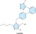
The nonpeptide angiotensin II antagonists are of much greater interest. Losartan (Figure 17-4), valsartan, eprosartan, irbesartan, candesartan, olmesartan, and telmesartan are orally active, potent, and specific competitive antagonists of angiotensin AT1 receptors. The efficacy of these drugs in hypertension is similar to that of ACE inhibitors, but they are associated with a lower incidence of cough. Like ACE inhibitors, nonpeptide angiotensin II antagonists slow the progression of diabetic nephropathy. The antagonists are also effective in the treatment of heart failure and provide a useful alternative when ACE inhibitors are not well tolerated. Like ACE inhibitors, they are well tolerated but should not be used by patients with nondiabetic renal disease or in pregnancy.
The current angiotensin II receptor antagonists are selective for the AT1 receptor. Since prolonged treatment with the drugs disinhibits renin secretion and increases circulating angiotensin II levels, there may be increased stimulation of AT2 receptors. This may be significant in view of the evidence that activation of the AT2 receptor causes vasodilation and other beneficial effects. AT2 receptor antagonists such as PD 123177 are available for research but have no clinical applications at this time.
The clinical benefits of AT1 receptor antagonists are similar to those of ACE inhibitors, and it is not clear if one group of drugs has significant advantages over the other. Combination therapy with both an ACE inhibitor and an angiotensin receptor antagonist has a number of potential advantages and is currently being investigated.
|
|
Figure 17-4. Structure of an angiotensin AT1 receptor antagonist. |
KININS
BIOSYNTHESIS OF KININS
Introduction
Kinins are potent vasodilator peptides formed enzymatically by the action of enzymes known as kallikreins or kininogenases acting on protein substrates called kininogens. The kallikrein-kinin system has several features in common with the renin-angiotensin system.
Kallikreins
Kallikreins are present in plasma and in several tissues, including the kidneys, pancreas, intestine, sweat glands, and salivary glands. Plasma prekallikrein can be activated to kallikrein by trypsin, Hageman factor, and possibly kallikrein itself. In general, the biochemical properties of tissue kallikreins are different from those of plasma kallikreins. Kallikreins can convert prorenin to active renin, but the physiologic significance of this action has not been established.
Kininogens
Kininogens¾the precursors of kinins and substrates of kallikreins¾are present in plasma, lymph, and interstitial fluid. Two kininogens are known to be present in plasma: a low-molecular-weight form (LMW kininogen) and a high-molecular-weight form (HMW kininogen). About 15-20% of the total plasma kininogen is in the HMW form. It is thought that LMW kininogen crosses capillary walls and serves as the substrate for tissue kallikreins, whereas HMW kininogen is confined to the bloodstream and serves as the substrate for plasma kallikrein.
FORMATION OF KININS IN PLASMA & TISSUES
The pathway for the formation and metabolism of kinins is shown in Figure 17-5. Three kinins have been identified in mammals: bradykinin, lysylbradykinin (also known as kallidin), and methionyllysylbradykinin. Each contains bradykinin in its structure.
Each kinin is formed from a kininogen by the action of a different enzyme. Bradykinin is released by plasma kallikrein, lysylbradykinin by tissue kallikrein, and methionyllysylbradykinin by pepsin and pepsin-like enzymes. The three kinins have been found in plasma and urine. Bradykinin is the predominant kinin in plasma, whereas lysylbradykinin is the major urinary form.
|
|
Figure 17-5. The kallikrein-kinin system. Kininase II is identical to converting enzyme peptidyl dipeptidase. |
ACTIONS OF KININS
Effects on the Cardiovascular System
Kinins produce marked vasodilation in several vascular beds, including the heart, kidney, intestine, skeletal muscle, and liver. In this respect, kinins are approximately 10 times more potent on a molar basis than histamine. The vasodilation may result from a direct inhibitory effect of kinins on arteriolar smooth muscle or may be mediated by the release of nitric oxide or vasodilator prostaglandins such as PGE2 and PGI2. In contrast, the predominant effect of kinins on veins is contraction; again, this may result from direct stimulation of venous smooth muscle or from the release of venoconstrictor prostaglandins such as PGF2a. Kinins also produce contraction of most visceral smooth muscle.
When injected intravenously, kinins produce a rapid but brief fall in blood pressure that is due to their arteriolar vasodilator action. Intravenous infusions of the peptide fail to produce a sustained decrease in blood pressure; prolonged hypotension can only be produced by progressively increasing the rate of infusion. The rapid reversibility of the hypotensive response to kinins is due primarily to reflex increases in heart rate, myocardial contractility, and cardiac output. In some species, bradykinin produces a biphasic change in blood pressure¾an initial hypotensive response followed by an increase above the preinjection level. The increase in blood pressure may be due to a reflex activation of the sympathetic nervous system, but under some conditions, bradykinin can directly release catecholamines from the adrenal medulla and stimulate sympathetic ganglia. Bradykinin also increases blood pressure when injected into the central nervous system, but the physiologic significance of this effect is not clear, since it is unlikely that kinins cross the blood-brain barrier. Kinins have no consistent effect on sympathetic or parasympathetic nerve endings.
The arteriolar dilation produced by kinins causes an increase in pressure and flow in the capillary bed, thus favoring efflux of fluid from blood to tissues. This effect may be facilitated by increased capillary permeability resulting from contraction of endothelial cells and widening of intercellular junctions, and by increased venous pressure secondary to constriction of veins. As a result of these changes, water and solutes pass from the blood to the extracellular fluid, lymph flow increases, and edema may result.
Effects on Endocrine & Exocrine Glands
As noted earlier, prekallikreins and kallikreins are present in several glands, including the pancreas, kidney, intestine, salivary glands, and sweat glands, and they can be released into the secretory fluids of these glands. The function of the enzymes in these tissues is not known. The enzymes (or active kinins) may diffuse from the organs to the blood and act as local modulators of blood flow. Since kinins have such marked effects on smooth muscle, they may also modulate the tone of salivary and pancreatic ducts and help regulate gastrointestinal motility. Kinins also influence the transepithelial transport of water, electrolytes, glucose, and amino acids, and may regulate the transport of these substances in the gastrointestinal tract and kidney. Finally, kallikreins may play a role in the physiologic activation of various pro-hormones, including proinsulin and prorenin.
Role in Inflammation
Kinins play an important role in the inflammatory process. Kallikreins and kinins can produce redness, local heat, swelling, and pain, and the production of kinins is increased in inflammatory lesions produced by a variety of methods.
Effects on Sensory Nerves
Kinins are potent pain-producing substances when applied to a blister base or injected intradermally. They elicit pain by stimulating nociceptive afferents in the skin and viscera.
KININ RECEPTORS & MECHANISMS OF ACTION
The biologic actions of kinins are mediated by specific receptors located on the membranes of the target tissues. Two types of kinin receptors, termed B1 and B2, have been defined based on the rank orders of agonist potencies. (Note that B here stands for bradykinin, not for b-adrenoceptor.) Bradykinin displays the highest affinity in most B2 receptor systems, followed by Lysbradykinin and then by Met-Lys-bradykinin. One exception is the B2 receptor that mediates contraction of venous smooth muscle; this appears to be most sensitive to Lys-bradykinin. Recent evidence suggests the existence of two B2-receptor subtypes, which have been termed B2A and B2B.
B1 receptors appear to have a very limited distribution in mammalian tissues and have few known functional roles. Studies with knockout mice that lack functional B1 receptors suggest that these receptors participate in the inflammatory response and may also be important in long-lasting kinin effects such as collagen synthesis and cell multiplication. By contrast, B2receptors have a widespread distribution that is consistent with the multitude of biologic effects that are mediated by this receptor type. B2 receptors are G protein-coupled and agonist binding sets in motion multiple signal transduction events, including calcium mobilization, chloride transport, formation of nitric oxide, and activation of phospholipase C, phospholipase A2, and adenylyl cyclase.
METABOLISM OF KININS
Kinins are metabolized rapidly (half-life < 15 seconds) by nonspecific exopeptidases or endopeptidases, commonly referred to as kininases. Two plasma kininases have been well characterized. Kininase I, apparently synthesized in the liver, is a carboxypeptidase that releases the carboxyl terminal arginine residue. Kininase II is present in plasma and vascular endothelial cells throughout the body. It is identical to angiotensin-converting enzyme (ACE, peptidyl dipeptidase), discussed above. Kininase II inactivates kinins by cleaving the carboxyl terminal dipeptide phenylalanyl-arginine. Like angiotensin I, bradykinin is almost completely hydrolyzed during a single passage through the pulmonary vascular bed.
DRUGS AFFECTING THE KALLIKREIN-KININ SYSTEM
Drugs that modify the activity of the kallikrein-kinin system are available, though none are in wide clinical use. Considerable effort has been directed toward developing kinin receptor antagonists, since such drugs have considerable therapeutic potential as anti-inflammatory and antinociceptive agents. Competitive antagonists of both B1 and B2 receptors are available for research use. Examples of B1 receptor antagonists are the peptides [Leu8-des-Arg9]bradykinin and Lys[Leu8-des Arg9] bradykinin. Nonpeptide B1 receptor antagonists are not yet available. The first B2 receptor antagonists to be discovered were also peptide derivatives of bradykinin. These first-generation antagonists were used extensively in animal studies of kinin receptor pharmacology. However, their half-life is short, and they are almost inactive on the human B2 receptor.
Icatibant is a second-generation B2 receptor antagonist. It is orally active, potent, and selective, has a long duration of action (> 60 minutes), and displays high B2-receptor affinity in humans and all other species in which it has been tested. Icatibant has been used extensively in animal studies to block exogenous and endogenous bradykinin and in human studies to evaluate the role of kinins in pain, hyperalgesia, and inflammation.
Recently, a third generation of B2-receptor antagonists was developed; examples are FR 173657, FR 172357, and NPC 18884. These antagonists block both human and animal B2receptors and are orally active. They have been reported to inhibit bradykinin-induced bronchoconstriction in guinea pigs, carrageenin-induced inflammatory responses in rats, and capsaicin-induced nociception in mice. These antagonists have promise for the treatment of inflammatory pain in humans.
SSR240612 is a new, potent, and orally active selective antagonist of B1 receptors in humans and several animal species. It exhibits analgesic and anti-inflammatory activities in mice and rats and is currently in preclinical development for the treatment of inflammatory and neurogenic pain.
The synthesis of kinins can be inhibited with the kallikrein inhibitor aprotinin. Actions of kinins mediated by prostaglandin generation can be blocked nonspecifically with inhibitors of prostaglandin synthesis such as aspirin. Conversely, the actions of kinins can be enhanced with ACE inhibitors, which block the degradation of the peptides. Indeed, as noted above, inhibition of bradykinin metabolism by ACE inhibitors contributes significantly to their antihypertensive action.
There is evidence that by acting on B2 receptors, bradykinin may play a beneficial, protective role in cardiovascular disease. Selective B2 agonists are available and have been shown to be effective in some animal models of human cardiovascular disease. These drugs may have potential for the treatment of hypertension and myocardial hypertrophy.
VASOPRESSIN
INTRODUCTION
Vasopressin (antidiuretic hormone, ADH) plays an important role in the long-term control of blood pressure through its action on the kidney to increase water reabsorption. This and other aspects of the physiology of vasopressin are discussed in Chapters 15 and 37 and will not be reviewed here.
Vasopressin also plays an important role in the short-term regulation of arterial pressure by its vasoconstrictor action. It increases total peripheral resistance when infused in doses less than those required to produce maximum urine concentration. Such doses do not normally increase arterial pressure because the vasopressor activity of the peptide is buffered by a reflex decrease in cardiac output. When the influence of this reflex is removed, eg, in shock, pressor sensitivity to vasopressin is greatly increased. Pressor sensitivity to vasopressin is also enhanced in patients with idiopathic orthostatic hypotension. Higher doses of vasopressin increase blood pressure even when baroreceptor reflexes are intact.
VASOPRESSIN RECEPTORS & ANTAGONISTS
Three subtypes of vasopressin receptors have been identified. V1a receptors mediate the vasoconstrictor action of vasopressin; V1b receptors potentiate the release of ACTH by pituitary corticotropes; and V2 receptors mediate the antidiuretic action. V1a effects are mediated by activation of phospholipase C, formation of inositol trisphosphate, and increased intracellular calcium concentration. V2 effects are mediated by activation of adenylyl cyclase.
Vasopressin-like peptides selective for either vasoconstrictor or antidiuretic activity have been synthesized. The most specific V1 vasoconstrictor agonist synthesized to date is [Phe2, Ile3, Orn8]vasotocin. Selective V2 antidiuretic analogs include 1-deamino[D-Arg8]arginine vasopressin (dDAVP) and 1-deamino[Val4, D-Arg8]arginine vasopressin (dVDAVP).
Specific antagonists of the vasoconstrictor action of vasopressin are also available. The peptide antagonist [1-(b-mercapto-b, b-cyclopentamethylenepropionic acid)-2-(O-methyl)tyrosine] arginine vasopressin also has antioxytocic activity but does not antagonize the antidiuretic action of vasopressin. Recently, nonpeptide, orally active V1a receptor antagonists have been discovered, examples being OPC-21268 and SR49059.
The vasopressor antagonists of vasopressin have been particularly useful in revealing the important role that vasopressin plays in blood pressure regulation in situations such as dehydration and hemorrhage. They have potential for the treatment of hypertension and heart failure. To date, most studies have focused on heart failure in which promising results have been obtained with V2 antagonists. However, V1a antagonists also have potential, and conivaptan (YM087), a drug with both V1a and V2 antagonist effects, has been approved for treatment of hyponatremia (see Chapter 15).
NATRIURETIC PEPTIDES
Synthesis & Structure
The atria and other tissues of mammals contain a family of peptides with natriuretic, diuretic, vasorelaxant, and other properties. The family consists of atrial natriuretic peptide (ANP), brain natriuretic peptide (BNP) and C-type natriuretic peptide (CNP). The structures of the three peptides are similar (Figure 17-6), but there are differences in their biologic effects. ANP is derived from the carboxyl terminal end of a common precursor termed preproANP. ANP is synthesized primarily in cardiac atrial cells, but small amounts are synthesized in ventricular cells. It is also synthesized by neurons in the central and peripheral nervous systems and in the lungs. ANP circulates as a 28-amino-acid peptide with a single disulfide bridge that forms a 17-residue ring.
Several factors increase the release of ANP from the heart, but the most important one appears to be atrial stretch via mechanosensitive ion channels. ANP release is also increased by volume expansion, head-out water immersion, changing from the standing to the supine position, and exercise. ANP release can also be increased by sympathetic stimulation via a1A-adrenoceptors, endothelins (see below) via the ETA-receptor subtype, glucocorticoids, and vasopressin. Finally, plasma ANP concentration increases in various pathologic states, including heart failure, primary aldosteronism, chronic renal failure, and inappropriate ADH secretion syndrome.
Administration of ANP produces prompt and marked increases in sodium excretion and urine flow. Glomerular filtration rate increases, with little or no change in renal blood flow, so that the filtration fraction increases. The ANP-induced natriuresis is apparently due to both the increase in glomerular filtration rate and a decrease in proximal tubular sodium reabsorption. ANP also inhibits the secretion of renin, aldosterone, and vasopressin; these changes may also increase sodium and water excretion. Finally, ANP decreases arterial blood pressure. This hypotensive action is due to vasodilation, which results from stimulated particulate guanylyl cyclase activity, increased cGMP levels, and decreased cytosolic free calcium concentration. ANP also reduces sympathetic tone to the peripheral vasculature and antagonizes the vasoconstrictor action of angiotensin II and other vasoconstrictors. These actions may contribute to the hypotensive action of the peptide.
There is considerable evidence that ANP participates in the physiologic regulation of sodium excretion and blood pressure. For example, suppression of ANP production or blockade of its action impairs the natriuretic response to volume expansion, and increases blood pressure.
BNP was originally isolated from porcine brain but, like ANP, it is synthesized primarily in the heart. It exists in two forms, having either 26 or 32 amino acids (Figure 17-6). Like ANP, the release of BNP appears to be volume-related; indeed, the two peptides may be co-secreted. BNP exhibits natriuretic, diuretic, and hypotensive activities similar to those of ANP but circulates at a lower concentration.
CNP consists of 22 amino acids (Figure 17-6). It is located predominantly in the central nervous system but is also present in several other tissues including the vascular endothelium, kidneys, and intestine. It has not been found in significant concentrations in the circulation. CNP has less natriuretic and diuretic activity than ANP and BNP but is a potent vasodilator. Its physiologic role is unclear.
|
|
Figure 17-6. Structures of atrial natriuretic peptide (ANP), brain natriuretic peptide (BNP), and C-type natriuretic peptide (CNP). Sequences common to the peptides are indicated in blue. |
Pharmacodynamics & Pharmacokinetics
The biologic actions of the natriuretic peptides are mediated through association with specific high-affinity receptors located on the surface of the target cells. Three receptor subtypes termed ANPA, ANPB, and ANPC have been identified. The ANPA receptor consists of a 120 kDa protein; its primary ligands are ANP and BNP. The ANPB receptor is similar in structure to the ANPA receptor, but its primary ligand appears to be CNP. The ANPA and ANPB receptors, but not the ANPC receptor, are coupled to guanylyl cyclase.
The natriuretic peptides have a short half-life in the circulation. They are metabolized in the kidneys, liver, and lungs by the neutral endopeptidase NEP 24.11. Inhibition of this endopeptidase results in increases in circulating levels of the natriuretic peptides, natriuresis, and diuresis. The peptides are also removed from the circulation by binding to ANPCreceptors in the vascular endothelium. This receptor binds the three natriuretic peptides with equal affinity. The receptor and bound peptide are internalized, the peptide is degraded enzymatically, and the receptor is returned to the cell surface.
Administration of BNP as nesiritide (see Chapter 13) in patients with severe heart failure increases sodium excretion and improves hemodynamics. However, the peptide has to be given by constant intravenous infusion and has caused fatal renal damage. A more promising approach may be the use of drugs that inhibit the neutral endopeptidase responsible for the breakdown of ANP. This is discussed below under Vasopeptidase Inhibitors. Patients with heart failure have high plasma levels of ANP and BNP; the latter has emerged as a diagnostic and prognostic marker in this condition.
VASOPEPTIDASE INHIBITORS
Vasopeptidase inhibitors are a new class of cardiovascular drugs that inhibit two metalloprotease enzymes, NEP 24.11 and ACE. They thus simultaneously increase the levels of natriuretic peptides and decrease the formation of angiotensin II. As a result, they enhance vasodilation, reduce vasoconstriction, and increase sodium excretion, in turn reducing peripheral vascular resistance and blood pressure.
Recently developed vasopeptidase inhibitors include omapatrilat, sampatrilat, and fasidotrilat. Omapatrilat, which has received the most attention, lowers blood pressure in animal models of hypertension as well as in hypertensive patients, and improves cardiac function in patients with heart failure. Unfortunately, omapatrilat causes a significant incidence of angioedema in addition to cough and dizziness and has not been approved for clinical use.
ENDOTHELINS
Introduction
The endothelium is the source of a variety of substances with vasodilator (PGI2 and nitric oxide) and vasoconstrictor activities. The latter include the endothelin family, potent vasoconstrictor peptides that were first isolated from aortic endothelial cells.
Biosynthesis, Structure, & Clearance
Three isoforms of endothelin have been identified: the originally described endothelin (ET-1) and two similar peptides, ET-2 and ET-3. Each isoform is a product of a different gene and is synthesized as a prepro form that is processed to a propeptide and then to the mature peptide. Processing to the mature peptides occurs through the action of endothelin-converting enzyme. Each endothelin is a 21-amino-acid peptide containing two disulfide bridges (Figure 17-7).
Endothelins are widely distributed in the body. ET-1 is the predominant endothelin secreted by the vascular endothelium. It is also produced by neurons and astrocytes in the central nervous system and in endometrial, renal mesangial, Sertoli, breast epithelial, and other cells. ET-2 is produced predominantly in the kidneys and intestine, whereas ET-3 is found in highest concentration in the brain but is also present in the gastrointestinal tract, lungs, and kidneys. Endothelins are present in the blood but in low concentration; they apparently act locally in a paracrine or autocrine fashion rather than as circulating hormones.
The expression of the ET-1 gene is increased by growth factors and cytokines, including transforming growth factor-b (TGF-b) and interleukin 1 (IL-1), vasoactive substances including angiotensin II and vasopressin, and mechanical stress. Expression is inhibited by nitric oxide, prostacyclin, and atrial natriuretic peptide.
Clearance of endothelins from the circulation is rapid and involves both enzymatic degradation by NEP 24.11 and clearance by the ETB receptor.
|
|
Figure 17-7. Structures of the endothelin peptides endothelin-1, endothelin-2, and endothelin-3. Sequences different in the three peptides are shown in blue. |
Actions
Endothelins exert widespread actions in the body. In particular, they cause dose-dependent vasoconstriction in most vascular beds. Intravenous administration of ET-1 causes a rapid and transient decrease in arterial blood pressure followed by a prolonged increase. The depressor response results from release of prostacyclin and nitric oxide from the vascular endothelium, whereas the pressor response is due to direct contraction of vascular smooth muscle. Endothelins also exert direct positive inotropic and chronotropic actions on the heart and are potent coronary vasoconstrictors. They act on the kidneys to cause vasoconstriction and decrease glomerular filtration rate and sodium and water excretion. In the respiratory system, they cause potent contraction of tracheal and bronchial smooth muscle. Endothelins interact with several endocrine systems, increasing the secretion of renin, aldosterone, vasopressin, and atrial natriuretic peptide. They exert a variety of actions on the central and peripheral nervous systems, the gastrointestinal system, the liver, the urinary tract, the male and female reproductive systems, the eye, the skeletal system, and the skin. Finally, ET-1 is a potent mitogen for vascular smooth muscle cells, cardiac myocytes, and glomerular mesangial cells.
Endothelin receptors are widespread in the body. Two endothelin receptor subtypes, termed ETA and ETB, have been cloned and sequenced. ETA receptors have a high affinity for ET-1 and a low affinity for ET-3 and are located on smooth muscle cells, where they mediate vasoconstriction. ETB receptors have approximately equal affinities for ET-1 and ET-3 and are located on vascular endothelial cells, where they mediate release of PGI2 and nitric oxide. Both receptor subtypes belong to the G protein-coupled seven-transmembrane domain family of receptors.
The signal transduction mechanisms triggered by binding of ET-1 to its receptors include stimulation of phospholipase C, formation of inositol trisphosphate, and release of calcium from the endoplasmic reticulum, which results in vasoconstriction. Stimulation of PGI2 and nitric oxide synthesis results in decreased intracellular calcium concentration and vasodilation.
INHIBITORS OF ENDOTHELIN SYNTHESIS & ACTION
The endothelin system can be blocked with receptor antagonists and drugs that block endothelin-converting enzyme. Endothelin ETA or ETB receptors can be blocked selectively, or both can be blocked with nonselective ETA-ETB antagonists.
Bosentan is a nonselective antagonist. This drug is active intravenously and orally, and blocks both the initial transient depressor (ETB) and the prolonged pressor (ETA) responses to intravenous endothelin. Many orally active endothelin receptor antagonists with increased selectivity have been developed and are available for research use.
The formation of endothelins can be blocked by inhibiting endothelin-converting enzyme with phosphoramidon. Phosphoramidon is not specific for endothelin-converting enzyme, but several potent and more selective inhibitors are now available. The therapeutic potential of these drugs may be similar to that of the endothelin receptor antagonists (see below).
PHYSIOLOGIC & PATHOLOGIC ROLES OF ENDOTHELIN: EFFECTS OF ENDOTHELIN ANTAGONISTS
Systemic administration of endothelin receptor antagonists or endothelin-converting enzyme inhibitors causes vasodilation and decreases arterial pressure in humans and experimental animals. Intra-arterial administration of the drugs also causes slow-onset forearm vasodilation in humans. These observations provide evidence that the endothelin system participates in the regulation of vascular tone, even under resting conditions.
There is increasing evidence that endothelins participate in a variety of cardiovascular diseases, including hypertension, cardiac hypertrophy, heart failure, atherosclerosis, coronary artery disease, and myocardial infarction. Endothelins have also been implicated in pulmonary diseases, including asthma and pulmonary hypertension, as well as in several renal diseases.
Endothelin antagonists have considerable potential in the treatment of these diseases. In clinical trials, bosentan and other nonselective antagonists as well as ETA-selective antagonists (sitaxentan, ambrisentan) have produced beneficial effects on hemodynamics and other symptoms in heart failure, pulmonary hypertension, and essential hypertension. Bosentan is currently approved for use in pulmonary hypertension (see Chapter 11), and a related antagonist, tezosentan, is under investigation for the treatment of acute heart failure.
Endothelin antagonists occasionally cause systemic hypotension, increased heart rate, facial flushing or edema, and headaches. Potential gastrointestinal effects include nausea, vomiting, and constipation. Because of their teratogenic effects, ET antagonists are contraindicated in pregnancy. Bosentan has been associated with fatal hepatotoxicity, and patients taking this drug must have monthly liver function tests. Negative pregnancy test results are required for women of child-bearing age to take this drug.
VASOACTIVE INTESTINAL PEPTIDE
Vasoactive intestinal peptide (VIP) is a 28-amino-acid peptide related structurally to secretin and glucagon. VIP is widely distributed in the central and peripheral nervous systems, where it functions as a neurotransmitter or neuromodulator. It is also present in the gastrointestinal tract, heart, lungs, kidneys, and thyroid gland. Many blood vessels are innervated by VIP neurons. VIP is found in blood but does not appear to function as a hormone.
VIP exerts significant effects on the cardiovascular system. It produces marked vasodilation in most vascular beds and in this regard is more potent on a molar basis than acetylcholine. In the heart, VIP causes coronary vasodilation and exerts positive inotropic and chronotropic effects. It may thus participate in the regulation of coronary blood flow, cardiac contraction, and heart rate.
The effects of VIP are mediated by G protein-coupled receptors; two subtypes, VPAC1 and VPAC2, have been cloned from human tissues. Both subtypes are widely distributed in the central nervous system and in the heart, blood vessels, and other tissues. Binding of VIP to its receptors results in activation of adenylyl cyclase and formation of cAMP, which is responsible for the vasodilation and many other effects of the peptide. Other actions may be mediated by nitric oxide and cGMP.
Specific VIP receptor agonists and antagonists are currently available for research use.
SUBSTANCE P
Substance P belongs to the tachykinin family of peptides, which share the common carboxyl terminal sequence Phe-X-Gly-Leu-Met. Other members of this family are neurokinin A and neurokinin B. Substance P is an undecapeptide, while neurokinins A and B are decapeptides.
Substance P is present in the central nervous system, where it is a neurotransmitter (see Chapter 21), and in the gastrointestinal tract, where it may play a role as a transmitter in the enteric nervous system and as a local hormone (see Chapter 6).
Substance P exerts a variety of incompletely understood central actions that implicate the peptide in behavior, anxiety, depression, nausea, and emesis. It is a potent arteriolar vasodilator, producing marked hypotension in humans and several animal species. The vasodilation is mediated by release of nitric oxide from the endothelium. Conversely, substance P causes contraction of venous, intestinal, and bronchial smooth muscle. It also stimulates secretion by the salivary glands and causes diuresis and natriuresis by the kidneys.
The actions of substance P and neurokinins A and B are mediated by three G protein-coupled tachykinin receptors designated NK1, NK2, and NK3. Substance P is the preferred ligand for the NK1 receptor, the predominant tachykinin receptor in the human brain. However, neurokinins A and B also possess considerable affinity for this receptor. In humans, most of the central and peripheral effects of substance P are mediated by NK1 receptors.
Several nonpeptide NK1 receptor antagonists have been developed. These compounds are highly selective and orally active, and enter the brain. Recent clinical trials have shown that these antagonists may be useful in treating depression and other disorders and in preventing chemotherapy-induced emesis. The first of these to be approved for the prevention of chemotherapy-induced nausea and vomiting is aprepitant (Chapter 63).
NEUROTENSIN
Neurotensin is a tridecapeptide that was first isolated from the central nervous system but subsequently was found to be present in the gastrointestinal tract and in the circulation. It is synthesized as part of a larger precursor that also contains neuromedin N, a six-amino-acid neurotensin-like peptide.
In the brain, processing of the precursor leads primarily to the formation of neurotensin and neuromedin N; these are released together from nerve endings. In the gut, processing leads mainly to the formation of neurotensin and a larger peptide that contains the neuromedin N sequence at the carboxyl terminal. Both peptides are secreted into the circulation after ingestion of food.
Like many other neuropeptides, neurotensin serves a dual function as a neurotransmitter or neuromodulator in the central nervous system and as a local hormone in the periphery. When administered centrally, neurotensin exerts potent effects including hypothermia, antinociception, and modulation of dopamine neurotransmission. When administered into the peripheral circulation, it causes vasodilation, hypotension, increased vascular permeability, increased secretion of several anterior pituitary hormones, hyperglycemia, inhibition of gastric acid and pepsin secretion, and inhibition of gastric motility.
In the central nervous system, there are close associations between neurotensin and dopamine systems, and neurotensin may be involved in clinical disorders involving dopamine pathways such as schizophrenia, Parkinson's disease, and drug abuse. Consistent with this, it has been shown that central administration of neurotensin produces effects in rodents similar to those produced by antipsychotic drugs.
Three subtypes of neurotensin receptors, designated NT1, NT2, and NT3, have been cloned. NT1 and NT2 receptors belong to the G protein-coupled superfamily with seven transmembrane domains; the NT3 receptor is a single transmembrane domain protein that belongs to a family of sorting proteins.
Neurotensin agonists that cross the blood-brain barrier have been developed and may have potential as therapeutic agents for diseases such as schizophrenia and Parkinson's disease.
Neurotensin receptors can be blocked with the nonpeptide antagonists SR142948A and SR48692. SR142948A is a potent antagonist of the hypothermia and analgesia produced by centrally administered neurotensin. It also blocks the cardiovascular effects of systemic neurotensin.
CALCITONIN GENE-RELATED PEPTIDE
Calcitonin gene-related peptide (CGRP) is a member of the calcitonin family of peptides, which also includes calcitonin, adrenomedullin and amylin. CGRP consists of 37 amino acids and displays approximately 30% structural homology with salmon calcitonin.
Like calcitonin, CGRP is present in large quantities in the C cells of the thyroid gland. It is also distributed widely in the central and peripheral nervous systems, in the cardiovascular system, the gastrointestinal tract, and the urogenital system. CGRP is found with substance P (see above) in some of these regions and with acetylcholine in others.
When CGRP is injected into the central nervous system, it produces a variety of effects, including hypertension and suppression of feeding. When injected into the systemic circulation, the peptide causes hypotension and tachycardia. The hypotensive action of CGRP results from the potent vasodilator action of the peptide; indeed, CGRP is the most potent vasodilator yet discovered. It dilates multiple vascular beds, but the coronary circulation is particularly sensitive.
The actions of CGRP are mediated by two receptors named CGRP1 and CGRP2. Peptide and nonpeptide antagonists of these receptors have been developed. Of the nonpeptide antagonists now available, the best characterized is BIBN4096BS, which has a high affinity and specificity for the human CGRP receptor.
Evidence is accumulating that release of CGRP from trigeminal nerves plays a central role in the pathophysiology of migraine. The peptide is released during migraine attacks, and successful treatment of migraine with a selective serotonin agonist normalizes cranial CGRP levels. BIBN4096BS has recently been shown to be an effective, well-tolerated treatment for migraine.
ADRENOMEDULLIN
Adrenomedullin was first discovered in human adrenal medullary pheochromocytoma tissue. It is a 52-amino acid peptide with a six-amino acid ring and a C-terminal amidation sequence. Like CGRP, adrenomedullin is a member of the calcitonin family of peptides.
Adrenomedullin is widely distributed in the body. The highest concentrations are found in the adrenal glands, hypothalamus, and anterior pituitary, but high levels are also present in the kidneys, lungs, cardiovascular system, and gastrointestinal tract. Adrenomedullin in plasma apparently originates in the heart and vasculature.
In animals, adrenomedullin dilates resistance vessels in the kidney, brain, lung, hind limbs, and mesentery, resulting in a marked, long-lasting hypotension. The hypotension in turn causes reflex increases in heart rate and cardiac output. These responses also occur during intravenous infusion of the peptide in healthy human subjects. Adrenomedullin also acts on the kidneys to increase sodium excretion, and it exerts several endocrine effects including inhibition of aldosterone and insulin secretion. Finally, it acts on the central nervous system to increase sympathetic outflow.
The diverse actions of adrenomedullin are mediated both by CGRP receptors and by specific adrenomedullin receptors termed AM1 and AM2. The major second messenger for both receptors is cAMP.
Circulating adrenomedullin levels increase during intense exercise. They also increase in a number of pathologic states, including essential hypertension, cardiac and renal failure, and septic shock. The roles of adrenomedullin in these states remain to be defined, but it is currently thought that the peptide functions as a physiologic antagonist of the actions of vasoconstrictors including endothelin 1 and angiotensin II. By virtue of these actions, adrenomedullin may protect against cardiovascular overload and injury.
NEUROPEPTIDE Y
Neuropeptide Y is a member of the family that also includes peptide YY and pancreatic polypeptide. Each peptide consists of 36 amino acids.
Neuropeptide Y is one of the most abundant neuropeptides in both the central and peripheral nervous systems. In the sympathetic nervous system, neuropeptide Y is frequently localized in noradrenergic neurons and apparently functions both as a vasoconstrictor and as a cotransmitter with norepinephrine. Peptide YY and pancreatic polypeptide are both gut endocrine peptides.
Neuropeptide Y produces a variety of central nervous system effects, including increased feeding (it is one of the most potent orexigenic molecules in the brain), hypotension, hypothermia, and respiratory depression. Other effects include vasoconstriction of cerebral blood vessels, positive chronotropic and inotropic actions on the heart, and hypertension. The peptide is a potent renal vasoconstrictor and suppresses renin secretion, but can cause diuresis and natriuresis. Prejunctional neuronal actions include inhibition of transmitter release from sympathetic and parasympathetic nerves. Vascular actions include direct vasoconstriction, potentiation of the action of vasoconstrictors, and inhibition of the action of vasodilators.
These diverse effects are mediated by multiple receptors designated Y1 through Y6. All receptors except Y3 have been cloned and shown to be G protein-coupled receptors linked to mobilization of Ca2+ and inhibition of adenylyl cyclase. Y1 and Y2 receptors are of major importance in the cardiovascular and other peripheral effects of the peptide. Y4 receptors have a high affinity for pancreatic polypeptide and may be a receptor for the pancreatic peptide rather than for neuropeptide Y. Y5 receptors are found mainly in the central nervous system and may be involved in the control of food intake. Y6 receptors do not appear to contribute significantly to the physiologic effects of neuropeptide Y in humans.
Selective nonpeptide neuropeptide Y receptor antagonists are now available for research. The first nonpeptide Y1 receptor antagonist, BIBP3226, is also the most thoroughly studied. It has a short half-life in vivo. In animal models, it blocks the vasoconstrictor and pressor responses to neuropeptide Y. Structurally related Y1 antagonists include BIB03304, and H409/22, which has been tested in humans. SR120107A and SR120819A are orally active Y1 antagonists and have a long duration of action. BIIE0246 is the first nonpeptide antagonist selective for the Y2 receptor.
These antagonists have been useful in analyzing the role of neuropeptide Y in cardiovascular regulation. It now appears that the peptide is not important in the regulation of hemodynamics under normal resting conditions, but may be of increased importance in cardiovascular disorders including hypertension and heart failure. Other studies have implicated neuropeptide Y in feeding disorders, seizures, anxiety, and diabetes, and Y1 and Y5 receptor antagonists have potential as antiobesity agents.
UROTENSIN
Urotensin II was originally identified in fish, but isoforms are now known to be present in mammalian species including the human, mouse, rat and pig. Human urotensin II is an 11-amino acid peptide. Major sites of urotensin II expression in humans include the brain, spinal cord, and kidneys. Urotensin II is also present in plasma, and the kidneys may be a major source of this circulating peptide.
In vitro, urotensin II is a potent constrictor of vascular smooth muscle; its activity depends on the type of blood vessel and the species from which it was obtained. Vasoconstriction occurs primarily in arterial vessels, where urotensin II can be more potent than endothelin 1, making it the most potent known vasoconstrictor. However, under some conditions, urotensin II may cause vasodilation. In vivo, urotensin II has complex hemodynamic effects, the most prominent being regional vasoconstriction and cardiac depression. In some ways, these effects resemble those produced by endothelin 1. Nevertheless, the extent to which the peptide is involved in the regulation of vascular tone and blood pressure in humans is not clear; recent studies have produced conflicting results.
The actions of urotensin II are mediated by a G protein-coupled receptor referred to as the UT receptor. UT receptors are widely distributed in the brain, spinal cord, heart, vascular smooth muscle, skeletal muscle, and pancreas. Some effects of the peptide including vasoconstriction are mediated by the phospholipase C, IP3-DAG signal transduction pathway.
Modifications of the disulfide bridge of urotensin II have yielded UT-receptor antagonists. A nonpeptide antagonist, palosuran, has also been developed.
Evidence is accumulating that urotensin II is involved in cardiovascular and other diseases. In particular, it has been reported that plasma urotensin II levels are increased in hypertension, heart failure, diabetes mellitus, and renal failure. UT-receptor antagonists will be valuable in studies of the pathophysiologic role of this peptide.
PREPARATIONS AVAILABLE1
Aprepitant (Emend)
Oral: 80, 125 mg capsules
Bosentan (Tracleer)
Oral: 62.5, 125 mg tablets
1Preparations of angiotensin-converting enzyme inhibitors and angiotensin receptor antagonists are found in Chapter 11; preparations of vasopressin are found in Chapter 37.
REFERENCES
Angiotensin
Batenburg WW et al: Angiotensin II type 2 receptor-mediated vasodilation. Focus on bradykinin, NO and endothelium-derived hyperpolarizing factor(s). Vascul Pharmacol 2005;42:109.
Danser AH, Deinum J: Renin, prorenin and the putative (pro)renin receptor. Hypertension 2005;46:1069.
Dinh DT et al: Angiotensin receptors: Distribution, signalling and function. Clin Sci 2001;100:481.
Fleming I, Kohlstedt K, Busse R: New fACEs to the renin-angiotensin system. Physiology (Bethesda) 2005;20:91.
Gradman AH et al: Aliskiren, a novel orally effective renin inhibitor, provides dose-dependent antihypertensive efficacy and placebo-like tolerability in hypertensive patients. Circulation 2005;111:1012.
Kelly DJ, Wilkinson-Berka JL, Gilbert RE: Renin inhibition: New potential for an old therapeutic target. Hypertension 2005;46:471.
McMurray JJ et al: Which inhibitor of the renin-angiotensin system should be used in chronic heart failure and acute myocardial infarction? Circulation 2004;110:3281.
Sica DA: Combination angiotensin-converting enzyme inhibitor and angiotensin receptor blocker therapy: Its role in clinical practice. J Clin Hypertens (Greenwich) 2003;5:414.
Watanabe T, Barker TA, Berk BC: Angiotensin II and the endothelium: Diverse signals and effects. Hypertension 2005;45:163.
Yan AT, Yan RT, Liu PP: Narrative review: Pharmacotherapy for chronic heart failure: Evidence from recent clinical trials. Ann Intern Med 2005;142:132.
Kinins
Couture R et al: Kinin receptors in pain and inflammation. Eur J Pharmacol 2001;429:161.
Ferreira J et al: The use of kinin B1 and B2 receptor knockout mice and selective antagonists to characterize the nociceptive responses caused by kinins at the spinal level.Neuropharmacology 2002;43:1188.
Leeb-Lundberg LM et al: International union of pharmacology. XLV. Classification of the kinin receptor family: From molecular mechanisms to pathophysiological consequences. Pharmacol Rev 2005;57:27.
Vasopressin
Goldsmith SR, Gheorghiade M: Vasopressin antagonism in heart failure. J Am Coll Cardiol 2005;46:1785.
Thibonnier M et al: The basic and clinical pharmacology of nonpeptide vasopressin receptor antagonists. Annu Rev Pharmacol Toxicol 2001;41:175.
Natriuretic Peptides
Boomsma F, van den Meiracker AH: Plasma A- and B-type natriuretic peptides: Physiology, methodology and clinical use. Cardiovasc Res 2001;51:442.
Munagala VK, Burnett JC, Jr, Redfield MM: The natriuretic peptides in cardiovascular medicine. Curr Probl Cardiol 2004;29:707.
Vasopeptidase Inhibitors
Campbell DJ: Vasopeptidase inhibition: A double-edged sword? Hypertension 2003;41:383.
Packer M et al: Comparison of omapatrilat and enalapril in patients with chronic heart failure: The Omapatrilat Versus Enalapril Randomized Trial of Utility in Reducing Events (OVERTURE). Circulation 2002;106:920.
Worthley MI, Corti R, Worthley SG: Vasopeptidase inhibitors: Will they have a role in clinical practice? Br J Clin Pharmacol 2004;57:27.
Endothelins
Channick RN et al: Endothelin receptor antagonists in pulmonary arterial hypertension. J Am Coll Cardiol 2004;43:62S.
Ertl G, Bauersachs J: Endothelin receptor antagonists in heart failure: Current status and future directions. Drugs 2004;64:1029.
Galie N, Manes A, Branzi A: The endothelin system in pulmonary arterial hypertension. Cardiovasc Res 2004;61:227.
Vasoactive Intestinal Peptide
Henning RJ, Sawmiller DR: Vasoactive intestinal peptide: Cardiovascular effects. Cardiovasc Res 2001;49:27.
Substance P
Aprepitant (emend) for prevention of nausea and vomiting due to cancer chemotherapy. Med Lett Drug Ther 2003;45:620.
Hokfelt T, Pernow B, Wahren J: Substance P: A pioneer amongst neuropeptides. J Intern Med 2001;249:27.
Neurotensin
Kinkead B, Nemeroff CB: Neurotensin, schizophrenia, and antipsychotic drug action. Int Rev Neurobiol 2004;59:327.
Calcitonin Gene-Related Peptide
Olesen J et al: Calcitonin gene-related peptide receptor antagonist BIBN 4096 BS for the acute treatment of migraine. N Engl J Med 2004;350:1104.
Poyner DR et al: International Union of Pharmacology. XXXII. The mammalian calcitonin gene-related peptides, adrenomedullin, amylin, and calcitonin receptors. Pharmacol Rev 2002;54:233.
Adrenomedullin
Hamid SA, Baxter GF: Adrenomedullin: Regulator of systemic and cardiac homeostasis in acute myocardial infarction. Pharmacol Ther 2005;105:95.
Smith DM et al: Adrenomedullin: Receptor and signal transduction. Biochem Soc Trans 2002;30:432.
Neuropeptide Y
DiBona GF: Neuropeptide Y. Am J Physiol 2002;282:R635.
Malmstrom RE: Pharmacology of neuropeptide Y receptor antagonists: Focus on cardiovascular functions. Eur J Pharmacol 2002;447:11.
Urotensin
Douglas SA, Dhanak D, Johns DG: From 'gills to pills': Urotensin-II as a regulator of mammalian cardiorenal function. Trends Pharmacol Sci 2004;25:76.
Ong KL, Lam KS, Cheung BM: Urotensin II: Its function in health and its role in disease. Cardiovasc Drugs Ther 2005;19:65.