INTRODUCTION
Histamine and serotonin (5-hydroxytryptamine) are biologically active amines that function as neurotransmitters and are found in non-neural tissues, have complex physiologic and pathologic effects through multiple receptor subtypes, and are often released locally. Together with endogenous peptides (see Chapter 17), prostaglandins and leukotrienes (see Chapter 18), and cytokines (see Chapter 56), they are sometimes called autacoids (Greek, "self-remedy") or local hormones in recognition of these properties.
Because of their broad and largely undesirable effects, neither histamine nor serotonin has any clinical application in the treatment of disease. However, compounds that selectively activate certain receptor subtypes or selectively antagonize the actions of these amines are of considerable clinical usefulness. This chapter therefore emphasizes the basic pharmacology of the agonist amines and the clinical pharmacology of the more selective agonist and antagonist drugs. The ergot alkaloids, compounds with partial agonist activity at serotonin and several other receptors, are discussed at the end of the chapter.
I. HISTAMINE
INTRODUCTION
Histamine was synthesized in 1907 and later isolated from mammalian tissues. Early hypotheses concerning the possible physiologic roles of tissue histamine were based on similarities between the effects of intravenously administered histamine and the symptoms of anaphylactic shock and tissue injury. Marked species variation is observed, but in humans histamine is an important mediator of immediate allergic and inflammatory reactions, although it plays only a modest role in anaphylaxis. Histamine plays an important role in gastric acid secretion (see Chapter 63) and functions as a neurotransmitter and neuromodulator (see Chapters 6, 21). Newer evidence indicates that histamine also plays a role in chemotaxis of white blood cells.
BASIC PHARMACOLOGY OF HISTAMINE
Chemistry & Pharmacokinetics
Histamine occurs in plants as well as in animal tissues and is a component of some venoms and stinging secretions.
Histamine is formed by decarboxylation of the amino acid l-histidine, a reaction catalyzed in mammalian tissues by the enzyme histidine decarboxylase. Once formed, histamine is either stored or rapidly inactivated. Very little histamine is excreted unchanged. The major metabolic pathways involve conversion to N-methylhistamine, methylimidazoleacetic acid, and imidazoleacetic acid (IAA). Certain neoplasms (systemic mastocytosis, urticaria pigmentosa, gastric carcinoid, and occasionally myelogenous leukemia) are associated with increased numbers of mast cells or basophils and with increased excretion of histamine and its metabolites.
Most tissue histamine is sequestered and bound in granules (vesicles) in mast cells or basophils; the histamine content of many tissues is directly related to their mast cell content. The bound form of histamine is biologically inactive, but many stimuli, as noted below, can trigger the release of mast cell histamine, allowing the free amine to exert its actions on surrounding tissues. Mast cells are especially rich at sites of potential tissue injury¾nose, mouth, and feet; internal body surfaces; and blood vessels, particularly at pressure points and bifurcations.
Non-mast cell histamine is found in several tissues, including the brain, where it functions as a neurotransmitter. Endogenous neurotransmitter histamine may play a role in many brain functions such as neuroendocrine control, cardiovascular regulation, thermal and body weight regulation, and arousal (see Chapter 21).
A second important nonneuronal site of histamine storage and release is the enterochromaffin-like (ECL) cells of the fundus of the stomach. ECL cells release histamine, one of the primary gastric acid secretagogues, to activate the acid-producing parietal cells of the mucosa (see Chapter 63).
Storage & Release of Histamine
The stores of histamine in mast cells can be released through several mechanisms.
A. IMMUNOLOGIC RELEASE
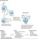
Immunologic processes account for the most important pathophysiologic mechanism of mast cell and basophil histamine release. These cells, if sensitized by IgE antibodies attached to their surface membranes, degranulate "explosively" when exposed to the appropriate antigen (see Figure 56-5, effector phase). This type of release also requires energy and calcium. Degranulation leads to the simultaneous release of histamine, adenosine triphosphate (ATP), and other mediators that are stored together in the granules. Histamine released by this mechanism is a mediator in immediate (type I) allergic reactions. Substances released during IgG- or IgM-mediated immune reactions that activate the complement cascade also release histamine from mast cells and basophils.
By a negative feedback control mechanism mediated by H2 receptors, histamine appears to modulate its own release and that of other mediators from sensitized mast cells in some tissues. In humans, mast cells in skin and basophils show this negative feedback mechanism; lung mast cells do not. Thus, histamine may act to limit the intensity of the allergic reaction in the skin and blood.
Endogenous histamine has a modulating role in a variety of inflammatory and immune responses. Upon injury to a tissue, released histamine causes local vasodilation and leakage of plasma containing mediators of acute inflammation (complement, C-reactive protein), and antibodies. Histamine has an active chemotactic attraction for inflammatory cells (neutrophils, eosinophils, basophils, monocytes, and lymphocytes). Histamine inhibits the release of lysosome contents and several T- and B-lymphocyte functions. Most of these actions are mediated by H2 or H4 receptors. Release of peptides from nerves in response to inflammation is also probably modulated by histamine, in this case acting through presynaptic H3 receptors.
B. CHEMICAL AND MECHANICAL RELEASE
Certain amines, including drugs such as morphine and tubocurarine, can displace histamine from the heparin-protein complex within cells. This type of release does not require energy and is not associated with mast cell injury or degranulation. Loss of granules from the mast cell also releases histamine, since sodium ions in the extracellular fluid rapidly displace the amine from the complex. Chemical and mechanical mast cell injury causes degranulation and histamine release. Compound 48/80, an experimental drug, selectively releases histamine from tissue mast cells by an exocytotic degranulation process requiring energy and calcium.
|
|
Figure 56-5. Mechanism of type I hypersensitivity. Initial exposure to allergen (sensitization phase) leads to production of IgE by plasma cells differentiated from allergen-specific B cells (not shown). The secreted IgE binds IgE-specific receptors (FceR) on blood basophils and tissue mast cells. Reexposure to allergen leads to cross-linking of membrane-bound IgE (effector phase). This cross-linking causes degranulation of cytoplasmic granules and release of mediators that induce vasodilation, smooth muscle contraction, and increased vascular permeability. These effects lead to the clinical symptoms characteristic of type I hypersensitivity. |
Pharmacodynamics
A. MECHANISM OF ACTION
Histamine exerts its biologic actions by combining with specific cellular receptors located on the surface membrane. The four different histamine receptors thus far characterized are designated H1-H4 and are described in Table 16-1. Unlike the other amine transmitter receptors discussed previously, no subfamilies have been found within these major types, although different splice variants of several receptor types have been found.
All four receptor types have been cloned and belong to the large superfamily of receptors having seven membrane-spanning regions and coupled with G proteins (GPCR). The structures of the H1 and H2 receptors differ significantly and appear to be more closely related to muscarinic and 5-HT1 receptors, respectively, than to each other. The H4 receptor has about 40% homology with the H3 receptor but does not seem to be closely related to any other histamine receptor. All four histamine receptors have been shown to have constitutive activity in some systems; thus, some "antihistamines" previously considered to be traditional pharmacologic antagonists must now be considered to be possible inverse agonists (Chapter 2). Indeed, many first- and second-generation H1 blockers (see below) are probably inverse agonists. Furthermore, a single molecule may be an agonist at one histamine receptor and an antagonist at another. For example, clobenprobit, an agonist at H4 receptors, is an antagonist or inverse agonist at H3 receptors (Table 16-1).
In the brain, H1 and H2 receptors are located on postsynaptic membranes, whereas H3 receptors are predominantly presynaptic. Activation of H1 receptors, which are present in endothelium, smooth muscle cells, and nerve endings, usually elicits an increase in phosphoinositol hydrolysis and an increase in intracellular calcium. Activation of H2 receptors, present in gastric mucosa, cardiac muscle cells, and some immune cells, increases intracellular cyclic adenosine monophosphate (cAMP) via Gs. Like the b2 adrenoceptor, under certain circumstances the H2 receptor may couple to Gq, activating the IP3-DAG (inositol 1,4,5-trisphosphate-diacylglycerol) cascade. Activation of H3 receptors decreases transmitter release from histaminergic and other neurons, probably mediated by a decrease in calcium influx through N-type calcium channels in nerve endings. H4 receptors are mainly found on leukocytes in the bone marrow and circulating blood. H4 receptors appear to have very important chemotactic effects on eosinophils and mast cells. In this role, they may play an important part in inflammation and allergy. They may also modulate production of these cell types and they may mediate, in part, the previously recognized effects of histamine on cytokine production.
B. TISSUE AND ORGAN SYSTEM EFFECTS OF HISTAMINE
Histamine exerts powerful effects on smooth and cardiac muscle, on certain endothelial and nerve cells, and on the secretory cells of the stomach. However, sensitivity to histamine varies greatly among species. Guinea pigs are exquisitely sensitive, humans, dogs, and cats somewhat less so, and mice and rats very much less so.
1. Nervous system¾ Histamine is a powerful stimulant of sensory nerve endings, especially those mediating pain and itching. This H1-mediated effect is an important component of the urticarial response and reactions to insect and nettle stings. Some evidence suggests that local high concentrations can also depolarize efferent (axonal) nerve endings (see paragraph 8, below). In the mouse, and probably in humans, respiratory neurons signaling inspiration and expiration are modulated by H1 receptors. Presynaptic H3 receptors play important roles in modulating transmitter release in the nervous system. H3 agonists reduce the release of acetylcholine, amine, and peptide transmitters in various areas of the brain and in peripheral nerves.
2. Cardiovascular system¾ In humans, injection or infusion of histamine causes a decrease in systolic and diastolic blood pressure and an increase in heart rate. The blood pressure changes are caused by the direct vasodilator action of histamine on arterioles and precapillary sphincters; the increase in heart rate involves both stimulatory actions of histamine on the heart and a reflex tachycardia. Flushing, a sense of warmth, and headache may also occur during histamine administration, consistent with the vasodilation. Vasodilation elicited by small doses of histamine is caused by H1-receptor activation and is mediated primarily by release of nitric oxide from the endothelium (see Chapter 19). The decrease in blood pressure is usually accompanied by a reflex tachycardia. Higher doses of histamine activate the H2-mediated cAMP process of vasodilation and direct cardiac stimulation. In humans, the cardiovascular effects of small doses of histamine can usually be antagonized by H1-receptor antagonists alone.
Histamine-induced edema results from the action of the amine on H1 receptors in the vessels of the microcirculation, especially the postcapillary vessels. The effect is associated with the separation of the endothelial cells, which permits the transudation of fluid and molecules as large as small proteins into the perivascular tissue. This effect is responsible for the urticaria (hives) that signals the release of histamine in the skin. Studies of endothelial cells suggest that actin and myosin within these cells contract, resulting in separation of the endothelial cells and increased permeability.
Direct cardiac effects of histamine include both increased contractility and increased pacemaker rate. These effects are mediated chiefly by H2 receptors. In human atrial muscle, histamine can also decrease contractility; this effect is mediated by H1 receptors. The physiologic significance of these cardiac actions is not clear. Some of the cardiovascular signs and symptoms of anaphylaxis are due to released histamine, though several other mediators are involved and appear to be more important than histamine in humans.
3. Bronchiolar smooth muscle¾ In both humans and guinea pigs, histamine causes bronchoconstriction mediated by H1 receptors. In the guinea pig, this effect is the cause of death from histamine toxicity, but in humans with normal airways, bronchoconstriction following small doses of histamine is not marked. However, patients with asthma are very sensitive to histamine. The bronchoconstriction induced in these patients probably represents a hyperactive neural response, since such patients also respond excessively to many other stimuli, and the response to histamine can be blocked by autonomic blocking drugs such as ganglionic blocking agents as well as by H1-receptor antagonists (see Chapter 20). Although methacholine provocation is more commonly used, tests using increasing doses of inhaled histamine have been used in the diagnosis of bronchial hyperreactivity in patients with suspected asthma or cystic fibrosis. Such individuals may be 100- to 1000-fold more sensitive to histamine (and methacholine) than are normal subjects. Curiously, a few species (eg, rabbit) respond to histamine with bronchodilation, reflecting the dominance of the H2 receptor in their airways.
4. Gastrointestinal tract smooth muscle¾ Histamine causes contraction of intestinal smooth muscle, and histamine-induced contraction of guinea pig ileum is a standard bioassay for this amine. The human gut is not as sensitive as that of the guinea pig, but large doses of histamine may cause diarrhea, partly as a result of this effect. This action of histamine is mediated by H1 receptors.
5. Other smooth muscle organs¾ In humans, histamine generally has insignificant effects on the smooth muscle of the eye and genitourinary tract. However, pregnant women suffering anaphylactic reactions may abort as a result of histamine-induced contractions, and in some species the sensitivity of the uterus is sufficient to form the basis for a bioassay.
6. Secretory tissue¾ Histamine has long been recognized as a powerful stimulant of gastric acid secretion and, to a lesser extent, of gastric pepsin and intrinsic factor production. The effect is caused by activation of H2 receptors on gastric parietal cells and is associated with increased adenylyl cyclase activity, cAMP concentration, and intracellular Ca2+ concentration. Other stimulants of gastric acid secretion such as acetylcholine and gastrin do not increase cAMP even though their maximal effects on acid output can be reduced¾but not abolished¾by H2-receptor antagonists. These actions are discussed in detail in Chapter 63. Histamine also stimulates secretion in the small and large intestine. In contrast, H3-selective histamine agonists inhibit acid secretion stimulated by food or pentagastrin in several species.
Histamine has much smaller effects on the activity of other glandular tissue at ordinary concentrations. Very high concentrations can cause adrenal medullary discharge.
7. Metabolic effects¾ Recent studies of H3-receptor knockout mice demonstrate that absence of this receptor results in animals with increased food intake, decreased energy expenditure, and obesity. They also show insulin resistance and increased blood levels of leptin and insulin. It is not yet known whether the H3 receptor has a similar role in humans.
8. The "triple response"¾ Intradermal injection of histamine causes a characteristic red spot, edema, and flare response that was first described over 60 years ago. The effect involves three separate cell types: smooth muscle in the microcirculation, capillary or venular endothelium, and sensory nerve endings. At the site of injection, a reddening appears owing to dilation of small vessels, followed soon by an edematous wheal at the injection site and a red irregular flare surrounding the wheal. The flare is said to be caused by an axon reflex. A sensation of itching may accompany these effects.
Similar local effects may be produced by injecting histamine liberators (compound 48/80, morphine, etc) intradermally or by applying the appropriate antigens to the skin of a sensitized person. Although most of these local effects can be blocked by prior administration of an H1-receptor-blocking agent, H2 and H3 receptors may also be involved.
9. Other effects possibly mediated by histamine receptors¾ In addition to the local stimulation of peripheral pain nerve endings via H3 and H1 receptors, histamine may play a role in nociception in the central nervous system. Burimamide, an early candidate for H2 blocking action, and newer analogs with no effect on H1, H2, or H3 receptors, have been shown to have significant analgesic action in rodents when administered into the central nervous system. The analgesia is said to be comparable to that produced by opioids, but tolerance, respiratory depression, and constipation have not been reported. Although the mechanism of this action is not known, these compounds may represent an important new class of analgesics.
Other Histamine Agonists
Small substitutions on the imidazole ring of histamine significantly modify the selectivity of the compounds for the histamine receptor subtypes. Some of these are listed in Table 16-1.
CLINICAL PHARMACOLOGY OF HISTAMINE
Clinical Uses
In pulmonary function laboratories, histamine aerosol has been used as a provocative test of bronchial hyperreactivity. Histamine has no other current clinical applications.
Toxicity & Contraindications
Adverse effects of histamine release, like those following administration of histamine, are dose-related. Flushing, hypotension, tachycardia, headache, wheals, bronchoconstriction, and gastrointestinal upset are noted. These effects are also observed after the ingestion of spoiled fish (scombroid fish poisoning), and there is evidence that histamine produced by bacterial action in the flesh of the fish is the major causative agent.
Histamine should not be given to patients with asthma (except as part of a carefully monitored test of pulmonary function) or to patients with active ulcer disease or gastrointestinal bleeding.
HISTAMINE ANTAGONISTS
The effects of histamine released in the body can be reduced in several ways. Physiologic antagonists, especially epinephrine, have smooth muscle actions opposite to those of histamine, but they act at different receptors. This is important clinically because injection of epinephrine can be lifesaving in systemic anaphylaxis and in other conditions in which massive release of histamine¾and other mediators¾occurs.
Release inhibitors reduce the degranulation of mast cells that results from immunologic triggering by antigen-IgE interaction. Cromolyn and nedocromil appear to have this effect (see Chapter 20) and are used in the treatment of asthma, though the molecular mechanism underlying their action is presently unknown. Beta2-adrenoceptor agonists also appear capable of reducing histamine release.
Histamine receptor antagonists represent a third approach to the reduction of histamine-mediated responses. For over 60 years, compounds have been available that competitively antagonize many of the actions of histamine on smooth muscle. However, not until the H2-receptor antagonist burimamide was described in 1972 was it possible to antagonize the gastric acid-stimulating activity of histamine. The development of selective H2-receptor antagonists has led to more effective therapy for peptic disease (see Chapter 63). Selective H3and H4 antagonists are not yet available for clinical use. However, potent and selective experimental H3-receptor antagonists, thioperamide and clobenpropit, have been developed.
II. H1-RECEPTOR ANTAGONISTS
INTRODUCTION
Compounds that competitively block histamine at H1 receptors have been used in the treatment of allergic conditions for many years, and many H1 antagonists are currently marketed in the USA. Many are available without prescription, both alone and in combination formulations such as "cold pills" and sleep aids (see Chapter 64).
BASIC PHARMACOLOGY OF H1-RECEPTOR ANTAGONISTS
Chemistry & Pharmacokinetics
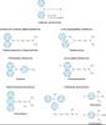
The H1 antagonists are conveniently divided into first-generation and second-generation agents. These groups are distinguished by the relatively strong sedative effects of most of the first-generation drugs. The first-generation agents are also more likely to block autonomic receptors. The relatively less sedating characteristic of the second-generation H1 blockers is due in part to their less complete distribution into the central nervous system. All of the H1 antagonists are stable amines with the general structure illustrated in Figure 16-1. Several chemical subgroups, and the structures of compounds representing different subgroups are shown in the figure. Doses of some of these drugs are given in Table 16-2.
These agents are rapidly absorbed following oral administration, with peak blood concentrations occurring in 1-2 hours. They are widely distributed throughout the body, and the first-generation drugs enter the central nervous system readily. Some of them are extensively metabolized, primarily by microsomal systems in the liver. Several of the second-generation agents are metabolized by the CYP3A4 system and thus are subject to important interactions when other drugs (such as ketoconazole) inhibit this subtype of P450 enzymes. Most of the drugs have an effective duration of action of 4-6 hours following a single dose, but meclizine and several second-generation agents are longer-acting, with a duration of action of 12-24 hours. The newer agents are considerably less lipid-soluble than the first-generation drugs and are substrates of the P-glycoprotein transporter in the blood-brain barrier; as a result they enter the central nervous system with difficulty or not at all. Many H1 antagonists have active metabolites. The active metabolites of hydroxyzine, terfenadine, and loratadine are available as drugs (cetirizine, fexofenadine, and desloratadine, respectively).
|
|
Figure 16-1. General structure of H1 antagonist drugs and examples of the major subgroups. |
Pharmacodynamics
A. HISTAMINE-RECEPTOR BLOCKADE
H1-receptor antagonists block the actions of histamine by reversible competitive antagonism at the H1 receptor. They have negligible potency at the H2 receptor and little at the H3receptor. For example, histamine-induced contraction of bronchiolar or gastrointestinal smooth muscle can be completely blocked by these agents, but the effects on gastric acid secretion and the heart are unmodified.
B. ACTIONS NOT CAUSED BY HISTAMINE RECEPTOR BLOCKADE
The first-generation H1-receptor antagonists have many actions not ascribable to blockade of the actions of histamine. The large number of these actions probably results from the similarity of the general structure (Figure 16-1) to the structure of drugs that have effects at muscarinic cholinoceptor, a adrenoceptor, serotonin, and local anesthetic receptor sites. Some of these actions are of therapeutic value and some are undesirable.
1. Sedation¾ A common effect of first-generation H1 antagonists is sedation, but the intensity of this effect varies among chemical subgroups (Table 16-2) and among patients as well. The effect is sufficiently prominent with some agents to make them useful as "sleep aids" (see Chapter 64) and unsuitable for daytime use. The effect resembles that of some antimuscarinic drugs and is considered very unlike the disinhibited sedation produced by sedative-hypnotic drugs. Compulsive use has not been reported. At ordinary dosages, children occasionally (and adults rarely) manifest excitation rather than sedation. At very high toxic dose levels, marked stimulation, agitation, and even convulsions may precede coma. Second-generation H1 antagonists have little or no sedative or stimulant actions. These drugs (or their active metabolites) also have far fewer autonomic effects than the first-generation antihistamines.
2. Antinausea and antiemetic actions¾ Several first-generation H1 antagonists have significant activity in preventing motion sickness (Table 16-2). They are less effective against an episode of motion sickness already present. Certain H1 antagonists, notably doxylamine (in Bendectin), were used widely in the past in the treatment of nausea and vomiting of pregnancy (see below).
3. Antiparkinsonism effects¾ Some of the H1 antagonists, especially diphenhydramine, have significant acute suppressant effects on the extrapyramidal symptoms associated with certain antipsychotic drugs. This drug is given parenterally for acute dystonic reactions to antipsychotics.
4. Anticholinoceptor actions¾ Many of the first-generation agents, especially those of the ethanolamine and ethylenediamine subgroups, have significant atropine-like effects on peripheral muscarinic receptors. This action may be responsible for some of the (uncertain) benefits reported for nonallergic rhinorrhea but may also cause urinary retention and blurred vision.
5. Adrenoceptor-blocking actions¾ Alpha-receptor-blocking effects can be demonstrated for many H1 antagonists, especially those in the phenothiazine subgroup, eg, promethazine. This action may cause orthostatic hypotension in susceptible individuals. Beta-receptor blockade is not observed.
6. Serotonin-blocking action¾ Strong blocking effects at serotonin receptors have been demonstrated for some first-generation H1 antagonists, notably cyproheptadine. This drug is promoted as an antiserotonin agent and is discussed with that drug group. Nevertheless, its structure resembles that of the phenothiazine antihistamines, and it is a potent H1-blocking agent.
7. Local anesthesia¾ Several first-generation H1 antagonists are potent local anesthetics. They block sodium channels in excitable membranes in the same fashion as procaine and lidocaine. Diphenhydramine and promethazine are actually more potent than procaine as local anesthetics. They are occasionally used to produce local anesthesia in patients allergic to conventional local anesthetic drugs. A small number of these agents also block potassium channels; this action is discussed below (see Toxicity).
8. Other actions¾ Certain H1 antagonists, eg, cetirizine, inhibit mast cell release of histamine and some other mediators of inflammation. This action is not due to H1-receptor blockade. The mechanism is not understood but could play a role in the beneficial effects of these drugs in the treatment of allergies such as rhinitis. A few H1 antagonists (eg, terfenadine, acrivastine) have been shown to inhibit the P-glycoprotein transporter found in cancer cells, the epithelium of the gut, and the capillaries of the brain. The significance of this effect is not known.
CLINICAL PHARMACOLOGY OF H1-RECEPTOR ANTAGONISTS
Clinical Uses
A. ALLERGIC REACTIONS
The H1 antihistaminic agents are often the first drugs used to prevent or treat the symptoms of allergic reactions. In allergic rhinitis and urticaria, in which histamine is the primary mediator, the H1 antagonists are the drugs of choice and are often quite effective. However, in bronchial asthma, which involves several mediators, the H1 antagonists are largely ineffective.
Angioedema may be precipitated by histamine release but appears to be maintained by peptide kinins that are not affected by antihistaminic agents. For atopic dermatitis, antihistaminic drugs such as diphenhydramine are used mostly for their sedative side effect, which reduces awareness of itching.
The H1 antihistamines used for treating allergic conditions such as hay fever are usually selected with the goal of minimizing sedative effects; in the USA, the drugs in widest use are the alkylamines and the second-generation nonsedating agents. However, the sedative effect and the therapeutic efficacy of different agents vary widely among individuals. In addition, the clinical effectiveness of one group may diminish with continued use, and switching to another group may restore drug effectiveness for as yet unexplained reasons.
The second-generation H1 antagonists are used mainly for the treatment of allergic rhinitis and chronic urticaria. Several double-blind comparisons with older agents (such as chlorpheniramine) indicated about equal therapeutic efficacy. However, sedation and interference with safe operation of machinery, which occur in about 50% of subjects taking first-generation antihistamines, occurred in only about 7% of subjects taking second-generation agents. The newer drugs are much more expensive.
B. MOTION SICKNESS AND VESTIBULAR DISTURBANCES
Scopolamine (Chapter 8) and certain first-generation H1 antagonists are the most effective agents available for the prevention of motion sickness. The antihistaminic drugs with the greatest effectiveness in this application are diphenhydramine and promethazine. Dimenhydrinate, which is promoted almost exclusively for the treatment of motion sickness, is a salt of diphenhydramine. The piperazines (cyclizine and meclizine) also have significant activity in preventing motion sickness and are less sedating than diphenhydramine in most patients. Dosage is the same as that recommended for allergic disorders (Table 16-2). Both scopolamine and the H1 antagonists are more effective in preventing motion sickness when combined with ephedrine or amphetamine.
It has been claimed that the antihistaminic agents effective in prophylaxis of motion sickness are also useful in Meniere's syndrome, but efficacy in the latter application is not established.
C. NAUSEA AND VOMITING OF PREGNANCY
Several H1-antagonist drugs have been studied for possible use in treating "morning sickness." The piperazine derivatives were withdrawn from such use when it was demonstrated that they have teratogenic effects in rodents. Doxylamine, an ethanolamine H1 antagonist, was promoted for this application as a component of Bendectin, a prescription medication that also contained pyridoxine. Possible teratogenic effects of doxylamine were widely publicized in the lay press after 1978 as a result of a few case reports of fetal malformation associated with maternal ingestion of Bendectin. However, several large prospective studies involving over 60,000 pregnancies, of which more than 3000 involved maternal Bendectin ingestion, disclosed no increase in the incidence of birth defects. However, because of the continuing controversy, adverse publicity, and lawsuits, the manufacturer of Bendectin withdrew the product from the market.
Toxicity
The wide spectrum of nonantihistaminic effects of the H1 antihistamines is described in the previous text. Several of these effects (sedation, antimuscarinic action) have been used for therapeutic purposes, especially in over-the-counter (OTC) remedies (see Chapter 64). Nevertheless, these two effects constitute the most common undesirable actions when these drugs are used to block histamine receptors.
Less common toxic effects of systemic use include excitation and convulsions in children, postural hypotension, and allergic responses. Drug allergy is relatively common after topical use of H1 antagonists. The effects of severe systemic overdosage of the older agents resemble those of atropine overdosage and are treated in the same way (see Chapters 8 and 59). Overdosage of astemizole or terfenadine may induce cardiac arrhythmias, but these drugs are no longer marketed in the USA; the same effect may be caused at normal dosage by interaction with enzyme inhibitors (see Drug Interactions, below).
Drug Interactions
Lethal ventricular arrhythmias occurred in several patients taking either of the early second-generation agents, terfenadine or astemizole, in combination with ketoconazole, itraconazole, or macrolide antibiotics such as erythromycin. These antimicrobial drugs inhibit the metabolism of many drugs by CYP3A4 and cause significant increases in blood concentrations of the antihistamines. The mechanism of this toxicity involves blockade of the HERG (IKr) potassium channels in the heart that are responsible for repolarization of the action potential (see Chapter 14). The result is prolongation of the action potential, and excessive prolongation leads to arrhythmias. Both terfenadine and astemizole were withdrawn from the United States market in recognition of these problems. Where still available, terfenadine and astemizole should be considered to be contraindicated in patients taking ketoconazole, itraconazole, or macrolides and in patients with liver disease. Grapefruit juice also inhibits CYP3A4 and has been shown to increase terfenadine's blood levels significantly.
For those H1 antagonists that cause significant sedation, concurrent use of other drugs that cause central nervous system depression produces additive effects and is contraindicated while driving or operating machinery. Similarly, the autonomic blocking effects of older antihistamines are additive with those of muscarinic and a-blocking drugs.
III. H2-RECEPTOR ANTAGONISTS
The development of H2-receptor antagonists was based on the observation that H1 antagonists had no effect on histamine-induced acid secretion in the stomach. Molecular manipulation of the histamine molecule resulted in drugs that blocked acid secretion and had no H1-agonist or antagonist effects. The high incidence of peptic ulcer disease and related gastrointestinal complaints created great interest in the therapeutic potential of these H2-receptor antagonists. Because of the ability of this class of drugs to reduce gastric acid secretion and their low toxicity, they are now among the most frequently used drugs in the USA and have become OTC items. These drugs are discussed in Chapter 63.
IV. H3- & H4-RECEPTOR ANTAGONISTS
Although no selective H3 or H4 ligands are presently available for clinical use, there is great interest in their therapeutic potential. H3-selective ligands may be of value in sleep disorders, obesity, and cognitive and psychiatric disorders. H4 blockers have potential in chronic inflammatory conditions such as asthma, in which eosinophils and mast cells play a prominent role.
V. SEROTONIN (5-HYDROXYTRYPTAMINE)
INTRODUCTION
Before the identification of 5-hydroxytryptamine (5-HT), it was known that when blood is allowed to clot, a vasoconstrictor (tonic) substance is released from the clot into the serum; this substance was called serotonin. Independent studies established the existence of a smooth muscle stimulant in intestinal mucosa; this was called enteramine. The synthesis of 5-hydroxytryptamine in 1951 permitted the identification of serotonin and enteramine as the same metabolite of 5-hydroxytryptophan.
Serotonin is an important neurotransmitter, a local hormone in the gut, a component of the platelet clotting process, and is thought to play a role in migraine headache. Serotonin is also one of the mediators of the signs and symptoms of carcinoid syndrome, an unusual manifestation of carcinoid tumor, a neoplasm of enterochromaffin cells. In patients whose tumor is not operable, a serotonin antagonist may constitute a useful treatment.
BASIC PHARMACOLOGY OF SEROTONIN
Chemistry & Pharmacokinetics
Like histamine, serotonin is widely distributed in nature, being found in plant and animal tissues, venoms, and stings. It is an indoleethylamine formed in biologic systems from the amino acid L-tryptophan by hydroxylation of the indole ring followed by decarboxylation of the amino acid (Figure 16-2). Hydroxylation at C5 is the rate-limiting step and can be blocked by p-chlorophenylalanine (PCPA; fenclonine) and by p-chloroamphetamine. These agents have been used experimentally to reduce serotonin synthesis in carcinoid syndrome but are too toxic for clinical use.
After synthesis, the free amine is stored or is rapidly inactivated, usually by oxidation catalyzed by the enzyme monoamine oxidase. In the pineal gland, serotonin serves as a precursor of melatonin, a melanocyte-stimulating hormone. In mammals (including humans), over 90% of the serotonin in the body is found in enterochromaffin cells in the gastrointestinal tract. In the blood, serotonin is found in platelets, which are able to concentrate the amine by means of an active serotonin transporter mechanism (SERT) similar to that in the membrane of serotonergic nerve endings. Once transported into the platelet or nerve ending, 5-HT is concentrated in vesicles by a vesicle-associated transporter (VAT) that is blocked by reserpine. Serotonin is also found in the raphe nuclei of the brainstem, which contain cell bodies of serotonergic neurons that synthesize, store, and release serotonin as a transmitter. Brain serotonergic neurons are involved in numerous diffuse functions such as mood, sleep, appetite, and temperature regulation, as well as the perception of pain, the regulation of blood pressure, and vomiting (see Chapter 21). Serotonin also appears to be involved in clinical conditions such as depression, anxiety, and migraine. Serotonergic neurons are also found in the enteric nervous system of the gastrointestinal tract and around blood vessels. In rodents (but not in humans), serotonin is found in mast cells.
The function of serotonin in enterochromaffin cells is not fully understood. These cells synthesize serotonin, store the amine in a complex with ATP and with other substances in granules, and release serotonin in response to mechanical and neuronal stimuli. Some of the released serotonin diffuses into blood vessels and is taken up and stored in platelets.
Stored serotonin can be depleted by reserpine in much the same manner as this drug depletes catecholamines from vesicles in adrenergic nerves (see Chapter 6).
Serotonin is metabolized by monoamine oxidase, and the intermediate product, 5-hydroxyindoleacetaldehyde, is further oxidized by aldehyde dehydrogenase to 5-hydroxyindoleacetic acid (5-HIAA). In humans consuming a normal diet, the excretion of 5-HIAA is a measure of serotonin synthesis. Therefore, the 24-hour excretion of 5-HIAA can be used as a diagnostic test for tumors that synthesize excessive quantities of serotonin, especially carcinoid tumor. A few foods (eg, bananas) contain large amounts of serotonin or its precursors and must be prohibited during such diagnostic tests.
|
|
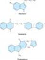
Figure 16-2. Structures of serotonin and two 5-HT-receptor blockers. |
Pharmacodynamics
A. MECHANISMS OF ACTION
Serotonin exerts many actions and, like histamine, has many species differences, making generalizations difficult. The actions of serotonin are mediated through a remarkably large number of cell membrane receptors. The serotonin receptors that have been characterized thus far are listed in Table 16-3. Seven families of 5-HT-receptor subtypes (those given numeric subscripts 1 through 7) have been identified, six involving G protein-coupled receptors of the usual 7-transmembrane serpentine type and one a ligand-gated ion channel. Among these receptor subtypes, a few still lack a well-documented physiologic function. Discovery of these functions awaits the development of more subtype-selective drugs or the knockout mutation of genes encoding these receptors from the mouse genome.
B. TISSUE AND ORGAN SYSTEM EFFECTS
1. Nervous system¾ Serotonin is present in a variety of sites in the brain. Its role as a neurotransmitter and its relation to the actions of drugs acting in the central nervous system are discussed in Chapters 21 and 30. Serotonin is also a precursor of melatonin in the pineal gland (see Box: Melatonin Pharmacology, and Chapter 65).
5-HT3 receptors in the gastrointestinal tract and in the vomiting center of the medulla participate in the vomiting reflex (see Chapter 63). They are particularly important in vomiting caused by chemical triggers such as cancer chemotherapy drugs. 5-HT1P and 5-HT4 receptors also play a role in enteric nervous system function.
Like histamine, serotonin is a potent stimulant of pain and itch sensory nerve endings and is responsible for some of the symptoms caused by insect and plant stings. In addition, serotonin is a powerful activator of chemosensitive endings located in the coronary vascular bed. Activation of 5-HT3 receptors on these afferent vagal nerve endings is associated with the chemoreceptor reflex (also known as the Bezold-Jarisch reflex). The reflex response consists of marked bradycardia and hypotension. The bradycardia is mediated by vagal outflow to the heart and can be blocked by atropine. The hypotension is a consequence of the decrease in cardiac output that results from bradycardia. A variety of other agents can activate the chemoreceptor reflex. These include nicotinic cholinoceptor agonists and some cardiac glycosides, eg, ouabain.
MELATONIN PHARMACOLOGY
Melatonin is N-acetyl-5-methoxytryptamine, a simple methoxylated and N-acetylated product of serotonin found in the pineal gland. It is produced and released primarily at night and has long been suspected of playing a role in diurnal cycles of animals and the sleep-wake behavior of humans.
Melatonin receptors have been characterized in the central nervous system and several peripheral tissues. In the brain, MT1 and MT2 receptors are found in membranes of neurons in the suprachiasmatic nucleus of the hypothalamus, an area associated¾from lesioning experiments¾with circadian rhythm. MT1 and MT2 are seven-transmembrane Gi protein-coupled receptors. The result of receptor binding is inhibition of adenylyl cyclase. A third receptor, MT3, is an enzyme; binding to this site has a poorly defined physiologic role, possibly related to intraocular pressure. Activation of the MT1 receptor results in sleepiness, whereas the MT2 receptor may be related to the light-dark synchronization of the biologic circadian clock.
Melatonin itself is promoted commercially as a sleep aid by the food supplement industry (see Chapter 65). Ramelteon is a selective MT1 and MT2 agonist that has recently been approved for the medical treatment of insomnia. This drug has no addiction liability (it is not a controlled substance), and it appears to be distinctly more efficacious than melatonin (but less efficacious than benzodiazepines) as a hypnotic. It is metabolized by P450 enzymes and should not be used in individuals taking CYP1A2 inhibitors. It has a half-life of 1-3 hours and an active metabolite with a half-life of up to 5 hours. The toxicity of ramelteon is as yet poorly defined, but prolactin levels were elevated in one clinical trial.
2. Respiratory system¾ Serotonin has a small direct stimulant effect on bronchiolar smooth muscle in normal humans. It also appears to facilitate acetylcholine release from bronchial vagal nerve endings. In patients with carcinoid syndrome, episodes of bronchoconstriction occur in response to elevated levels of the amine or peptides released from the tumor. Serotonin may also cause hyperventilation as a result of the chemoreceptor reflex or stimulation of bronchial sensory nerve endings.
3. Cardiovascular system¾ Serotonin directly causes the contraction of vascular smooth muscle, mainly through 5-HT2 receptors. In humans, serotonin is a powerful vasoconstrictor except in skeletal muscle and heart, where it dilates blood vessels. At least part of this 5-HT-induced vasodilation requires the presence of vascular endothelial cells. When the endothelium is damaged, coronary vessels constrict. As noted previously, serotonin can also elicit reflex bradycardia by activation of 5-HT3 receptors on chemoreceptor nerve endings. A triphasic blood pressure response is often seen following injection of serotonin in experimental animals. Initially, there is a decrease in heart rate, cardiac output, and blood pressure caused by the chemoreceptor response. Following this decrease, blood pressure increases as a result of vasoconstriction. The third phase is again a decrease in blood pressure attributed to vasodilation in vessels supplying skeletal muscle. Pulmonary and renal vessels seem especially sensitive to the vasoconstrictor action of serotonin.
Serotonin also constricts veins, and venoconstriction with a resulting increased capillary filling appears to be responsible for the flush that is observed following serotonin administration or release from a carcinoid tumor. Serotonin has small direct positive chronotropic and inotropic effects on the heart, which are probably of no clinical significance. However, prolonged elevation of the blood level of serotonin (which occurs in carcinoid syndrome) is associated with pathologic alterations in the endocardium (subendocardial fibroplasia), which may result in valvular or electrical malfunction.
Serotonin causes blood platelets to aggregate by activating surface 5-HT2 receptors. This response, in contrast to aggregation induced during clot formation, is not accompanied by the release of serotonin stored in the platelets. The physiologic role of this effect is unclear.
4. Gastrointestinal tract¾ Serotonin is a powerful stimulant of gastrointestinal smooth muscle, increasing tone and facilitating peristalsis. This action is caused by the direct action of serotonin on 5-HT2 smooth muscle receptors plus a stimulating action on ganglion cells located in the enteric nervous system (see Chapter 6). Activation of 5-HT4 receptors in the enteric nervous system causes increased acetylcholine release and thereby mediates a motility-enhancing or "prokinetic" effect of selective serotonin agonists such as cisapride. These agents are useful in several gastrointestinal disorders (see Chapter 63). Overproduction of serotonin (and other substances) in carcinoid tumor is associated with severe diarrhea. Serotonin has little effect on secretions, and what effects it has are generally inhibitory.
5. Skeletal muscle¾ 5-HT2 receptors are present on skeletal muscle membranes, but their physiologic role is not understood.Serotonin syndrome is a condition precipitated when MAO inhibitors are given with serotonin agonists, especially antidepressants of the selective serotonin reuptake inhibitor class (SSRIs; see Chapter 30). Although associated with skeletal muscle contractions and the hyperthermia resulting from excessive muscle contraction, serotonin syndrome is probably the result of a central nervous system effect of these drugs (see Table 16-4 and Box: Serotonin Syndrome and Similar Syndromes).
SEROTONIN SYNDROME AND SIMILAR SYNDROMES
Excess synaptic serotonin causes a serious, potentially fatal syndrome that is diagnosed on the basis of a history of administration of a serotonergic drug within recent weeks and physical findings (Table 16-4). It has some characteristics in common with neuroleptic malignant syndrome (NMS) and malignant hyperthermia (MH), but its pathophysiology and management are quite different.
As suggested by the drugs that precipitate it, serotonin syndrome occurs when overdose with a single drug, or concurrent use of several drugs, results in excess serotonergic activity in the central nervous system. It is predictable and not idiosyncratic, but milder forms may easily be misdiagnosed. In experimental animal models, many of the signs of the syndrome can be reversed by administration of 5-HT2 antagonists; however, other 5-HT receptors may be involved as well. Dantrolene is of no value, unlike the treatment of MH. NMS is idiosyncratic rather than predictable and appears to be associated with hypersensitivity to the parkinsonism-inducing effects of D2-blocking antipsychotics in certain individuals. MH is associated with a genetic defect in the RyR1 calcium channel of skeletal muscle sarcoplasmic reticulum that permits uncontrolled calcium release from the SR when precipitating drugs are given (Chapter 27).
CLINICAL PHARMACOLOGY OF SEROTONIN
Serotonin Agonists
Serotonin has no clinical applications as a drug. However, several receptor subtype-selective agonists have proved to be of value. Buspirone, a 5-HT1A agonist, has received wide attention for its usefulness as an effective nonbenzodiazepine anxiolytic (see Chapter 22). Dexfenfluramine, another selective 5-HT agonist, was widely used as an appetite suppressant but was withdrawn because of toxicity. Appetite suppression appears to be associated with agonist action at 5-HT2C receptors in the central nervous system. Sumatriptan and its congeners are agonists effective in the treatment of acute migraine and cluster headache attacks (see below).
5-HT1D/1B Agonists & Migraine Headache
The 5-HT1D/1B agonists (triptans) are used almost exclusively in migraine headache. Migraine in its "classic" form is characterized by an aura of variable duration that may involve nausea, vomiting, and visual scotomas or even hemianopsia and speech abnormalities; followed by a severe throbbing unilateral headache that lasts for a few hours to 1-2 days. "Common" migraine lacks the aura phase, but the headache is similar. Although the symptom pattern varies among patients, the severity of migraine headache justifies vigorous therapy in the great majority of cases.
Migraine involves the trigeminal nerve distribution to intracranial (and possibly extracranial) arteries. These nerves release peptide neurotransmitters, especially calcitonin gene-related peptide (CGRP; see Chapter 17), an extremely powerful vasodilator. Substance P and neurokinin A may also be involved. Extravasation of plasma and plasma proteins into the perivascular space appears to be a common feature of animal migraine models and biopsy specimens from migraine patients and probably represents the effect of the neuropeptides on the vessels. The mechanical stretching caused by this perivascular edema may be the immediate cause of activation of pain nerve endings in the dura. The onset of headache is sometimes associated with a marked increase in amplitude of temporal artery pulsations, and relief of pain by administration of effective therapy is sometimes accompanied by diminution of the arterial pulsations.
The mechanisms of action of drugs used in migraine are poorly understood, in part because they include such a wide variety of drug groups and actions. In addition to the triptans, these include ergot alkaloids, nonsteroidal anti-inflammatory analgesic agents, b-adrenoceptor blockers, calcium channel blockers, tricyclic antidepressants and SSRIs, and several antiseizure agents. Furthermore, some of these drug groups are effective only for prophylaxis and not for the acute attack.
Two primary hypotheses have been proposed to explain the actions of these drugs. First, the triptans, the ergot alkaloids, and antidepressants may activate 5-HT1D/1B receptors on presynaptic trigeminal nerve endings to inhibit the release of vasodilating peptides, and antiseizure agents may suppress excessive firing of these nerve endings. Second, the vasoconstrictor actions of direct 5-HT agonists (the triptans and ergot) may prevent vasodilation and stretching of the pain endings. It is possible that both mechanisms contribute in the case of some drugs. Sumatriptan and its congeners are currently first-line therapy for acute severe migraine attacks in most patients. However, they should not be used in patients at risk for coronary artery disease. Anti-inflammatory analgesics such as aspirin and ibuprofen are often helpful in controlling the pain of migraine. Rarely, parenteral opioids may be needed in refractory cases. For patients with very severe nausea and vomiting, parenteral metoclopramide may be helpful.
Propranolol, amitriptyline, and some calcium channel blockers have been found to be effective for the prophylaxis of migraine in some patients. They are of no value in the treatment of acute migraine. The anticonvulsants valproic acid and topiramate (see Chapter 24) have recently been found to have good prophylactic efficacy in many migraine patients. Flunarizine, a calcium channel blocker used in Europe, has been reported in clinical trials to effectively reduce the severity of the acute attack and to prevent recurrences. Verapamil appears to have modest efficacy as prophylaxis against migraine.
Sumatriptan and the other triptans are selective agonists for 5-HT1D and 5-HT1B receptors; the similarity of the triptan structure to that of 5-HT can be seen in the structure below. These receptor types are found in cerebral and meningeal vessels and mediate vasoconstriction. They are also found on neurons and probably function as presynaptic inhibitory receptors.
The efficacy of triptan 5-HT1 agonists in migraine is equal to or greater than that of other acute drug treatments, eg, parenteral, oral, or rectal ergot alkaloids. The pharmacokinetics of the triptans are set forth in Table 16-5. Most adverse effects are mild and include altered sensations (tingling, warmth, etc), dizziness, muscle weakness, neck pain, and for parenteral sumatriptan, injection site reactions. Chest discomfort occurs in 1-5% of patients, and chest pain has been reported, probably because of the ability of these drugs to cause coronary vasospasm. They are therefore contraindicated in patients with coronary artery disease and in patients with angina. Another disadvantage is the fact that their duration of effect (especially that of almotriptan, sumatriptan, rizatriptan, and zolmitriptan, Table 16-5) is often shorter than the duration of the headache. As a result, several doses may be required during a prolonged migraine attack, but their adverse effects limit the maximum safe daily dosage. In addition, these drugs are extremely expensive. Naratriptan and eletriptan are contraindicated in patients with severe hepatic or renal impairment or peripheral vascular syndromes; frovatriptan in patients with peripheral vascular disease; and zolmitriptan in patients with Wolff-Parkinson-White syndrome.
Other Serotonin Agonists in Clinical Use
Cisapride, a 5-HT4 agonist, was used in the treatment of gastroesophageal reflux and motility disorders. Because of toxicity, it is now available only for compassionate use in the USA. Tegaserod, a newer 5-HT4 partial agonist, is used for irritable bowel syndrome with constipation. These drugs are discussed in Chapter 63.
Compounds such as fluoxetine and other SSRIs, which modulate serotonergic transmission by blocking reuptake of the transmitter, are among the most widely prescribed drugs for the management of depression and similar disorders. These drugs are discussed in Chapter 30.
SEROTONIN ANTAGONISTS
The actions of serotonin, like those of histamine, can be antagonized in several ways. Such antagonism is clearly desirable in those rare patients who have carcinoid tumor and may also be valuable in certain other conditions.
As noted above, serotonin synthesis can be inhibited by p-chlorophenylalanine and p-chloroamphetamine. However, these agents are too toxic for general use. Storage of serotonin can be inhibited by the use of reserpine, but the sympatholytic effects of this drug (see Chapter 11) and the high levels of circulating serotonin that result from release prevent its use in carcinoid. Therefore, receptor blockade is the major approach to therapeutic limitation of serotonin effects.
SEROTONIN-RECEPTOR ANTAGONISTS
A wide variety of drugs with actions at other receptors (a adrenoceptors, H1-histamine receptors, etc) are also serotonin receptor-blocking agents. Phenoxybenzamine (see Chapter 10) has a long-lasting blocking action at 5-HT2 receptors. In addition, the ergot alkaloids discussed in the last portion of the chapter are partial agonists at serotonin receptors.
Cyproheptadine resembles the phenothiazine antihistaminic agents in chemical structure and has potent H1-receptor-blocking as well as 5-HT2-blocking actions. The actions of cyproheptadine are predictable from its H1 histamine and serotonin receptor affinities. It prevents the smooth muscle effects of both amines but has no effect on the gastric secretion stimulated by histamine. It also has significant antimuscarinic effects and causes sedation.
The major clinical applications of cyproheptadine are in the treatment of the smooth muscle manifestations of carcinoid tumor and in cold-induced urticaria. The usual dosage in adults is 12-16 mg/d in three or four divided doses. It is of some value in serotonin syndrome, but because it is available only in tablet form, it must be crushed and administered by stomach tube in unconscious patients.
Ketanserin (Figure 16-2) blocks 5-HT1c and 5-HT2 receptors and has little or no reported antagonist activity at other 5-HT or H1 receptors. However, this drug potently blocks vascular a1 adrenoceptors. The drug blocks 5-HT2 receptors on platelets and antagonizes platelet aggregation promoted by serotonin. The mechanism involved in ketanserin's hypotensive action is not clear but probably involves a1 adrenoceptors more than 5-HT2 receptor blockade. Ketanserin is available in Europe for the treatment of hypertension and vasospastic conditions but has not been approved in the USA.
Ritanserin, another 5-HT2 antagonist, has little or no a-blocking action. It has been reported to alter bleeding time and to reduce thromboxane formation, presumably by altering platelet function.
Ondansetron (Figure 16-2) is the prototypical 5-HT3 antagonist. This drug and its analogs are very important in the prevention of nausea and vomiting associated with surgery and cancer chemotherapy. They are discussed in Chapter 63.
Considering the diverse effects attributed to serotonin and the heterogeneous nature of 5-HT receptors, other selective 5-HT antagonists may prove to be clinically useful.
VI. THE ERGOT ALKALOIDS
INTRODUCTION
Ergot alkaloids are produced by Claviceps purpurea, a fungus that infects grain¾especially rye¾under damp growing or storage conditions. This fungus synthesizes histamine, acetylcholine, tyramine, and other biologically active products in addition to a score or more of unique ergot alkaloids. These alkaloids affect a adrenoceptors, dopamine receptors, 5-HT receptors, and perhaps other receptor types. Similar alkaloids are produced by fungi parasitic to a number of other grass-like plants.
The accidental ingestion of ergot alkaloids in contaminated grain can be traced back more than 2000 years from descriptions of epidemics of ergot poisoning (ergotism). The most dramatic effects of poisoning are dementia with florid hallucinations; prolonged vasospasm, which may result in gangrene; and stimulation of uterine smooth muscle, which in pregnancy may result in abortion. In medieval times, ergot poisoning was called St. Anthony's fire after the saint whose help was sought in relieving the burning pain of vasospastic ischemia. Identifiable epidemics have occurred sporadically up to modern times (see Box: Ergot Poisoning: Not Just an Ancient Disease) and mandate continuous surveillance of all grains used for food. Poisoning of grazing animals is common in many areas because the fungi may grow on pasture grasses.
In addition to the effects noted above, the ergot alkaloids produce a variety of other central nervous system and peripheral effects. Detailed structure-activity analysis and appropriate semisynthetic modifications have yielded a large number of agents with documented or potential clinical value.
ERGOT POISONING: NOT JUST AN ANCIENT DISEASE
As noted in the text, epidemics of ergotism, or poisoning by ergot-contaminated grain, are known to have occurred sporadically in ancient times and through the Middle Ages. It is easy to imagine the social chaos that might result if fiery pain, gangrene, hallucinations, convulsions, and abortions occurred simultaneously throughout a community in which all or most of the people believed in witchcraft, demonic possession, and the visitation of supernatural punishments upon humans for their misdeeds. Such beliefs are uncommon in most cultures today. However, ergotism has not disappeared. A most convincing demonstration of ergotism occurred in the small French village of Pont-Saint-Esprit in 1951. It was described in the British Medical Journal in 1951 (Gabbai et al, 1951) and in a later book-length narrative account (Fuller, 1968). Several hundred individuals suffered symptoms of hallucinations, convulsions, and ischemia¾and several died¾after eating bread made from contaminated flour. Similar events have occurred even more recently when poverty, famine, or incompetence resulted in the consumption of contaminated grain. Ergot toxicity caused by excessive self-medication with pharmaceutical ergot preparations is still occasionally reported.
BASIC PHARMACOLOGY OF ERGOT ALKALOIDS
Chemistry & Pharmacokinetics
Two major families of compounds that incorporate the tetracyclic ergoline nucleus may be identified; the amine alkaloids and the peptide alkaloids (Table 16-6). Drugs of therapeutic and toxicologic importance are found in both groups.
The ergot alkaloids are variably absorbed from the gastrointestinal tract. The oral dose of ergotamine is about ten times larger than the intramuscular dose, but the speed of absorption and peak blood levels after oral administration can be improved by administration with caffeine (see below). The amine alkaloids are also absorbed from the rectum and the buccal cavity and after administration by aerosol inhaler. Absorption after intramuscular injection is slow but usually reliable. Bromocriptine and the amine derivative cabergoline are well absorbed from the gastrointestinal tract.
The ergot alkaloids are extensively metabolized in the body. The primary metabolites are hydroxylated in the A ring, and peptide alkaloids are also modified in the peptide moiety.
Pharmacodynamics
A. MECHANISM OF ACTION
As suggested, the ergot alkaloids act on several types of receptors. Their effects include agonist, partial agonist, and antagonist actions at a-adrenoceptors and serotonin receptors (especially 5-HT1A and 5-HT1D; less for 5-HT1C, 5-HT2, and 5-HT3); and agonist or partial agonist actions at central nervous system dopamine receptors (Table 16-7). Furthermore, some members of the ergot family have a high affinity for presynaptic receptors, whereas others are more selective for postjunctional receptors. There is a powerful stimulant effect on the uterus that seems to be most closely associated with agonist or partial agonist effects at 5-HT2 receptors. Structural variations increase the selectivity of certain members of the family for specific receptor types.
B. ORGAN SYSTEM EFFECTS
1. Central nervous system¾ As indicated by traditional descriptions of ergotism, certain of the naturally occurring alkaloids are powerful hallucinogens. Lysergic acid diethylamide (LSD; "acid") is a synthetic ergot compound that clearly demonstrates this action. The drug has been used in the laboratory as a potent peripheral 5-HT2 antagonist, but good evidence suggests that its behavioral effects are mediated by agonist effects at prejunctional or postjunctional 5-HT2 receptors in the central nervous system. In spite of extensive research, no clinical value has been discovered for LSD's dramatic central nervous system effects. Abuse of this drug has waxed and waned but is still widespread. It is discussed in Chapter 32.
Dopamine receptors in the central nervous system play important roles in extrapyramidal motor control and the regulation of pituitary prolactin release. The actions of the peptide ergoline bromocriptine on the extrapyramidal system are discussed in Chapter 28. Of all the currently available ergot derivatives, bromocriptine, cabergoline, and pergolide have the highest selectivity for the pituitary dopamine receptors. These drugs directly suppress prolactin secretion from pituitary cells by activating regulatory dopamine receptors (Chapter 37). They compete for binding to these sites with dopamine itself and with other dopamine agonists such as apomorphine.
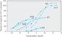
2. Vascular smooth muscle¾ The action of ergot alkaloids on vascular smooth muscle is drug-, species-, and vessel-dependent, so few generalizations are possible. Ergotamine and related compounds potently constrict most human blood vessels in a predictable, and prolonged manner (Figure 16-3). This response is partially blocked by conventional a-blocking agents. However, ergotamine's effect is also associated with "epinephrine reversal" (see Chapter 10) and with blockade of the response to other a agonists. This dual effect represents partial agonist action (Table 16-7). Because ergotamine dissociates very slowly from the a receptor, it produces very long-lasting agonist and antagonist effects at this receptor. There is little or no effect at b adrenoceptors.
While much of the vasoconstriction elicited by ergot alkaloids can be ascribed to partial agonist effects at a adrenoceptors, some may be the result of effects at 5-HT receptors. Ergotamine, ergonovine, and methysergide all have partial agonist effects at 5-HT2 vascular receptors. The remarkably specific antimigraine action of the ergot derivatives was originally thought to be related to their actions on vascular serotonin receptors. Current hypotheses, however, emphasize their action on prejunctional neuronal 5-HT receptors.
After overdosage with ergotamine and similar agents, vasospasm is severe and prolonged (see Toxicity, below). This vasospasm is not easily reversed by a antagonists, serotonin antagonists, or combinations of both.
Ergotamine is typical of the ergot alkaloids that have a strong vasoconstrictor spectrum of action. The hydrogenation of ergot alkaloids at the 9 and 10 positions (Table 16-6) yields dihydro derivatives that have reduced serotonin partial agonist effects and increased selective a receptor-blocking actions.
3. Uterine smooth muscle¾ The stimulant action of ergot alkaloids on the uterus, as on vascular smooth muscle, appears to combine a agonist, serotonin, and other effects. Furthermore, the sensitivity of the uterus to the stimulant effects of ergot increases dramatically during pregnancy, perhaps because of increasing dominance of a1 receptors as pregnancy progresses. As a result, the uterus at term is more sensitive to ergot than earlier in pregnancy and far more sensitive than the nonpregnant organ.
In very small doses, ergot preparations can evoke rhythmic contraction and relaxation of the uterus. At higher concentrations, these drugs induce powerful and prolonged contracture. Ergonovine is more selective than other ergot alkaloids in affecting the uterus and is the agent of choice in obstetric applications of these drugs.
4. Other smooth muscle organs¾ In most patients, the ergot alkaloids have little or no significant effect on bronchiolar or urinary smooth muscle. The gastrointestinal tract, on the other hand, is quite sensitive in most patients. Nausea, vomiting, and diarrhea may be induced even by low doses in some patients. The effect is consistent with action on the central nervous system emetic center and on gastrointestinal serotonin receptors.
|
|
Figure 16-3. Effects of ergot derivatives on contraction of isolated segments of human basilar artery strips removed at surgery. All of the ergot derivatives are partial agonists, and all are more potent than the full agonists, norepinephrine and serotonin. (NE, norepinephrine; 5-HT, serotonin; ERG, ergotamine; MT, methylergometrine; DHE, dihydroergotamine; MS, methysergide.) (Modified and reproduced, with permission, from Muller-Schweinitzer E. In: 5-Hydroxytryptamine Mechanisms in Primary Headaches. Oleson J, Saxena PR [editors]. Raven Press, 1992.) |
CLINICAL PHARMACOLOGY OF ERGOT ALKALOIDS
Clinical Uses
A. MIGRAINE
Ergot derivatives are highly specific for migraine pain; they are not analgesic for any other condition. Although the triptan drugs discussed above are preferred by most clinicians and patients, traditional therapy with ergotamine can also be quite effective when given during the prodrome of an attack; it becomes progressively less effective if delayed. Ergotamine tartrate is available for oral, sublingual, rectal suppository, and inhaler use. It is often combined with caffeine (100 mg caffeine for each 1 mg ergotamine tartrate) to facilitate absorption of the ergot alkaloid.
The vasoconstriction induced by ergotamine is long-lasting and cumulative when the drug is taken repeatedly, as in a severe migraine attack. Therefore, patients must be carefully informed that no more than 6 mg of the oral preparation may be taken for each attack and no more than 10 mg per week. For very severe attacks, ergotamine tartrate, 0.25-0.5 mg, may be given intravenously or intramuscularly. Dihydroergotamine, 0.5-1 mg intravenously, is favored by some clinicians for treatment of intractable migraine. Intranasal dihydroergotamine may also be effective.
B. HYPERPROLACTINEMIA
Increased serum levels of the anterior pituitary hormone prolactin are associated with secreting tumors of the gland and also with the use of centrally acting dopamine antagonists, especially the D2-blocking antipsychotic drugs. Because of negative feedback effects, hyperprolactinemia is associated with amenorrhea and infertility in women as well as galactorrhea in both sexes.
Bromocriptine is extremely effective in reducing the high levels of prolactin that result from pituitary tumors and has even been associated with regression of the tumor in some cases. The usual dosage of bromocriptine is 2.5 mg two or three times daily. Cabergoline is similar but more potent. Bromocriptine has also been used in the same dosage to suppress physiologic lactation. However, serious postpartum cardiovascular toxicity has been reported in association with the latter use of bromocriptine or pergolide, and this application is discouraged (Chapter 37).
C. POSTPARTUM HEMORRHAGE
The uterus at term is extremely sensitive to the stimulant action of ergot, and even moderate doses produce a prolonged and powerful spasm of the muscle quite unlike natural labor. Therefore, ergot derivatives should be used only for control of late uterine bleeding and should never be given before delivery. Oxytocin is the preferred agent for control of postpartum hemorrhage, but if this peptide agent is ineffective, ergonovine maleate, 0.2 mg usually given intramuscularly, can be tried. It is usually effective within 1-5 minutes and is less toxic than other ergot derivatives for this application. It is given at the time of delivery of the placenta or immediately afterward if bleeding is significant.
D. DIAGNOSIS OF VARIANT ANGINA
Ergonovine given intravenously produces prompt vasoconstriction during coronary angiography to diagnose variant angina if reactive segments of the coronaries are present.
E. SENILE CEREBRAL INSUFFICIENCY
Dihydroergotoxine, a mixture of dihydro-a-ergocryptine and three similar dihydrogenated peptide ergot alkaloids (ergoloid mesylates), has been promoted for many years for the relief of senility and more recently for the treatment of Alzheimer's dementia. There is no useful evidence that this drug has significant benefit.
Toxicity & Contraindications
The most common toxic effects of the ergot derivatives are gastrointestinal disturbances, including diarrhea, nausea, and vomiting. Activation of the medullary vomiting center and of the gastrointestinal serotonin receptors is involved. Since migraine attacks are often associated with these symptoms before therapy is begun, these adverse effects are rarely contraindications to the use of ergot.
A more dangerous toxic effect of overdosage with agents like ergotamine and ergonovine is prolonged vasospasm. As described above, this sign of vascular smooth muscle stimulation may result in gangrene and require amputation. Bowel infarction has also been reported and may require resection. Peripheral vascular vasospasm caused by ergot is refractory to most vasodilators, but infusions of large doses of nitroprusside or nitroglycerin have been successful in some cases.
Chronic therapy with methysergide was associated with connective tissue proliferation in the retroperitoneal space, the pleural cavity, and the endocardial tissue of the heart. These changes occurred insidiously over months and presented as hydronephrosis (from obstruction of the ureters) or a cardiac murmur (from distortion of the valves of the heart). In some cases, valve damage required surgical replacement. As a result, this drug was withdrawn. Similar fibrotic change has resulted from the chronic use of 5-HT agonists promoted in the past for weight loss (fenfluramine, desfenfluramine).
Other toxic effects of the ergot alkaloids include drowsiness and, in the case of methysergide, occasional instances of central stimulation and hallucinations. In fact, methysergide was sometimes used as a substitute for LSD by members of the "drug culture."
Contraindications to the use of ergot derivatives consist of the obstructive vascular diseases and collagen diseases.
There is no evidence that ordinary use of ergotamine for migraine is hazardous in pregnancy. However, most clinicians counsel restraint in the use of the ergot derivatives by pregnant patients.
PREPARATIONS AVAILABLE
ANTIHISTAMINES (H1 BLOCKERS)*
Azelastine
Nasal (Astelin): 137 mcg/puff nasal spray
Ophthalmic (Optivar): 0.5 mg/mL solution
Brompheniramine (generic, Brovex)
Oral: 6, 12 mg extended release tablets; 12 mg chewable tablets; 8, 12 mg/5 mL suspension
Buclizine (Bucladin-S Softabs)
Oral: 50 mg tablets
Carbinoxamine (Histex, Pediatex)
Oral: 4 mg tablets; 8 mg timed-release tablets; 10 mg extended release capsules; 1.5, 4 mg/5 mL liquid; 3.6 mg/5 mL oral suspension
Cetirizine (Zyrtec)
Oral: 5, 10 mg tablets; 5, 10 mg chewable tablets; 5 mg/5 mL syrup
Chlorpheniramine (generic, Chlor-Trimeton)
Oral: 2 mg chewable tablets; 4 mg tablets; 2 mg/5 mL syrup
Oral sustained-release: 8, 12, 16 mg tablets; 8, 12 mg capsules
Clemastine (generic, Tavist)
Oral: 1.34, 2.68 mg tablets; 0.67 mg/5 mL syrup
Cyclizine (Marezine)
Oral: 50 mg tablets
Cyproheptadine (generic)
Oral: 4 mg tablets; 2 mg/5 mL syrup
Desloratadine (Clarinex)
Oral: 5 mg regular or rapidly disintegrating tablets; 2.5 mg/5 mL syrup
Dexchlorpheniramine (generic)
Oral: 4, 6 mg extended release tablets; 2 mg/5 mL syrup
Dimenhydrinate (Dramamine, others)
Oral: 50 mg tablets; 50 mg chewable tablets; 12.5/5 mL, 12.5 mg/4 mL, 15.62 mg/5 mL liquid
Parenteral: 50 mg/mL for IM or IV injection
Diphenhydramine (generic, Benadryl)
Oral: 12.5 mg chewable tablets; 25, 50 mg tablets, capsules; 12.5 mg/5 mL elixir and syrup
Parenteral: 50 mg/mL for injection
Emedastine (Emadine)
Ophthalmic: 0.05% solution
Epinastine (Elestat)
Ophthalmic: 0.05% solution
Fexofenadine (Allegra)
Oral: 30, 60, 180 mg tablets; 60 mg capsules
Hydroxyzine (generic, Vistaril)
Oral: 10, 25, 50 mg tablets; 25, 50, 100 mg capsules; 10 mg/5 mL syrup; 25 mg/5 mL suspension
Parenteral: 25, 50 mg/mL for injection
Ketotifen (Zaditor)
Ophthalmic: 0.025% solution
Levocabastine (Livostin)
Ophthalmic: 0.05% solution
Loratadine (generic, Claritin, Tavist)
Oral: 10 mg tablets; 10 mg rapidly disintegrating tablets; 1 mg/mL syrup
Meclizine (generic, Antivert)
Oral: 12.5, 25, 50 mg tablets; 25 mg capsules; 25 mg chewable tablets
Olopatadine (Patanol)
Ophthalmic: 0.1% solution
Phenindamine (Nolahist)
Oral: 25 mg tablets
Promethazine (generic, Phenergan)
Oral: 25, 50 mg tablets; 6.25 mg/5 mL syrups
Parenteral: 25, 50 mg/mL for injection
Rectal: 12.5, 25, 50 mg suppositories
Triprolidine (Zymine)
Oral: 1.25 mg/5 mL liquid
H2 BLOCKERS
See Chapter 63.
5-HT AGONISTS
Almotriptan (Axert)
Oral: 6.25, 12.5 mg tablets
Eletriptan (Relpax)
Oral: 24.2, 48.5 mg tablets (equivalent to 20, 40 mg base)
Frovatriptan (Frova)
Oral: 2.5 mg tablets
Naratriptan (Amerge)
Oral: 1, 2.5 mg tablets
Rizatriptan
Oral: 5, 10 mg tablets (Maxalt); 5, 10 mg orally disintegrating tablets (Maxalt-MLT)
Sumatriptan (Imitrex)
Oral: 25, 50, 100 mg tablets
Nasal: 5, 20 mg unit dose spray devices
Parenteral: 6 mg/0.5 mL in SELFdose autoinjection units for subcutaneous injection
Zolmitriptan (Zomig)
Oral: 2.5, 5 mg tablets
Nasal: 5 mg
5-HT ANTAGONISTS
See Chapter 63.
MELATONIN RECEPTOR AGONISTS
Ramelteon (Rozarem)
Oral: 8 mg tablets
ERGOT ALKALOIDS
Dihydroergotamine
Nasal (Migranal): 4 mg/mL nasal spray
Parenteral (D.H.E. 45): 1 mg/mL for injection
Ergonovine (Ergotrate)
Parenteral: 0.2 mg/mL for injection
Oral: 0.2 mg tablets
Ergotamine mixtures (generic, Cafergot)
Oral: 1 mg ergotamine/100 mg caffeine tablets
Rectal: 2 mg ergotamine/100 mg caffeine suppositories
Ergotamine tartrate (Ergomar)
Sublingual: 2 mg sublingual tablets
Methylergonovine (Methergine)
Oral: 0.2 mg tablets
Parenteral: 0.2 mg/mL for injection
*Several other antihistamines are available only in combination products with, for example, phenylephrine.
Dimenhydrinate is the chlorotheophylline salt of diphenhydramine.
REFERENCES
Histamine
Bakker RA: Histamine H3-receptor isoforms. Inflamm Res 2004;53:509.
Barnes PJ: Histamine and serotonin. Pulm Pharmacol Ther 2001;14:329.
Izzo AA et al: The role of histamine H1, H2, and H3 receptors on enteric ascending synaptic transmission in the guinea pig ileum. J Pharmacol Exp Ther 1998;287:952.
Leurs R et al: Therapeutic potential of histamine H3-receptor agonists and antagonists. Trends Pharmacol Sci 1998;19:177.
Lieberman P: Anaphylaxis. Med Clin North Am 2006;90:77.
Schneider E et al: Trends in histamine research: New functions during immune responses and hematopoiesis. Trends Immunol 2002;23:255.
Shin N et al: Molecular modeling and site-specific mutagenesis of the histamine-binding site of the histamine H4 receptor. Mol Pharmacol 2002;62:38.
Simons FE: Advances in H1-antihistamines. N Engl J Med 2004;351:2203.
Takahashi K et al: Targeted disruption of H3 receptors results in changes in brain histamine tone leading to an obese phenotype. J Clin Invest 2002;110:1791.
Serotonin
Boyer EW, Shannon M: The serotonin syndrome. N Engl J Med 2005;352:1112.
Durham PL, Russo AF: New insights into the molecular actions of serotonergic antimigraine drugs. Pharmacol Ther 2002;94:77.
Egermayer P: Epidemics of vascular toxicity and pulmonary hypertension: What can be learned? J Intern Med 2000;247:11.
Michel K et al: Subpopulations of gastric myenteric neurons are differentially activated via distinct serotonin receptors: Projection, neurochemical coding, and functional implications. J Neurosci 1997;17:8009.
Raymond JR et al: Multiplicity of mechanisms of serotonin receptor signal transduction. Pharmacol Therap 2001;92:179.
Ergot Alkaloids: Historical
Fuller JG: The Day of St. Anthony's Fire. Macmillan, 1968; Signet, 1969.
Gabbai Dr, Lisbonne Dr, Pourquier Dr: Ergot poisoning at Pont St. Esprit. Br Med J 1951;Sept. 15:650.
Ergot Alkaloids: Pharmacology
DeGroot ANJA et al: Ergot alkaloids. Current status and review of clinical pharmacology and therapeutic use compared with other oxytocics in obstetrics and gynaecology. Drugs 1998;56:523.
Dierckx RA et al: Intraarterial sodium nitroprusside infusion in the treatment of severe ergotism. Clin Neuropharmacol 1986;9:542.
Dildy GA: Postpartum hemorrhage: New management options. Clin Obstet Gynecol 2002;45:330.
Lake AE, Saper JR: Chronic headache. New advances in treatment strategies. Neurology 2002;59:S8
Mantegani S, Brambilla E, Varasi M: Ergoline derivatives: receptor affinity and selectivity. Farmaco 1999;54:288.
Porter JK, Thompson FN Jr: Effects of fescue toxicosis on reproduction in livestock. J Animal Sci 1992;70:1594.
Snow V et al: Pharmacologic management of acute attacks of migraine and prevention of migraine headache. Ann Intern Med 2002;137:840.