INTRODUCTION
Asthma is characterized clinically by recurrent bouts of coughing, shortness of breath, chest tightness, and wheezing; physiologically by widespread, reversible narrowing of the bronchial airways and a marked increase in bronchial responsiveness to inhaled stimuli; and pathologically by lymphocytic, eosinophilic inflammation of the bronchial mucosa. It is also characterized pathologically by remodeling of the bronchial mucosa, with deposition of collagen beneath the epithelium's lamina reticularis and hyperplasia of the cells of all structural elements¾vessels, smooth muscle, and secretory glands and goblet cells.
In mild asthma, symptoms occur only occasionally, as on exposure to allergens or certain pollutants, on exercise, or after a viral upper respiratory infection. More severe forms of asthma are associated with frequent attacks of wheezing dyspnea, especially at night, and may be associated with chronic airway narrowing, causing chronic respiratory impairment. These consequences of asthma are regarded as largely preventable, because effective treatments for relief of acute bronchoconstriction ("short term relievers") and for reduction in symptoms and prevention of attacks ("long-term controllers") are available (but underutilized).
The causes of airway narrowing in acute asthmatic attacks include contraction of airway smooth muscle, inspissation of thick, viscid mucus plugs in the airway lumen, and thickening of the bronchial mucosa from edema, cellular infiltration, and hyperplasia of secretory, vascular, and smooth muscle cells. Of these causes of airway obstruction, contraction of smooth muscle is most easily reversed by current therapy; reversal of the edema and cellular infiltration requires sustained treatment with anti-inflammatory agents.
Short-term relief is thus most effectively achieved by agents that relax airway smooth muscle, of which b-adrenoceptor stimulants (see Chapter 9) are the most effective and most widely used. Theophylline, a methylxanthine drug, and antimuscarinic agents (see Chapter 8) are also used for reversal of airway constriction.
Long-term control is most effectively achieved with an anti-inflammatory agent such as an inhaled corticosteroid. It can also be achieved, though less effectively, with a leukotriene pathway antagonist or an inhibitor of mast cell degranulation, such as cromolyn or nedocromil. Finally, clinical trials have established the efficacy of treatment for asthma with a humanized monoclonal antibody, omalizumab, which is specifically targeted against IgE, the antibody responsible for allergic sensitization.
The distinction between "short-term relievers" and "long-term controllers" has become blurred. Theophylline, regarded as a bronchodilator, inhibits some lymphocyte functions and modestly reduces airway mucosal inflammation. Inhaled corticosteroids, regarded as long-term controllers, produce modest immediate bronchodilation. Two recently released long-acting b-adrenoceptor stimulants, salmeterol and formoterol, appear to be effective in improving asthma control when added to inhaled corticosteroid treatment.
This chapter presents the basic pharmacology of the methylxanthines, cromolyn, leukotriene pathway inhibitors, and monoclonal anti-IgE antibody¾agents whose medical use is almost exclusively for pulmonary disease. The other classes of drugs listed above are discussed in relation to the therapy of asthma.
PATHOGENESIS OF ASTHMA
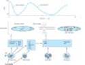
The classic immunologic model of asthma presents it as a disease mediated by reaginic immune globulin (IgE). Foreign materials that provoke IgE production are described as "allergens"; the most common are proteins from house dust mite, cockroach, cat dander, molds, and pollens. The tendency to produce IgE antibodies is genetically determined; asthma and other allergic diseases cluster in families. Once produced, IgE antibodies bind to mast cells in the airway mucosa (Figure 20-1). On reexposure to a specific allergen, antigen-antibody interaction on the surface of the mast cells triggers both the release of mediators stored in the cells' granules and the synthesis and release of other mediators. The histamine, tryptase, leukotrienes C4 and D4, and prostaglandin D2, when released, diffuse through the airway mucosa triggering the muscle contraction and vascular leakage responsible for the acute bronchoconstriction of the "early asthmatic response." This response is often followed in 4-6 hours by a second, more sustained phase of bronchoconstriction, the "late asthmatic response," which is associated with an influx of inflammatory cells into the bronchial mucosa and with an increase in bronchial responsiveness that may last for several weeks after a single inhalation of allergen. The mediators responsible for this late response are thought to be cytokines characteristically produced by TH2 lymphocytes, especially interleukins 5, 9, and 13. The cytokines are thought to attract and activate eosinophils, stimulate IgE production by B lymphocytes, and directly stimulate mucus production by bronchial epithelial cells. It is not clear whether lymphocytes or mast cells in the airway mucosa are the primary source of the mediators responsible for the late inflammatory response, but the benefits of corticosteroid therapy are attributed to their inhibition of cytokine production in the airways.
The allergen challenge model does not reproduce all the features of asthma. Most asthma attacks are not triggered by inhalation of allergens. They are triggered by viral respiratory infection. Some adults with asthma have no evidence of allergic sensitivity to allergens, and even in people with allergic sensitivity, the severity of symptoms correlates poorly with levels of allergen in the atmosphere. Moreover, bronchospasm can be provoked by nonallergenic stimuli such as distilled water, exercise, cold air, sulfur dioxide, and rapid respiratory maneuvers.
This tendency to develop bronchospasm on encountering stimuli that do not affect healthy nonasthmatic airways is characteristic of asthma and is sometimes called "nonspecific bronchial hyperreactivity" to distinguish it from bronchial responsiveness to specific antigens. Bronchial reactivity is assessed by measuring the fall in forced expiratory volume in 1 second (FEV1) provoked by inhaling serially increasing concentrations of aerosolized methacholine. The exaggerated reactivity of the airways appears to be fundamental to asthma's pathogenesis, because it is nearly ubiquitous in patients with asthma and its degree correlates with the clinical severity of the disease.
The mechanisms underlying bronchial hyperreactivity are somehow related to inflammation of the airway mucosa. The agents that increase bronchial reactivity, such as ozone exposure, allergen inhalation, and infection with respiratory viruses, also cause airway inflammation. The increase in reactivity due to allergen inhalation is associated with an increase in both eosinophils and polymorphonuclear leukocytes in bronchial lavage fluid. The increase in reactivity that is associated with the late asthmatic response to allergen inhalation (Figure 20-1) is sustained and, because it is prevented by treatment with an inhaled corticosteroid, is thought to be caused by airway inflammation.
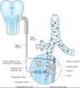
Whatever the mechanisms responsible for bronchial hyperreactivity, bronchoconstriction itself seems to result not simply from the direct effect of the released mediators but also from their activation of neural or humoral pathways. Evidence for the importance of neural pathways stems largely from studies of laboratory animals. The bronchospasm provoked in dogs by inhalation of histamine is reduced by pretreatment with an inhaled topical anesthetic agent, by transection of the vagus nerves, and by pretreatment with atropine. Studies of asthmatic humans, however, have shown that treatment with atropine causes only a reduction in¾not abolition of¾the bronchospastic responses to antigens and to nonantigenic stimuli. It is possible that activity in another neural pathway, such as the nonadrenergic, noncholinergic system, contributes to bronchomotor responses stimuli (Figure 20-2).
The hypothesis suggested by these studies¾that asthmatic bronchospasm results from a combination of release of mediators and an exaggeration of responsiveness to their effects¾predicts that asthma may be effectively treated by drugs with different modes of action. Asthmatic bronchospasm might be reversed or prevented, for example, by drugs that reduce the amount of IgE bound to mast cells (anti-IgE antibody), prevent mast cell degranulation (cromolyn or nedocromil, sympathomimetic agents, calcium channel blockers), block the action of the products released (antihistamines and leukotriene receptor antagonists), inhibit the effect of acetylcholine released from vagal motor nerves (muscarinic antagonists), or directly relax airway smooth muscle (sympathomimetic agents, theophylline).
The second approach to the treatment of asthma is aimed not only at preventing or reversing acute bronchospasm but at reducing the level of bronchial responsiveness. Because increased responsiveness appears to be linked to airway inflammation and because airway inflammation is a feature of late asthmatic responses, this strategy is implemented both by reducing exposure to the allergens that provoke inflammation and by prolonged therapy with anti-inflammatory agents, especially inhaled corticosteroids.
|
|
Figure 20-1. Conceptual model for the immunopathogenesis of asthma. Exposure to allergen causes synthesis of IgE, which binds to mast cells in the airway mucosa. On reexposure to allergen, antigen-antibody interaction on mast cell surfaces triggers release of mediators of anaphylaxis: histamine, tryptase, prostaglandin D2 (PGD2), leukotriene C4, and platelet-activating factor (PAF). These agents provoke contraction of airway smooth muscle, causing the immediate fall in FEV1. Reexposure to allergen also causes the synthesis and release of a variety of cytokines: interleukins 4 and 5, granulocyte-macrophage colony stimulating factor (GM-CSF), tumor necrosis factor (TNF), and tissue growth factor (TGF) from T cells and mast cells. These cytokines in turn attract and activate eosinophils and neutrophils, whose products include eosinophil cationic protein (ECP), major basic protein (MBP), proteases, and platelet-activating factor. These mediators cause the edema, mucus hypersecretion, smooth muscle contraction, and increase in bronchial reactivity associated with the late asthmatic response, indicated by a fall in FEV1 2-8 hours after the exposure. |
|
|
|
Figure 20-2. Mechanisms of response to inhaled irritants. The airway is represented microscopically by a cross-section of the wall with branching vagal sensory endings lying adjacent to the lumen. Afferent pathways in the vagus nerves travel to the central nervous system; efferent pathways from the central nervous system travel to efferent ganglia. Postganglionic fibers release acetylcholine (ACh), which binds to muscarinic receptors on airway smooth muscle. Inhaled materials may provoke bronchoconstriction by several possible mechanisms. First, they may trigger the release of chemical mediators from mast cells. Second, they may stimulate afferent receptors to initiate reflex bronchoconstriction or to release tachykinins (eg, substance P) that directly stimulate smooth muscle contraction. |
|
I. BASIC PHARMACOLOGY OF AGENTS USED IN THE TREATMENT OF ASTHMA
INTRODUCTION
The drugs most used for management of asthma are adrenoceptor agonists, or sympathomimetic agents (used as "relievers" or bronchodilators) and inhaled corticosteroids (used as "controllers" or anti-inflammatory agents). Their basic pharmacology is presented elsewhere (see Chapters 9 and 39). In this chapter, we review their pharmacology relevant to asthma.
SYMPATHOMIMETIC AGENTS
Introduction
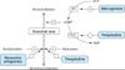
The adrenoceptor agonists have several pharmacologic actions that are important in the treatment of asthma. They relax airway smooth muscle and inhibit release of bronchoconstricting mediators from mast cells. They may also inhibit microvascular leakage and increase mucociliary transport by increasing ciliary activity. As in other tissues, the b agonists stimulate adenylyl cyclase and increase the formation of intracellular cAMP (Figure 20-3).
The best-characterized action of the adrenoceptor agonists in the airways is relaxation of airway smooth muscle. Although there is no evidence for significant sympathetic innervation of human airway smooth muscle, ample evidence exists for the presence of adrenoceptors on airway smooth muscle. In general, stimulation of b2 receptors relaxes airway smooth muscle, inhibits mediator release, and causes tachycardia and skeletal muscle tremor as side effects.
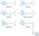
The sympathomimetic agents that have been widely used in the treatment of asthma include epinephrine, ephedrine, isoproterenol, and albuterol and other b2-selective agents (Figure 20-4). Because epinephrine and isoproterenol increase the rate and force of cardiac contraction (mediated mainly by b1 receptors), they are reserved for special situations (see below).
In general, adrenoceptor agonists are best delivered by inhalation because this results in the greatest local effect on airway smooth muscle with the least systemic toxicity. Aerosol deposition depends on the particle size, the pattern of breathing (tidal volume and rate of airflow), and the geometry of the airways. Even with particles in the optimal size range of 2-5 mm, 80-90% of the total dose of aerosol is deposited in the mouth or pharynx. Particles under 1-2 um remain suspended and may be exhaled. Deposition can be increased by holding the breath in inspiration.
Epinephrine is an effective, rapidly acting bronchodilator when injected subcutaneously (0.4 mL of 1:1000 solution) or inhaled as a microaerosol from a pressurized canister (320 mcg per puff). Maximal bronchodilation is achieved 15 minutes after inhalation and lasts 60-90 minutes. Because epinephrine stimulates a and b1 as well as b2 receptors, tachycardia, arrhythmias, and worsening of angina pectoris are troublesome adverse effects. The cardiovascular effects of epinephrine are of value for treating the acute vasodilation and shock as well as the bronchospasm of anaphylaxis, but its use in asthma has been displaced by other, more b2-selective agents.
Ephedrine was used in China for more than 2000 years before its introduction into Western medicine in 1924. Compared with epinephrine, ephedrine has a longer duration, oral activity, more pronounced central effects, and much lower potency. Because of the development of more efficacious and b2-selective agonists, ephedrine is now used infrequently in treating asthma.
Isoproterenol is a potent bronchodilator; when inhaled as a microaerosol from a pressurized canister, 80-120 mcg isoproterenol causes maximal bronchodilation within 5 minutes. Isoproterenol has a 60- to 90-minute duration of action. An increase in the asthma mortality rate that occurred in the United Kingdom in the mid 1960s was attributed to cardiac arrhythmias resulting from the use of high doses of inhaled isoproterenol. It is now rarely used for asthma.
|
|
Figure 20-3. Bronchodilation is promoted by cAMP. Intracellular levels of cAMP can be increased by b-adrenoceptor agonists, which increase the rate of its synthesis by adenylyl cyclase (AC); or by phosphodiesterase (PDE) inhibitors such as theophylline, which slow the rate of its degradation. Bronchoconstriction can be inhibited by muscarinic antagonists and possibly by adenosine antagonists. |
|
|
|
Figure 20-4. Structures of isoproterenol and several b2-selective analogs. |
|
Beta2-Selective Drugs
The b2-selective adrenoceptor agonist drugs are the most widely used sympathomimetics for the treatment of asthma at present (Figure 20-4). These agents differ structurally from epinephrine in having a larger substitution on the amino group and in the position of the hydroxyl groups on the aromatic ring. They are effective after inhaled or oral administration and have a long duration of action.
Albuterol, terbutaline, metaproterenol, and pirbuterol are available as metered-dose inhalers. Given by inhalation, these agents cause bronchodilation equivalent to that produced by isoproterenol. Bronchodilation is maximal within 15-30 minutes and persists for 3-4 hours. All can be diluted in saline for administration from a hand-held nebulizer. Because the particles generated by a nebulizer are much larger than those from a metered-dose inhaler, much higher doses must be given (2.5-5.0 mg vs 100-400 mcg) but are no more effective. Nebulized therapy should thus be reserved for patients unable to coordinate inhalation from a metered-dose inhaler.
Albuterol and terbutaline are also available in tablet form. One tablet two or three times daily is the usual regimen; the principal adverse effects of skeletal muscle tremor, nervousness, and occasional weakness may be reduced by starting the patient on half-strength tablets for the first 2 weeks of therapy, but this route of administration presents no advantage over inhaled treatment.
Of these agents, only terbutaline is available for subcutaneous injection (0.25 mg). The indications for this route are similar to those for subcutaneous epinephrine¾severe asthma requiring emergency treatment when aerosolized therapy is not available or has been ineffective¾but it should be remembered that terbutaline's longer duration of action means that cumulative effects may be seen after repeated injections.
A new generation of long-acting b2-selective agonists includes salmeterol and formoterol. Both drugs are potent selective b2 agonists that achieve their long duration of action (12 hours or more) as a result of high lipid solubility. This permits them to dissolve in the smooth muscle cell membrane in high concentrations or, possibly, attach to "mooring" molecules in the vicinity of the adrenoceptor. These drugs appear to interact with inhaled corticosteroids to improve asthma control. They are not recommended as the sole therapy for asthma.
Toxicities
The use of sympathomimetic agents by inhalation at first raised fears about possible cardiac arrhythmias and about hypoxemia acutely and tachyphylaxis or tolerance when given repeatedly. It is true that the vasodilating action of b2-agonist treatment may increase perfusion of poorly ventilated lung units, transiently decreasing arterial oxygen tension (PaO2). This effect is usually small, however, and may occur with any bronchodilator drug; the significance of such an effect depends on the initial PaO2 of the patient. Administration of supplemental oxygen, routine in treatment of an acute severe attack of asthma, eliminates any concern over this effect. The other concern, that b-agonist treatment may cause lethal cardiac arrhythmias appears unsubstantiated. In patients presenting for emergency treatment of severe asthma, irregularities in cardiac rhythm improve with the improvements in gas exchange effected by bronchodilator treatment.
The concept that b-agonist drugs cause worsening of clinical asthma by inducing tachyphylaxis to their own action remains unestablished. Most studies have shown only a small change in the bronchodilator response to b stimulation after prolonged treatment with b-agonist drugs, but some studies have shown a loss in the ability of b-agonist treatment to inhibit the response to subsequent challenge with exercise, methacholine, or antigen challenge (referred to as a loss of bronchoprotective action).
Fears that heavy use of b-agonist inhalers could actually increase morbidity and mortality have not been borne out by careful epidemiologic investigations. Heavy use most often indicates that the patient should be receiving more effective prophylactic therapy with corticosteroids.
Although it is true that b2-adrenoceptor agonists appear to be safe and effective bronchodilators for most patients, there is some evidence that the risk of adverse effects from chronic treatment with long-acting b agonists may be greater for some individuals, possibly as a function of genetic variants for the b receptor. Two retrospective and one prospective study have shown differences between patients homozygous for glycine versus arginine at the B-16 locus of the b receptor. Among patients homozygous for arginine, a genotype found in 16% of the Caucasian population in the USA, but more commonly in African Americans, asthma control deteriorated with regular use of albuterol or salmeterol, whereas asthma control improved with this treatment among those homozygous for glycine at the same locus. These findings need to be replicated in larger studies, but it is tempting to speculate that a genetic variant may underlie the report of an increase in asthma mortality from regular use of a long-acting b agonist in studies involving very large numbers of patients.
METHYLXANTHINE DRUGS
Introduction
The three important methylxanthines are theophylline, theobromine, and caffeine. Their major source is beverages (tea, cocoa, and coffee, respectively). The importance of theophylline as a therapeutic agent in the treatment of asthma has waned as the greater effectiveness of inhaled adrenoceptor agents for acute asthma and of inhaled anti-inflammatory agents for chronic asthma has been established, but theophylline's very low cost is an important advantage for economically disadvantaged patients in societies in which health care resources are limited.
Chemistry
As shown below, theophylline is 1,3-dimethylxanthine; theobromine is 3,7-dimethylxanthine; and caffeine is 1,3,7-trimethylxanthine. A theophylline preparation commonly used for therapeutic purposes is aminophylline, a theophylline-ethylenediamine complex. The pharmacokinetics of theophylline are discussed below (see Clinical Use of Methylxanthines). The metabolic products, partially demethylated xanthines (not uric acid), are excreted in the urine.
Mechanism of Action
Several mechanisms have been proposed for the actions of methylxanthines, but none has been firmly established. At high concentrations, they can be shown in vitro to inhibit several members of the phosphodiesterase (PDE) enzyme family (Figure 20-3). Because the phosphodiesterases hydrolyze cyclic nucleotides, this inhibition results in higher concentrations of intracellular cAMP and, in some tissues, cGMP. Cyclic AMP is responsible for a myriad of cellular functions including, but not limited to, stimulation of cardiac function, relaxation of smooth muscle, and reduction in the immune and inflammatory activity of specific cells.
Of the various isoforms of phosphodiesterase that have been identified, PDE4 appears to be the most directly involved in actions of methylxanthines on airway smooth muscle and on inflammatory cells. The inhibition of PDE4 in inflammatory cells reduces their release of cytokines and chemokines, which in turn results in a decrease in immune cell migration and activation.
In an effort to reduce toxicity while maintaining therapeutic efficacy, more selective inhibitors of different isoforms of PDE4 have been developed, particularly for the treatment of chronic obstructive pulmonary disease (COPD). Several are now in advanced stages of clinical development, (eg, roflumilast, cilomilast, tofimilast), but at the time of writing, none has been approved for clinical use by the FDA. Although several appear promising, none is entirely free of the major adverse effect of this class of drugs, nausea and vomiting.
Another proposed mechanism is inhibition of cell surface receptors for adenosine. These receptors modulate adenylyl cyclase activity, and adenosine has been shown to provoke contraction of isolated airway smooth muscle and histamine release from airway mast cells. It has been shown, however, that xanthine derivatives devoid of adenosine antagonism (eg, enprofylline) may be potent in inhibiting bronchoconstriction in asthmatic subjects.
Pharmacodynamics of Methylxanthines
The methylxanthines have effects on the central nervous system, kidney, and cardiac and skeletal muscle as well as smooth muscle. Of the three agents, theophylline is most selective in its smooth muscle effects, whereas caffeine has the most marked central nervous system effects.
A. CENTRAL NERVOUS SYSTEM EFFECTS
In low and moderate doses, the methylxanthines¾especially caffeine¾cause mild cortical arousal with increased alertness and deferral of fatigue. The caffeine contained in beverages¾eg, 100 mg in a cup of coffee¾is sufficient to cause nervousness and insomnia in sensitive individuals and slight bronchodilation in patients with asthma. The larger doses necessary for more effective bronchodilation commonly cause nervousness and tremor in some patients. Very high doses, from accidental or suicidal overdose, cause medullary stimulation and convulsions and may lead to death.
B. CARDIOVASCULAR EFFECTS
The methylxanthines have positive chronotropic and inotropic effects. At low concentrations, these effects appear to result from inhibition of presynaptic adenosine receptors in sympathetic nerves increasing catecholamine release at nerve endings. The higher concentrations (> 10 umol/L, 2 mg/L) associated with inhibition of phosphodiesterase and increases in cAMP may result in increased influx of calcium. At much higher concentrations (> 100 umol/L), sequestration of calcium by the sarcoplasmic reticulum is impaired.
The clinical expression of these effects on cardiovascular function varies among individuals. Ordinary consumption of coffee and other methylxanthine-containing beverages usually produces slight tachycardia, an increase in cardiac output, and an increase in peripheral resistance, raising blood pressure slightly. In sensitive individuals, consumption of a few cups of coffee may result in arrhythmias. In large doses, these agents also relax vascular smooth muscle except in cerebral blood vessels, where they cause contraction.
Methylxanthines decrease blood viscosity and may improve blood flow under certain conditions. The mechanism of this action is not well defined, but the effect is exploited in the treatment of intermittent claudication with pentoxifylline, a dimethylxanthine agent. However, no evidence suggests that this therapy is superior to other approaches.
C. EFFECTS ON GASTROINTESTINAL TRACT
The methylxanthines stimulate secretion of both gastric acid and digestive enzymes. However, even decaffeinated coffee has a potent stimulant effect on secretion, which means that the primary secretagogue in coffee is not caffeine.
D. EFFECTS ON KIDNEY
The methylxanthines¾especially theophylline¾are weak diuretics. This effect may involve both increased glomerular filtration and reduced tubular sodium reabsorption. The diuresis is not of sufficient magnitude to be therapeutically useful.
E. EFFECTS ON SMOOTH MUSCLE
The bronchodilation produced by the methylxanthines is the major therapeutic action in asthma. Tolerance does not develop, but adverse effects, especially in the central nervous system, may limit the dose (see below). In addition to their effect on airway smooth muscle, these agents¾in sufficient concentration¾inhibit antigen-induced release of histamine from lung tissue; their effect on mucociliary transport is unknown.
F. EFFECTS ON SKELETAL MUSCLE
The respiratory actions of the methylxanthines may not be confined to the airways, for they also strengthen the contractions of isolated skeletal muscle in vitro and improve contractility and reverse fatigue of the diaphragm in patients with COPD. This effect on diaphragmatic performance¾rather than an effect on the respiratory center¾may account for theophylline's ability to improve the ventilatory response to hypoxia and to diminish dyspnea even in patients with irreversible airflow obstruction.
Clinical Use of Methylxanthines
Of the xanthines, theophylline is the most effective bronchodilator, and it has been shown repeatedly both to relieve airflow obstruction in acute asthma and to reduce the severity of symptoms and time lost from work or school in patients with chronic asthma. Theophylline base is only slightly soluble in water, so it has been administered as several salts containing varying amounts of theophylline base. Most preparations are well absorbed from the gastrointestinal tract, but absorption of rectal suppositories is unreliable.
Improvements in theophylline preparations have come from alterations in the physical state of the drugs rather than from new chemical formulations. For example, the increased surface area of anhydrous theophylline in a microcrystalline form facilitates solubilization for complete and rapid absorption after oral administration. Numerous sustained-release preparations (see Preparations Available) are available and can produce therapeutic blood levels for 12 hours or more. These preparations offer the advantages of less frequent drug administration, less fluctuation of theophylline blood levels, and, in many cases, more effective treatment of nocturnal bronchospasm.
Theophylline should be used only where methods to measure theophylline blood levels are available because it has a narrow therapeutic window, and its therapeutic and toxic effects are related to its blood level. Improvement in pulmonary function is correlated with plasma concentration in the range of 5-20 mg/L. Anorexia, nausea, vomiting, abdominal discomfort, headache, and anxiety occur at concentrations of 15 mg/L in some patients and become common at concentrations greater than 20 mg/L. Higher levels (> 40 mg/L) may cause seizures or arrhythmias; these may not be preceded by gastrointestinal or neurologic warning symptoms.
The plasma clearance of theophylline varies widely. Theophylline is metabolized by the liver, so usual doses may lead to toxic concentrations of the drug in patients with liver disease. Conversely, clearance may be increased through the induction of hepatic enzymes by cigarette smoking or by changes in diet. In normal adults, the mean plasma clearance is 0.69 mL/kg/min. Children clear theophylline faster than adults (1-1.5 mL/kg/min). Neonates and young infants have the slowest clearance (see Chapter 60). Even when maintenance doses are altered to correct for the above factors, plasma concentrations vary widely.
Theophylline improves long-term control of asthma when taken as the sole maintenance treatment or when added to inhaled corticosteroids. It is inexpensive, and it can be taken orally. Its use, however, also requires occasional measurement of plasma levels; it often causes unpleasant minor side effects (especially insomnia); and accidental or intentional overdose can result in severe toxicity or death. For oral therapy with the prompt-release formulation, the usual dose is 3-4 mg/kg of theophylline every 6 hours. Changes in dosage result in a new steady-state concentration of theophylline in 1-2 days, so the dosage may be increased at intervals of 2-3 days until therapeutic plasma concentrations are achieved (10-20 mg/L) or until adverse effects develop.
ANTIMUSCARINIC AGENTS
Introduction
Observation of the use of leaves from Datura stramonium for asthma treatment in India led to the discovery of atropine, a potent competitive inhibitor of acetylcholine at postganglionic "muscarinic" receptors, as a bronchodilator. Interest in the potential value of antimuscarinic agents increased with demonstration of the importance of the vagus nerves in bronchospastic responses of laboratory animals and by the development of a potent atropine analog that is poorly absorbed after aerosol administration and that is therefore relatively free of systemic atropine-like effects.
Mechanism of Action
Muscarinic antagonists competitively inhibit the effect of acetylcholine at muscarinic receptors (see Chapter 8). In the airways, acetylcholine is released from efferent endings of the vagus nerves, and muscarinic antagonists block the contraction of airway smooth muscle and the increase in secretion of mucus that occurs in response to vagal activity (Figure 20-2). Very high concentrations¾well above those achieved even with maximal therapy¾are required to inhibit the response of airway smooth muscle to nonmuscarinic stimulation. This selectivity of muscarinic antagonists accounts for their usefulness as investigative tools in examining the role of parasympathetic pathways in bronchomotor responses but limits their usefulness in preventing bronchospasm. In the doses given, antimuscarinic agents inhibit only that portion of the response mediated by muscarinic receptors, which varies by stimulus, and which further appears to vary among individuals in responses to the same stimulus.
Clinical Use of Muscarinic Antagonists
Antimuscarinic agents are effective bronchodilators. When given intravenously, atropine, the prototypical muscarinic antagonist, causes bronchodilation at a lower dose than that needed to cause an increase in heart rate. The selectivity of atropine's effect can be increased further by administering the drug by inhalation or by use of a more selective quaternary ammonium derivative of atropine, ipratropium bromide. Ipratropium can be delivered in high doses by this route because it is poorly absorbed into the circulation and does not readily enter the central nervous system. Studies with this agent have shown that the degree of involvement of parasympathetic pathways in bronchomotor responses varies among subjects. In some, bronchoconstriction is inhibited effectively; in others, only modestly. The failure of higher doses of the muscarinic antagonist to further inhibit the response in these individuals indicates that mechanisms other than parasympathetic reflex pathways must be involved.
Even in the subjects least protected by this antimuscarinic agent, however, the bronchodilation and partial inhibition of provoked bronchoconstriction are of potential clinical value, and antimuscarinic agents are valuable for patients intolerant of inhaled b-agonist agents. Although antimuscarinic drugs appear to be slightly less effective than b-agonist agents in reversing asthmatic bronchospasm, the addition of ipratropium enhances the bronchodilation produced by nebulized albuterol in acute severe asthma.
Ipratropium appears to be at least as effective in patients with COPD that includes a partially reversible component. A longer-acting, selective antimuscarinic agent, tiotropium, is approved as a treatment for COPD. This drug is also taken by inhalation, and a single dose of 18 mcg has 24-hour duration of action. Daily inhalation of tiotropium has been shown not only to improve functional capacity of patients with COPD, but also to reduce the frequency of exacerbations of their condition.
CORTICOSTEROIDS
Mechanism of Action
Corticosteroids have been used to treat asthma since 1950 and are presumed to act by their broad anti-inflammatory efficacy, mediated in part by inhibition of production of inflammatory cytokines (see Chapter 39). They do not relax airway smooth muscle directly but reduce bronchial reactivity and reduce the frequency of asthma exacerbations if taken regularly. Their effect on airway obstruction may be due in part to their contraction of engorged vessels in the bronchial mucosa and their potentiation of the effects of b-receptor agonists, but their most important action is inhibition of the lymphocytic, eosinophilic mucosal inflammation of asthmatic airways.
Clinical Use of Corticosteroids
Clinical studies of corticosteroids consistently show them to be effective in improving all indices of asthma control¾severity of symptoms, tests of airway caliber and bronchial reactivity, frequency of exacerbations, and quality of life. Because of severe adverse effects when given chronically, oral and parenteral corticosteroids are reserved for patients who require urgent treatment, ie, those who have not improved adequately with bronchodilators or who experience worsening symptoms despite maintenance therapy. Regular or "controller" therapy is maintained with aerosol corticosteroids.
Urgent treatment is often begun with an oral dose of 30-60 mg prednisone per day or an intravenous dose of 1 mg/kg methylprednisolone every 6 hours; the daily dose is decreased after airway obstruction has improved. In most patients, systemic corticosteroid therapy can be discontinued in a week or 10 days, but in other patients symptoms may worsen as the dose is decreased to lower levels. Because adrenal suppression by corticosteroids is related to dose and because secretion of endogenous corticosteroids has a diurnal variation, it is customary to administer corticosteroids early in the morning after endogenous ACTH secretion has peaked. For prevention of nocturnal asthma, however, oral or inhaled corticosteroids are most effective when given in the late afternoon.
Aerosol treatment is the most effective way to avoid the systemic adverse effects of corticosteroid therapy. The introduction of corticosteroids such as beclomethasone, budesonide, flunisolide, fluticasone, mometasone, and triamcinolone has made it possible to deliver corticosteroids to the airways with minimal systemic absorption. An average daily dose of four puffs twice daily of beclomethasone (400 mcg/d) is equivalent to about 10-15 mg/d of oral prednisone for the control of asthma, with far fewer systemic effects. Indeed, one of the cautions in switching patients from oral to inhaled corticosteroid therapy is to taper oral therapy slowly to avoid precipitation of adrenal insufficiency. In patients requiring continued prednisone treatment despite inhalation of standard doses of an aerosol corticosteroid, higher doses appear to be more effective; inhaled dosages up to 2000 mcg/d of fluticasone are effective in weaning patients from chronic prednisone therapy. Although these high doses of inhaled steroids may cause adrenal suppression, the risks of systemic toxicity from chronic use appear negligible compared with those of the oral corticosteroid therapy they replace.
A special problem caused by inhaled topical corticosteroids is the occurrence of oropharyngeal candidiasis. The risk of this complication can be reduced by having patients gargle water and spit after each inhaled treatment. Hoarseness can also result from a direct local effect of inhaled corticosteroids on the vocal cords. These agents are remarkably free of other short-term complications in adults but may increase the risks of osteoporosis and cataracts over the long term. In children, inhaled corticosteroid therapy has been shown to slow the rate of growth, but this effect appears to be transient: Asthma itself delays puberty, and there is no evidence that inhaled corticosteroid therapy in childhood influences adult height.
A novel approach to minimizing the risk of toxicity from systemic absorption of an inhaled corticosteroid underlay the development of ciclesonide. This investigational corticosteroid is inhaled as a prodrug activated by cleavage by esterases in bronchial epithelial cells. When absorbed into the circulation, the active product is tightly bound to serum proteins, and so has little access to glucocorticoid receptors in skin, eye, and bone, minimizing its risk of causing cutaneous thinning, cataracts, osteoporosis, or temporary slowing of growth.
Chronic use of inhaled corticosteroids effectively reduces symptoms and improves pulmonary function in patients with mild asthma. Such use also reduces or eliminates the need for oral corticosteroids in patients with more severe disease. In contrast to b-stimulant agents and theophylline, chronic use of inhaled corticosteroids reduces bronchial reactivity. Because of the efficacy and safety of inhaled corticosteroids, they are now routinely prescribed for patients who require more than occasional inhalations of a b agonist for relief of symptoms. This therapy is continued for 10-12 weeks and then withdrawn to determine whether more prolonged therapy is needed. Inhaled corticosteroids are not curative. In most patients, the manifestations of asthma return within a few weeks after stopping therapy even if they have been taken in high doses for 2 years or longer.
CROMOLYN & NEDOCROMIL
Introduction
Cromolyn sodium (disodium cromoglycate) and nedocromil sodium are stable but extremely insoluble salts (see structures below). When used as aerosols (by nebulizer or metered-dose inhaler), they effectively inhibit both antigen- and exercise-induced asthma, and chronic use (four times daily) slightly reduces the overall level of bronchial reactivity. However, these drugs have no effect on airway smooth muscle tone and are ineffective in reversing asthmatic bronchospasm; they are only of value when taken prophylactically.
Cromolyn is poorly absorbed from the gastrointestinal tract and must be inhaled as a microfine powder or aerosolized solution. Nedocromil also has a very low bioavailability and is available only in metered-dose aerosol form.
Mechanism of Action
Cromolyn and nedocromil differ structurally but are thought to share a common mechanism of action: an alteration in the function of delayed chloride channels in the cell membrane, inhibiting cell activation. This action on airway nerves is thought to be responsible for nedocromil's inhibition of cough; on mast cells, for inhibition of the early response to antigen challenge; and on eosinophils, for inhibition of the inflammatory response to inhalation of allergens. The inhibitory effect on mast cells appears to be specific for cell type, since cromolyn has little inhibitory effect on mediator release from human basophils. It may also be specific for different organs, since cromolyn inhibits mast cell degranulation in human and primate lung but not in skin. This in turn may reflect known differences in mast cells found in different sites, as in their neutral protease content.
Until recently, the idea that cromolyn inhibits mast cell degranulation was so well accepted that the inhibition of a response by cromolyn was thought to indicate the involvement of mast cells in the response. This simplistic idea has been overturned in part by the finding that cromolyn and nedocromil inhibit the function of cells other than mast cells and in part by the finding that nedocromil inhibits appearance of the late response even when given after the early response to antigen challenge, ie, after mast cell degranulation has occurred.
Clinical Use of Cromolyn & Nedocromil
In short-term clinical trials, pretreatment with cromolyn or nedocromil blocks the bronchoconstriction caused by allergen inhalation, by exercise, by sulfur dioxide, and by a variety of causes of occupational asthma. This acute protective effect of a single treatment makes cromolyn useful for administration shortly before exercise or before unavoidable exposure to an allergen.
When taken regularly (two to four puffs two to four times daily) by patients with perennial (nonseasonal) asthma, both agents modestly but significantly reduce symptomatic severity and the need for bronchodilator medications. These drugs are neither as potent nor as predictably effective as inhaled corticosteroids. In general, young patients with extrinsic asthma are most likely to respond favorably. At present, the only way of determining whether a patient will respond is by a therapeutic trial for 4 weeks. The addition of nedocromil to a standard dose of an inhaled corticosteroid appears to improve asthma control.
Cromolyn solution is also useful in reducing symptoms of allergic rhinoconjunctivitis. Applying the solution by nasal spray or eye drops several times a day is effective in about 75% of patients, even during the peak pollen season.
Because the drugs are so poorly absorbed, adverse effects of cromolyn and nedocromil are minor and are localized to the sites of deposition. These include such minor symptoms as throat irritation, cough, and mouth dryness, and, rarely, chest tightness, and wheezing. Some of these symptoms can be prevented by inhaling a b2-adrenoceptor agonist before cromolyn or nedocromil treatment. Serious adverse effects are rare. Reversible dermatitis, myositis, or gastroenteritis occurs in less than 2% of patients, and a very few cases of pulmonary infiltration with eosinophilia and anaphylaxis have been reported. This lack of toxicity accounts for cromolyn's widespread use in children, especially those at ages of rapid growth. For children who have difficulty coordinating the use of the inhaler device, cromolyn may be given by aerosol of a 1% solution.
LEUKOTRIENE PATHWAY INHIBITORS
Because of the evidence of leukotriene involvement in many inflammatory diseases (see Chapter 18) and in anaphylaxis, considerable effort has been expended on the development of drugs that block the synthesis of these arachidonic acid derivatives or their receptors. Leukotrienes result from the action of 5-lipoxygenase on arachidonic acid and are synthesized by a variety of inflammatory cells in the airways, including eosinophils, mast cells, macrophages, and basophils. Leukotriene B4 (LTB4) is a potent neutrophil chemoattractant, and LTC4 and LTD4 exert many effects known to occur in asthma, including bronchoconstriction, increased bronchial reactivity, mucosal edema, and mucus hypersecretion. Early studies established that antigen challenge of sensitized human lung tissue results in the generation of leukotrienes, whereas other studies of human subjects have shown that inhalation of leukotrienes causes not only bronchoconstriction but also an increase in bronchial reactivity to histamine that persists for several days.
Two approaches to interrupting the leukotriene pathway have been pursued: inhibition of 5-lipoxygenase, thereby preventing leukotriene synthesis; and inhibition of the binding of LTD4to its receptor on target tissues, thereby preventing its action. Efficacy in blocking airway responses to exercise and to antigen challenge has been shown for drugs in both categories: zileuton, a 5-lipoxygenase inhibitor, and zafirlukast and montelukast, LTD4-receptor antagonists. All have been shown to improve asthma control and to reduce the frequency of asthma exacerbations in outpatient clinical trials. Their effects on symptoms, airway caliber, bronchial reactivity, and airway inflammation are less marked than the effects of inhaled corticosteroids, but they are more nearly equal in reducing the frequency of exacerbations. Their principal advantage is that they are taken orally; some patients¾especially children¾comply poorly with inhaled therapies. Montelukast is approved for children as young as 6 years of age.
Some patients appear to have particularly favorable responses, but no clinical features allow identification of "responders" before a trial of therapy. In the USA, zileuton is approved for use in an oral dosage of 400-800 mg for administration 2-4 times daily; zafirlukast, 20 mg twice daily; and montelukast, 10 mg (for adults) or 4 mg (for children) once daily.
Trials with leukotriene inhibitors have demonstrated an important role for leukotrienes in aspirin-induced asthma. It has long been known that 5-10% of asthmatics are exquisitely sensitive to aspirin, so that ingestion of even a very small dose causes profound bronchoconstriction and symptoms of systemic release of histamine, such as flushing and abdominal cramping. Because this reaction to aspirin is not associated with any evidence of allergic sensitization to aspirin or its metabolites and because it is produced by any of the nonsteroidal anti-inflammatory agents, it is thought to result from inhibition of prostaglandin synthetase (cyclooxygenase), shifting arachidonic acid metabolism from the prostaglandin to the leukotriene pathway. Support for this idea was provided by the demonstration that leukotriene pathway inhibitors impressively reduce the response to aspirin challenge and improve overall control of asthma on a day-to-day basis.
Of these agents, zileuton is the least prescribed because of the former requirement of four-times-daily dosing (a formulation for twice-daily use has recently been developed) and because of occasional liver toxicity. The receptor antagonists appear to be safe to use. Reports of Churg-Strauss syndrome (a systemic vasculitis accompanied by worsening asthma, pulmonary infiltrates, and eosinophilia) appear to have been coincidental, with the syndrome unmasked by the reduction in prednisone dosage made possible by the addition of zafirlukast or montelukast.
OTHER DRUGS IN THE TREATMENT OF ASTHMA
Anti-IgE Monoclonal Antibodies
An entirely new approach to the treatment of asthma exploits advances in molecular biology to target IgE antibody. From a collection of monoclonal antibodies raised in mice against IgE antibody itself, a monoclonal antibody was selected that appeared to be targeted against the portion of IgE that binds to its receptors (FCe-R1 and FCe-R2 receptors) on mast cells and other inflammatory cells. Omalizumab (an anti-IgE monoclonal antibody) inhibits the binding of IgE to mast cells but does not activate IgE already bound to these cells and thus does not provoke mast cell degranulation. It may also inhibit IgE synthesis by B lymphocytes. The murine antibody has been genetically humanized by replacing all but a small fraction of its amino acids with those found in human proteins, and it does not appear to cause sensitization when given to human subjects.
Studies of omalizumab in asthmatic volunteers showed that its administration over 10 weeks lowered plasma IgE to undetectable levels and significantly reduced the magnitude of both the early and the late bronchospastic responses to antigen challenge. Clinical trials have shown that repeated intravenous or subcutaneous injection of anti-IgE MAb lessens asthma severity and reduces the corticosteroid requirement in patients with moderate to severe disease, especially those with a clear environmental antigen precipitating factor, and improves nasal and conjunctival symptoms in patients with perennial or seasonal allergic rhinitis. Omalizumab's most important effect is reduction of the frequency and severity of asthma exacerbations, even while enabling a reduction in corticosteroid requirements. Combined analysis of several clinical trials has shown that the patients most likely to respond are, fortunately, those with the greatest need, ie, patients with a history of repeated exacerbations, a high requirement for corticosteroid treatment, and poor pulmonary function. Similarly, the exacerbations most prevented are the ones most important to prevent: Omalizumab treatment reduced exacerbations requiring hospitalization by 88%. These benefits justify the high cost of this treatment in selected individuals with severe disease characterized by frequent exacerbations.
Possible Future Therapies
The rapid advance in the scientific description of the immunopathogenesis of asthma has spurred the development of many new therapies targeting different sites in the immune cascade. These include monoclonal antibodies directed against cytokines (IL-4, IL-5, IL-13), antagonists of cell adhesion molecules, protease inhibitors, and immunomodulators aimed at shifting CD4 lymphocytes from the TH2 to the TH1 phenotype or at selective inhibition of the subset of TH2 lymphocytes directed against particular antigens. There is evidence that asthma may be aggravated¾or even caused¾by chronic airway infection with Chlamydia pneumoniae or Mycoplasma pneumoniae. This may explain the reports of benefit from treatment with macrolide antibiotics and, if confirmed, would stimulate the development of new diagnostic methods and antimicrobial therapies.
II. CLINICAL PHARMACOLOGY OF DRUGS USED IN THE TREATMENT OF ASTHMA
INTRODUCTION
Asthma is best thought of as a disease in two time domains. In the present domain, it is important for the distress it causes¾cough, nocturnal awakenings, and shortness of breath that interferes with the ability to exercise or to pursue desired activities. For mild asthma, occasional inhalation of a bronchodilator may be all that is needed. For more severe asthma, treatment with a long-term controller, like an inhaled corticosteroid, is necessary to relieve symptoms and restore function. The second domain of asthma is the risk it presents of future events, such as exacerbations, or of progressive loss of pulmonary function. A patient's satisfaction with his or her ability to control symptoms and maintain function by frequent use of an inhaled b2 agonist does not mean that the risk of future events is also controlled. In fact, use of two or more canisters of an inhaled b agonist per month is a marker of increased risk of asthma fatality.
The challenges of assessing severity and adjusting therapy for these two domains of asthma are different. For relief of distress in the present domain, the key information can be obtained by asking specific questions about the frequency and severity of symptoms, the frequency of use of an inhaled b2 agonist for relief of symptoms, the frequency of nocturnal awakenings, and the ability to exercise. Estimating the risk for future exacerbations is more difficult. In general, patients with poorly controlled symptoms in the present have a heightened risk of exacerbations in the future, but some patients seem unaware of the severity of their underlying airflow obstruction (sometimes described as "poor perceivers") and can be identified only by measurement of pulmonary function, as by spirometry. Reductions in the FEV1 correlate with heightened risk of attacks of asthma in the future. Other possible markers of heightened risk are unstable pulmonary function (large variations in FEV1 from visit to visit, large change with bronchodilator treatment), extreme bronchial reactivity, or high numbers of eosinophils in sputum or of nitric oxide in exhaled air. Assessment of these features may identify patients who need increases in therapy for protection against exacerbations.
BRONCHODILATORS
Bronchodilators, such as inhaled albuterol, are rapidly effective, safe, and inexpensive. Patients with only occasional symptoms of asthma require no more than an inhaled b2-receptor agonist taken on an as-needed basis. If symptoms require this "rescue" therapy more than twice a week, if nocturnal symptoms occur more than twice a month, or if the FEV1 is less than 80% predicted, additional treatment is needed. The treatment first recommended is a low dose of an inhaled corticosteroid, although treatment with a leukotriene receptor antagonist or with cromolyn may be used. Theophylline is now largely reserved for patients in whom symptoms remain poorly controlled despite the combination of regular treatment with an inhaled anti-inflammatory agent and as-needed use of a b2 agonist. If the addition of theophylline fails to improve symptoms or if adverse effects become bothersome, it is important to check the plasma level of theophylline to be sure it is in the therapeutic range (10-20 mg/L).
An important caveat for patients with mild asthma is that although the risk of a severe, life-threatening attack is lower than in patients with severe asthma, it is not zero. All patients with asthma should be instructed in a simple action plan for severe, frightening attacks: to take up to four puffs of albuterol every 20 minutes over 1 hour. If they do not note clear improvement after the first four puffs, they should take the additional treatments while on their way to an Emergency Department or some other higher level of care.
MUSCARINIC ANTAGONISTS
Inhaled muscarinic antagonists have so far earned a limited place in the treatment of asthma. When adequate doses are given, their effect on baseline airway resistance is nearly as great as that of the sympathomimetic drugs. The airway effects of antimuscarinic and sympathomimetic drugs given in full doses have been shown to be additive only in patients with severe airflow obstruction who present for emergency care. Antimuscarinic agents appear to be of greater value in COPD¾perhaps more so than in asthma. They are also useful as alternative therapies for patients intolerant of b2-adrenoceptor agonists.
Although it was predicted that muscarinic antagonists would dry airway secretions and interfere with mucociliary clearance, direct measurements of fluid volume secretion from single airway submucosal glands in animals show that atropine decreases baseline secretory rates only slightly. The drugs do, however, inhibit the increase in mucus secretion caused by vagal stimulation. No cases of inspissation of mucus have been reported following administration of these drugs.
CORTICOSTEROIDS
If asthmatic symptoms occur frequently or if significant airflow obstruction persists despite bronchodilator therapy, inhaled corticosteroids should be started. For patients with severe symptoms or severe airflow obstruction (eg, FEV1 < 50% predicted), initial treatment with a combination of inhaled and oral corticosteroid (eg, 30 mg/d of prednisone for 3 weeks) treatment is appropriate. Once clinical improvement is noted, usually after 7-10 days, the oral dose should be discontinued or reduced to the minimum necessary to control symptoms.
An issue for inhaled corticosteroid treatment is patient compliance. Analysis of prescription renewals shows that corticosteroids are taken regularly by a minority of patients. This may be a function of a general "steroid phobia" fostered by emphasis in the lay press over the hazards of long-term oral corticosteroid therapy and by ignorance over the difference between corticosteroids and anabolic steroids, taken to enhance muscle strength by now-infamous athletes. This fear of corticosteroid toxicity makes it hard to persuade patients whose symptoms have improved after starting the treatment that they should continue it for protection against attacks. This context accounts for the interest in a recent report that instructing patients with mild but persistent asthma to initiate inhaled corticosteroid therapy only when their symptoms worsened was as effective in maintaining pulmonary function and preventing attacks as taking it twice each day.
In patients with more severe asthma, whose symptoms are inadequately controlled by a standard dose of an inhaled corticosteroid, two options may be considered: to double the dose of inhaled corticosteroid or to add a long-acting inhaled b2-receptor agonist (salmeterol or formoterol). Many studies have shown this combination therapy to be more effective than doubling the dose of the inhaled corticosteroid, but the FDA has issued a warning that the use of a long-acting b agonist is associated with a very small but statistically significant increase in the risk of death or near death from an asthma attack, especially in African Americans. This warning has not so far had much effect on prescriptions for a fixed-dose combination of inhaled fluticasone (a corticosteroid) and salmeterol (a long-acting b agonist), probably because their combination in a single inhaler offers several advantages. Combination inhalers are convenient; they ensure that the long-acting b agonist will not be taken as monotherapy (known not to protect against attacks); and they produce prompt, sustained improvements in clinical symptoms and pulmonary function and reduce the frequency of exacerbations requiring oral corticosteroid treatment. In patients prescribed such combination treatment, it is important to provide explicit instructions that a standard, short-acting inhaled b2 agonist, such as albuterol, be used as needed for relief of acute symptoms.
CROMOLYN & NEDOCROMIL; LEUKOTRIENE ANTAGONISTS
Cromolyn or nedocromil by inhalation, or a leukotriene-receptor antagonist as an oral tablet, may be considered as alternatives to inhaled corticosteroid treatment in patients with symptoms occurring more than twice a week or who are wakened from sleep by asthma more than twice a month. Neither treatment is as effective as even a low dose of an inhaled corticosteroid, but both prevent the issue of "steroid phobia" described above.
Cromolyn and nedocromil may also be useful in patients whose symptoms occur seasonally or after clear-cut inciting stimuli such as exercise or exposure to animal danders or irritants. In patients whose symptoms are continuous or occur without an obvious inciting stimulus, the value of these drugs can be established only with a therapeutic trial of inhaled drug four times a day for 4 weeks. If the patient responds to this therapy, the dose can then be optimized.
Treatment with a leukotriene-receptor antagonist, particularly montelukast, is widely prescribed, especially by primary care providers. Taken orally, leukotriene-receptor antagonists are easy to use and appear to be taken more regularly than inhaled corticosteroids. They are rarely associated with troublesome side effects. Maintenance therapy with a leukotriene antagonist or with cromolyn or nedocromil appears to be roughly as effective as maintenance therapy with theophylline. Because of concerns over the possible long-term toxicity of systemic absorption of inhaled corticosteroids, this maintenance therapy has become widely used for treating children in the USA.
ANTI-IGE MONOCLONAL ANTIBODY
Treatment with omalizumab, the monoclonal humanized anti-IgE antibody, is reserved for patients with chronic severe asthma inadequately controlled by high-dose inhaled corticosteroid plus long-acting b-agonist combination treatment (eg, fluticasone 500 mcg plus salmeterol 50 mcg inhaled twice daily). This treatment reduces lymphocytic, eosinophilic bronchial inflammation and effectively reduces the frequency and severity of exacerbations. It is reserved for patients with demonstrated IgE-mediated sensitivity (by positive skin test or radioallergosorbent test [RAST] to common allergens) and an IgE level within a range that can be reduced sufficiently by twice weekly subcutaneous injection.
OTHER ANTI-INFLAMMATORY THERAPIES
Some reports suggest that agents commonly used to treat rheumatoid arthritis may also be used to treat patients with chronic steroid-dependent asthma. The development of an alternative treatment is important, because chronic treatment with oral corticosteroids may cause osteoporosis, cataracts, glucose intolerance, worsening of hypertension, and cushingoid changes in appearance. Initial studies suggested that oral methotrexate or gold salt injections were beneficial in prednisone-dependent asthmatics, but subsequent studies did not confirm this promise. In contrast, the benefit from treatment with cyclosporine seems real. However, this drug's great toxicity makes this finding only a source of hope that other immunomodulatory therapies will ultimately be developed for the small proportion of patients whose asthma can be managed only with high oral doses of prednisone. An immunomodulatory therapy recently reported to improve asthma is injection of etanercept, a TNF-a antagonist used for treatment of ankylosing spondylitis and severe rheumatoid arthritis.
MANAGEMENT OF ACUTE ASTHMA
The treatment of acute attacks of asthma in patients reporting to the hospital requires close, continuous clinical assessment and repeated objective measurement of lung function. For patients with mild attacks, inhalation of a b2-receptor agonist is as effective as subcutaneous injection of epinephrine. Both of these treatments are more effective than intravenous administration of aminophylline (a soluble salt of theophylline). Severe attacks require treatment with oxygen, frequent or continuous administration of aerosolized albuterol, and systemic treatment with prednisone or methylprednisolone (0.5 mg/kg every 6 hours). Even this aggressive treatment is not invariably effective, and patients must be watched closely for signs of deterioration. General anesthesia, intubation, and mechanical ventilation of asthmatic patients cannot be undertaken lightly but may be lifesaving if respiratory failure supervenes.
PROSPECTS FOR PREVENTION
The high prevalence of asthma in the developed world and its rapid increases in the developing world call for a strategy for primary prevention. Strict antigen avoidance during infancy, once thought to be sensible, has now been shown to be ineffective. In fact, growing up in a household where cats and dogs are kept as pets may protect against developing asthma. The best hope seems to lie in understanding the importance of microbial exposures during infancy in shaping a balanced immune response, and one study showing that feeding Lactobacillus caseii to infants born to allergic parents reduced the rate of allergic dermatitis at age 2 years offers reason for hope.
PREPARATIONS AVAILABLE
SYMPATHOMIMETICS USED IN ASTHMA
Albuterol (generic, Proventil, Ventolin)
Inhalant: 90 mcg/puff aerosol; 0.083, 0.5, 0.63% solution for nebulization
Oral: 2, 4 mg tablets; 2 mg/5 mL syrup
Oral sustained-release: 4, 8 mg tablets
Albuterol/Ipratropium (Combivent, DuoNeb)
Inhalant: 103 mcg albuterol + 18 mcg ipratropium/ puff; 3 mg albuterol + 0.5 mg ipratropium/3 mL solution for nebulization
Bitolterol (Tornalate)
Inhalant: 0.2% solution for nebulization
Ephedrine (generic)
Oral: 25 mg capsules
Parenteral: 50 mg/mL for injection
Epinephrine (generic, Adrenalin)
Inhalant: 1, 10 mg/mL for nebulization; 0.22 mg/spray epinephrine base aerosol
Parenteral: 1:10,000 (0.1 mg/mL), 1:1000 (1 mg/mL)
Formoterol (Foradil)
Inhalant: 12 mcg/unit inhalant powder
Isoetharine (generic)
Inhalant: 1% solution for nebulization
Isoproterenol (generic, Isuprel)
Inhalant: 0.5, 1% for nebulization; 80, 131 mcg/puff aerosols
Parenteral: 0.02, 0.2 mg/mL for injection
Levalbuterol (Xenopex)
Inhalant: 0.31, 0.63, 1.25 mg/3 mL solution
Metaproterenol (Alupent, generic)
Inhalant: 0.65 mg/puff aerosol in 7, 14 g containers; 0.4, 0.6, 5% for nebulization
Pirbuterol (Maxair)
Inhalant: 0.2 mg/puff aerosol in 80 and 300 dose containers
Salmeterol (Serevent)
Inhalant aerosol: 25 mcg salmeterol base/puff in 60 and 120 dose containers
Inhalant powder: 50 mcg/unit
Salmeterol/Fluticasone (Advair Diskus)
Inhalant: 100, 250, 500 mcg fluticasone + 50 mcg salmeterol/unit
Terbutaline (generic, Brethine)
Oral: 2.5, 5 mg tablets
Parenteral: 1 mg/mL for injection
AEROSOL CORTICOSTEROIDS (SEE ALSO CHAPTER 39.)
Beclomethasone (QVAR)
Aerosol: 40, 80 mcg/puff in 100 dose containers
Budesonide (Pulmicort)
Aerosol powder (Turbuhaler): 160 mcg/activation
Inhalation suspension (Respules): 0.25, 0.5 mg/2 mL
Flunisolide (AeroBid, Aerospan)
Aerosol: 80, 250 mcg/puff in 80, 100, and 120 dose containers
Fluticasone (Flovent)
Aerosol: 44, 110, and 220 mcg/puff in 120 dose container; powder, 50, 100, 250 mcg/activation
Fluticasone/Salmeterol (Advair Diskus)
Inhalant: 100, 250, 500 mcg fluticasone + 50 mcg salmeterol/unit
Mometasone (Asmanex Twisthaler)
Inhalant: 220 mcg/actuation in 14, 30, 60, 120 dose units
Triamcinolone (Azmacort)
Aerosol: 100 mcg/puff in 240 dose container
LEUKOTRIENE INHIBITORS
Montelukast (Singulair)
Oral: 10 mg tablets; 4, 5 mg chewable tablets; 4 mg/packet granules
Zafirlukast (Accolate)
Oral: 10, 20 mg tablets
Zileuton (Zyflo)
Oral: 600 mg tablets
CROMOLYN SODIUM & NEDOCROMIL SODIUM
Cromolyn sodium
Pulmonary aerosol (generic, Intal): 800 mcg/puff in 200 dose container; 20 mg/2 mL for nebulization (for asthma)
Nasal aerosol (Nasalcrom): 5.2 mg/puff (for hay fever)
Oral (Gastrocrom): 100 mg/5 mL concentrate (for gastrointestinal allergy)
Nedocromil sodium (Tilade)
Pulmonary aerosol: 1.75 mg/puff in 104 metered-dose container
METHYLXANTHINES: THEOPHYLLINE & DERIVATIVES
Aminophylline (theophylline ethylenediamine, 79% theophylline) (generic)
Oral: 105 mg/5 mL liquid; 100, 200 mg tablets
Rectal: 250, 500 mg suppositories
Parenteral: 250 mg/10 mL for injection
Theophylline (generic, Elixophyllin, Slo-Phyllin, Uniphyl, Theo-Dur, Theo-24, others)
Oral: 100, 125, 200, 250, 300 mg tablets; 100, 200 mg capsules; 26.7, 50 mg/5 mL elixirs, syrups, and solutions
Oral sustained-release, 8-12 hours: 50, 60, 75, 100, 125, 200, 250, 300 mg capsules
Oral sustained-release, 8-24 hours: 100, 200, 300, 450 mg tablets
Oral timed-release, 12 hours: 125, 130, 250, 260, 300 mg capsules
Oral timed-release, 12-24 hours: 100, 200, 300 tablets
Oral timed-release, 24 hours: 100, 200, 300 mg tablets and capsules; 400, 600 mg tablets
Parenteral: 200, 400, 800 mg/container, theophylline and 5% dextrose for injection
OTHER METHYLXANTHINES
Dyphylline (generic)
Oral: 200, 400 mg tablets; 33.3 mg/5 mL elixir
Oxtriphylline (generic, Choledyl)
Oral: equivalent to 64, 127, 254, 382 mg theophylline tablets; 32, 64 mg/5 mL syrup
Pentoxifylline (generic, Trental)
Oral: 400 mg tablets and controlled-release tablets
Note: Pentoxifylline is labeled for use in intermittent claudication only.
ANTIMUSCARINIC DRUGS USED IN ASTHMA
Ipratropium (generic, Atrovent)
Aerosol: 17 (freon-free), 18 mcg/puff in 200 metered-dose inhaler; 0.02% (500 mcg/vial) for nebulization
Nasal spray: 21, 42 mcg/spray
Tiotropium (Spiriva)
Aerosol: 18 mcg/puff in 6 packs
ANTIBODY
Omalizumab (Xolair)
Powder for SC injection, 202.5 mg
REFERENCES
Pathophysiology of Airway Disease
Mazzone SB, Canning BJ: Central nervous system control of the airways: Pharmacological implications. Curr Opin Pharmacol 2002;2:220.
Spina D, Page CP: Pharmacology of airway irritability. Curr Opin Pharmacol 2002;2:264.
Methylxanthines
Nakano J et al: Aminophilline suppresses the release of chemical mediators in treatment of acute asthma. Respir Med 2006;100:542.
Page CP: Recent advances in our understanding of the use of theophylline in the treatment of asthma. J Clin Pharmacol 1999;39:237.
Cromolyn & Nedocromil
Guevara J et al: Inhaled corticosteroids versus sodium cromoglycate in children and adults with asthma. Cochrane Database Syst Rev 2006;2:CD003558.
Yoshihara S et al: Effects of early intervention with inhaled sodium cromoglycate in childhood asthma. Lung 2006;184:63.
Corticosteroids
Boushey HA et al: Daily versus as-needed corticosteroids for mild persistent asthma. N Engl J Med 2005;352:1519.
Suissa S et al: Low-dose inhaled corticosteroids and the prevention of death from asthma. N Engl J Med 2000;343:332.
Wilson AM et al: Anti-inflammatory effects of once daily low dose inhaled ciclesonide in mild to moderate asthmatic patients. Allergy 2006;61:537.
Beta Agonists
Kelly HW, Harkins MS, Boushey H: The role of inhaled long-acting beta-2 agonists in the management of asthma. J Natl Med Assoc 2006;98:8.
Maneechotesuwan K et al: Formoterol attenuates neutrophilic airway inflammation in asthma. Chest 2005;128:1936.
Antimuscarinic Drugs
Lee AM, Jacoby DB, Fryer AD: Selective muscarinic receptor antagonists for airway diseases. Curr Opin Pharmacol 2001;1:223.
Leukotriene Pathway Inhibitors
Biernacki WA et al: Effect of montelukast on exhaled leukotrienes and quality of life in asthmatic patients. Chest 2005;128:1958.
Calhoun WJ: Anti-leukotrienes for asthma. Curr Opin Pharmacol 2001;1:230.
Krawiec ME, Wenzel SE: Leukotriene inhibitors and non-steroidal therapies in the treatment of asthma. Exp Opin Invest Drugs 2001;2:47.
Other Drugs For Asthma
Barnes J: Novel signal transduction modulators for the treatment of airway diseases. Pharmacol Ther 2006;109:238.
Bryan SA et al: Novel therapy for asthma. Exp Opin Invest Drugs 2000;9:25.
Leckie MJ et al: Effects of an interleukin-5 blocking monoclonal antibody on eosinophils, airway hyper-responsiveness, and the late asthmatic response. Lancet 2000;456:2144.
Lock SH et al: Double-blind, placebo-controlled study of cyclosporin A as a corticosteroid-sparing agent in corticosteroid-dependent asthma. Am J Respir Crit Care Med 1996;153:509.
Patacchini R, Maggi CA: Peripheral tachykinin receptors as targets for new drugs. Eur J Pharmacol 2001;429:13.
Walker S et al: Anti-IgE for chronic asthma in adults and children. Cochrane Database Syst Rev 2006;2:CD003559.
Clinical Management Of Airway Disease
Barnes PJ: Drugs for asthma. Br J Pharmacol 2006;147(Suppl 1):S297.
Tattersfield AE et al: Asthma. Lancet 2002;360:1313.