Introduction
Drugs acting in the central nervous system (CNS) were among the first to be discovered by primitive humans and are still the most widely used group of pharmacologic agents. In addition to their use in therapy, many drugs acting on the CNS are used without prescription to increase one's sense of well-being.
The mechanisms by which various drugs act in the CNS have not always been clearly understood. In the last three decades, however, dramatic advances have been made in the methodology of CNS pharmacology. It is now possible to study the action of a drug on individual cells and even single ion channels within synapses. The information obtained from such studies is the basis for several major developments in studies of the CNS.
First, it is clear that nearly all drugs with CNS effects act on specific receptors that modulate synaptic transmission. A very few agents such as general anesthetics and alcohol may have nonspecific actions on membranes (although these exceptions are not fully accepted), but even these nonreceptor-mediated actions result in demonstrable alterations in synaptic transmission.
Second, drugs are among the most important tools for studying all aspects of CNS physiology, from the mechanism of convulsions to the laying down of long-term memory. As described below, agonists that mimic natural transmitters (and in many cases are more selective than the endogenous substances) and antagonists are extremely useful in such studies. The Box, Natural Toxins: Tools for Characterizing Ion Channels, describes a few of these substances.
Third, unraveling the actions of drugs with known clinical efficacy has led to some of the most fruitful hypotheses regarding the mechanisms of disease. For example, information on the action of antipsychotic drugs on dopamine receptors has provided the basis for important hypotheses regarding the pathophysiology of schizophrenia. Studies of the effects of a variety of agonists and antagonists on g-aminobutyric acid (GABA) receptors has resulted in new concepts pertaining to the pathophysiology of several diseases, including anxiety and epilepsy.
This chapter provides an introduction to the functional organization of the CNS and its synaptic transmitters as a basis for understanding the actions of the drugs described in the following chapters.
NATURAL TOXINS: TOOLS FOR CHARACTERIZING ION CHANNELS
Evolution is tireless in the development of natural toxins. A vast number of variations are possible with even a small number of amino acids in peptides, and peptides make up only one of a broad array of toxic compounds. For example, the predatory marine snail genus Conus is estimated to include at least 500 different species. Each species kills or paralyzes its prey with a venom that contains 50-200 different peptides or proteins. Furthermore, there is little duplication of peptides among Conus species. Other animals with useful toxins include snakes, frogs, spiders, bees, wasps, and scorpions. Plant species with toxic (or therapeutic) substances are too numerous to mention here; they are referred to in many chapters of this book.
Since many toxins act on ion channels, they provide a wealth of chemical tools for studying the function of these channels. In fact, much of our current understanding of the properties of ion channels comes from studies utilizing only a small percentage of the highly potent and selective toxins that are now available. The toxins typically target voltage-sensitive ion channels, but a number of very useful toxins block ionotropic neurotransmitter receptors. Table 21-1 lists some of the toxins most commonly used in research, their mode of action, and their source.
Methods for the Study of CNS Pharmacology
Like many areas of science, major progress in the study of CNS drugs has depended on the development of new experimental techniques. The first detailed description of synaptic transmission was made possible by the invention of glass microelectrodes, which permit intracellular recording. The development of the brain slice technique permitted an analysis of the physiology and pharmacology of synapses. Detailed electrophysiologic studies of the action of drugs on both voltage- and transmitter-operated channels were further facilitated by the introduction of the patch clamp technique, which permits the recording of current through single channels. Histochemical, immunologic, and radioisotopic methods have made it possible to map the distribution of specific transmitters, their associated enzyme systems, and their receptors. Molecular cloning has had a major impact on our understanding of CNS receptors. These techniques make it possible to determine the precise molecular structure of the receptors and their associated channels. Finally, mice with mutated genes for specific receptors or enzymes (knockout mice) can provide important information regarding the physiologic and pharmacologic roles of these components.
ION CHANNELS & NEUROTRANSMITTER RECEPTORS
The membranes of nerve cells contain two types of channels defined on the basis of the mechanisms controlling their gating (opening and closing): voltage-gated and ligand-gated channels (Figure 21-1A and B). Voltage-gated channels respond to changes in the membrane potential of the cell. The voltage-gated sodium channel described in Chapter 14 for the heart is an example of the first type of channel. In nerve cells, these channels are concentrated on the initial segment and the axon and are responsible for the fast action potential, which transmits the signal from cell body to nerve terminal. There are many types of voltage-sensitive calcium and potassium channels on the cell body, dendrites, and initial segment, which act on a much slower time scale and modulate the rate at which the neuron discharges. For example, some types of potassium channels opened by depolarization of the cell result in slowing of further depolarization and act as a brake to limit further action potential discharge.
Ligand-gated channels, also called ionotropic receptors, are opened by the binding of neurotransmitters to the channel. The receptor is formed of subunits, and the channel is an integral part of the receptor complex (see Figures 22-6 and 27-1). These channels are insensitive or only weakly sensitive to membrane potential. Activation of these channels typically results in a brief (a few milliseconds to tens of milliseconds) opening of the channel. Ligand-gated channels are responsible for fast synaptic transmission typical of hierarchical pathways in the CNS (see below).
It is now well established that the traditional view of completely separate voltage-gated and ligand-gated channels requires substantial modifications. As discussed in Chapter 2, most neurotransmitters, in addition to binding to ionotropic receptors, also bind to G protein-coupled receptors, often referred to as metabotropic receptors. Metabotropic receptors, via G proteins, modulate voltage-gated channels. This interaction can occur entirely within the membrane and is then referred to as a membrane delimited pathway (Figure 21-1C). In this case, the G protein (often the bg subunit) interacts directly with the voltage-gated ion channel. In general, two types of voltage-gated ion channels are the targets of this type of signaling: calcium channels and potassium channels. When G proteins interact with calcium channels, they inhibit channel function. This mechanism accounts for the presynaptic inhibition that occurs when presynaptic metabotropic receptors are activated. In contrast, when these receptors are postsynaptic, they activate (cause the opening of) potassium channels, resulting in a slow postsynaptic inhibition. Metabotropic receptors can also modulate voltage-gated channels less directly by the generation of diffusible second messengers (Figure 21-1D). A classic example of this type of action is provided by the b adrenoceptor, which generates cAMP via the activation of adenylyl cyclase (see Chapter 2). Whereas membrane-delimited actions occur within microdomains in the membrane, second messenger-mediated effects can occur over considerable distances. Finally, an important consequence of the involvement of G proteins in receptor signaling is that, in contrast to the brief effect of ionotropic receptors, the effects of metabotropic receptor activation can last tens of seconds to minutes. Metabotropic receptors predominate in the diffuse neuronal systems in the CNS (see below).
|
|
Figure 21-1. Types of ion channels and neurotransmitter receptors in the CNS. A shows a voltage-gated channel in which a voltage sensor component of the protein controls the gating (broken arrow) of the channel. B shows a ligand-gated channel in which the binding of the neurotransmitter to the ionotropic channel receptor controls the gating (broken arrow) of the channel. C shows a G protein-coupled (metabotropic) receptor, which when bound, activates a G protein that then interacts directly with an ion channel. D shows a G protein-coupled receptor, which when bound, activates a G protein that then activates an enzyme. The activated enzyme generates a diffusible second messenger, eg, cAMP, which interacts with an ion channel. |
|
|
|
Figure 22-6. A model of the GABAA receptor-chloride ion channel macromolecular complex (others could be proposed). A heterooligomeric glycoprotein, the complex consists of five or more membrane-spanning subunits. Multiple forms of a, b, and g subunits are arranged in different pentameric combinations so that GABAA receptors exhibit molecular heterogeneity. GABA appears to interact with a or b subunits triggering chloride channel opening with resulting membrane hyperpolarization. Binding of benzodiazepines to g subunits or to an area of the a unit influenced by the g unit facilitates the process of channel opening but does not directly initiate chloride current. (Modified and reproduced, with permission, from Zorumski CF, Isenberg KE: Insights into the structure and function of GABA receptors: Ion channels and psychiatry. Am J Psychiatry 1991;148:162.) |
|
|
|
Figure 27-1. The adult nicotinic acetylcholine receptor (nAChR) is an intrinsic membrane protein with five distinct subunits (a2bdg) A: Cartoon of the one of five subunits of the AChR in the end plate surface of adult mammalian muscle. Each subunit contains four helical domains labeled M1 to M4. The M2 domains line the channel pore. B: Cartoon of the full AChR. The N termini of two subunits cooperate to form two distinct binding pockets for acetylcholine (ACh). These pockets occur at the a-b and the d-a subunit interfaces. |
THE SYNAPSE & SYNAPTIC POTENTIALS
The communication between neurons in the CNS occurs through chemical synapses in the vast majority of cases. (A few instances of electrical coupling between neurons have been documented, and such coupling may play a role in synchronizing neuronal discharge. However, it is unlikely that these electrical synapses are an important site of drug action.) The events involved in synaptic transmission can be summarized as follows.
An action potential in the presynaptic fiber propagates into the synaptic terminal and activates voltage-sensitive calcium channels in the membrane of the terminal (see Figure 6-3). The calcium channels responsible for the release of transmitter are generally resistant to the calcium channel-blocking agents discussed in Chapter 12 (verapamil, etc) but are sensitive to blockade by certain marine toxins and metal ions (see Tables 12-4 and 21-1). Calcium flows into the terminal, and the increase in intraterminal calcium concentration promotes the fusion of synaptic vesicles with the presynaptic membrane. The transmitter contained in the vesicles is released into the synaptic cleft and diffuses to the receptors on the postsynaptic membrane. Binding of the transmitter to its receptor causes a brief change in membrane conductance (permeability to ions) of the postsynaptic cell. The time delay from the arrival of the presynaptic action potential to the onset of the postsynaptic response is approximately 0.5 ms. Most of this delay is consumed by the release process, particularly the time required for calcium channels to open.
The first systematic analysis of synaptic potentials in the CNS was in the early 1950s by Eccles and associates, who recorded intracellularly from spinal motoneurons. When a microelectrode enters a cell, there is a sudden change in the potential recorded by the electrode, which is typically about -70 mV (Figure 21-2). This is the resting membrane potential of the neuron. Two types of pathways¾excitatory and inhibitory¾impinge on the motoneuron.
When an excitatory pathway is stimulated, a small depolarization or excitatory postsynaptic potential (EPSP) is recorded. This potential is due to the excitatory transmitter acting on an ionotropic receptor, causing an increase in sodium and potassium permeability. Changing the stimulus intensity to the pathway and, therefore, the number of presynaptic fibers activated, results in a graded change in the size of the depolarization. When a sufficient number of excitatory fibers are activated, the EPSP depolarizes the postsynaptic cell to threshold, and an all-or-none action potential is generated.
When an inhibitory pathway is stimulated, the postsynaptic membrane is hyperpolarized owing to the selective opening of Cl- channels, producing an inhibitory postsynaptic potential (IPSP) (Figure 21-3). However, because the equilibrium potential for Cl- is only slightly more negative than the resting potential (~ -65 mV), the hyperpolarization is small and contributes only modestly to the inhibitory action. The opening of the Cl- channel during the IPSP makes the neuron "leaky" so that changes in membrane potential are more difficult to achieve. This shunting effect decreases the change in membrane potential during the excitatory postsynaptic potential (EPSP). As a result an EPSP that evoked an action potential under resting conditions fails to evoke an action potential during the IPSP (Figure 21-3). A second type of inhibition is termed presynaptic inhibition. It was first described for sensory fibers entering the spinal cord, where excitatory synaptic terminals receive synapses called axoaxonic synapses (described later). When activated, axoaxonic synapses reduce the amount of transmitter released from the terminals of sensory fibers. It is interesting that presynaptic inhibitory receptors are present on virtually all presynaptic terminals in the brain even though axoaxonic synapses appear to be restricted to the spinal cord. In the brain, transmitter spills over to neighboring synapses to activate the presynaptic receptors.
|
|
Figure 6-3. Schematic illustration of a generalized cholinergic junction (not to scale). Choline is transported into the presynaptic nerve terminal by a sodium-dependent choline transporter (CHT). This transporter can be inhibited by hemicholinium drugs. In the cytoplasm, acetylcholine is synthesized from choline and acetyl Co-A (AcCoA) by the enzyme choline acetyltransferase (ChAT). ACh is then transported into the storage vesicle by a second carrier, the vesicle-associated transporter (VAT), which can be inhibited by vesamicol. Peptides (P), adenosine triphosphate (ATP), and proteoglycan are also stored in the vesicle. Release of transmitter occurs when voltage-sensitive calcium channels in the terminal membrane are opened, allowing an influx of calcium. The resulting increase in intracellular calcium causes fusion of vesicles with the surface membrane and exocytotic expulsion of ACh and cotransmitters into the junctional cleft (see text). This step can be blocked by botulinum toxin. Acetylcholine's action is terminated by metabolism by the enzyme acetylcholinesterase. Receptors on the presynaptic nerve ending regulate transmitter release. (SNAPs, synaptosome-associated proteins; VAMPs, vesicle-associated membrane proteins.) |
|
|
|
Figure 21-2. Excitatory synaptic potentials and spike generation. The figure shows entry of a microelectrode into a postsynaptic cell and subsequent recording of a resting membrane potential of -70 mV. Stimulation of an excitatory pathway (E) generates transient depolarization. Increasing the stimulus strength (second E) increases the size of the depolarization, so that the threshold for spike generation is reached. |
|
|
|
Figure 21-3. Interaction of excitatory and inhibitory synapses. On the left, a suprathreshold stimulus is given to an excitatory pathway (E) and an action potential is evoked. On the right, this same stimulus is given shortly after activating an inhibitory pathway (I), which results in an inhibitory postsynaptic potential (IPSP) that prevents the excitatory potential from reaching threshold. |
SITES OF DRUG ACTION
Virtually all of the drugs that act in the CNS produce their effects by modifying some step in chemical synaptic transmission. Figure 21-4 illustrates some of the steps that can be altered. These transmitter-dependent actions can be divided into presynaptic and postsynaptic categories.
Drugs acting on the synthesis, storage, metabolism, and release of neurotransmitters fall into the presynaptic category. Synaptic transmission can be depressed by blockade of transmitter synthesis or storage. For example, reserpine depletes monoamine synapses of transmitter by interfering with intracellular storage. Blockade of transmitter catabolism inside the nerve terminal can increase transmitter concentrations and has been reported to increase the amount of transmitter released per impulse. Drugs can also alter the release of transmitter. The stimulant amphetamine induces the release of catecholamines from adrenergic synapses (Chapters 6 and 32). Capsaicin causes the release of the peptide substance P from sensory neurons, and tetanus toxin blocks the release of transmitters. After a transmitter has been released into the synaptic cleft, its action is terminated either by uptake or by degradation. For most neurotransmitters, there are uptake mechanisms into the synaptic terminal and also into surrounding neuroglia. Cocaine, for example, blocks the uptake of catecholamines at adrenergic synapses and thus potentiates the action of these amines. However, acetylcholine is inactivated by enzymatic degradation, not reuptake. Anticholinesterases block the degradation of acetylcholine and thereby prolong its action. No uptake mechanism has been found for any of the numerous CNS peptides, and it has yet to be demonstrated whether specific enzymatic degradation terminates the action of peptide transmitters.
In the postsynaptic region, the transmitter receptor provides the primary site of drug action. Drugs can act either as neurotransmitter agonists, such as the opioids, which mimic the action of enkephalin, or they can block receptor function. Receptor antagonism is a common mechanism of action for CNS drugs. An example is strychnine's blockade of the receptor for the inhibitory transmitter glycine. This block, which underlies strychnine's convulsant action, illustrates how the blockade of inhibitory processes results in excitation. Drugs can also act directly on the ion channel of ionotropic receptors. For example, barbiturates can enter and block the channel of many excitatory ionotropic receptors. In the case of metabotropic receptors, drugs can act at any of the steps downstream of the receptor. Perhaps the best example is provided by the methylxanthines, which can modify neurotransmitter responses mediated through the second-messenger cAMP. At high concentrations, the methylxanthines elevate the level of cAMP by blocking its metabolism and thereby prolong its action.
The selectivity of CNS drug action is based almost entirely on the fact that different transmitters are used by different groups of neurons. Furthermore, these transmitters are often segregated into neuronal systems that subserve broadly different CNS functions. Without such segregation, it would be impossible to selectively modify CNS function even if one had a drug that operated on a single neurotransmitter system. That such segregation does occur has provided neuroscientists with a powerful pharmacologic approach for analyzing CNS function and treating pathologic conditions.
|
|
Figure 21-4. Sites of drug action. Schematic drawing of steps at which drugs can alter synaptic transmission. (1) Action potential in presynaptic fiber; (2) synthesis of transmitter; (3) storage; (4) metabolism; (5) release; (6) reuptake; (7) degradation; (8) receptor for the transmitter; (9) receptor-induced increase or decrease in ionic conductance. |
IDENTIFICATION OF CENTRAL NEUROTRANSMITTERS
Introduction
Because drug selectivity is based on the fact that different pathways utilize different transmitters, a primary goal of neuropharmacologists is to identify the transmitters in CNS pathways. Establishing that a chemical substance is a transmitter has been far more difficult for central synapses than for peripheral synapses. The following criteria have been established for transmitter identification.
Localization
Approaches that have been used to prove that a suspected transmitter resides in the presynaptic terminal of the pathway under study include biochemical analysis of regional concentrations of suspected transmitters and immunocytochemical techniques for enzymes and peptides.
Release
To determine whether the substance is released from a particular region, local collection (in vivo) of the extra-cellular fluid can sometimes be accomplished. In addition, slices of brain tissue can be electrically or chemically stimulated in vitro and the released substances measured. To determine whether the release is relevant to synaptic transmission, it is important to establish that the release is calcium-dependent.
Synaptic Mimicry
Finally, application of the suspected substance should produce a response that mimics the action of the transmitter released by nerve stimulation. Furthermore, application of a selective antagonist should block the response. Microiontophoresis, which permits highly localized drug administration, has been a valuable technique in assessing the action of suspected transmitters. Because of the complexity of the CNS, specific pharmacologic antagonism of a synaptic response provides a particularly powerful technique for transmitter identification.
CELLULAR ORGANIZATION OF THE BRAIN
Introduction
Most of the neuronal systems in the CNS can be divided into two broad categories: hierarchical systems and nonspecific or diffuse neuronal systems.
Hierarchical Systems
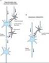
Hierarchical systems include all the pathways directly involved in sensory perception and motor control. The pathways are generally clearly delineated, being composed of large myelinated fibers that can often conduct action potentials at a rate in excess of 50 m/s. The information is typically phasic and occurs in bursts of action potentials. In sensory systems, the information is processed sequentially by successive integrations at each relay nucleus on its way to the cortex. A lesion at any link will incapacitate the system. Within each nucleus and in the cortex, there are two types of cells: relay or projection neurons and local circuit neurons (Figure 21-5A). The projection neurons that form the interconnecting pathways transmit signals over long distances. The cell bodies are relatively large, and their axons emit collaterals that arborize extensively in the vicinity of the neuron. These neurons are excitatory, and their synaptic influences, which involve ionotropic receptors, are very short-lived. The excitatory transmitter released from these cells is, in most instances, glutamate. Local circuit neurons are typically smaller than projection neurons, and their axons arborize in the immediate vicinity of the cell body. The majority of these neurons are inhibitory, and they release either GABA or glycine. They synapse primarily on the cell body of the projection neurons but can also synapse on the dendrites of projection neurons as well as with each other. Two common types of pathways for these neurons (Figure 21-5A) include recurrent feedback pathways and feed-forward pathways. A special class of local circuit neurons in the spinal cord forms axoaxonic synapses on the terminals of sensory axons (Figure 21-5B). In some sensory pathways such as the retina and olfactory bulb, local circuit neurons may actually lack an axon and release neurotransmitter from dendritic synapses in a graded fashion in the absence of action potentials.
Although there is a great variety of synaptic connections in these hierarchical systems, the fact that a limited number of transmitters are utilized by these neurons indicates that any major pharmacologic manipulation of this system will have a profound effect on the overall excitability of the CNS. For instance, selectively blocking GABAA receptors with a drug such as picrotoxin results in generalized convulsions. Thus, although the mechanism of action of picrotoxin is specific in blocking the effects of GABA, the overall functional effect appears to be quite nonspecific, because GABA-mediated synaptic inhibition is so widely utilized in the brain.
|
|
Figure 21-5. Pathways in the central nervous system. A shows parts of three relay neurons (color) and two types of inhibitory pathways, recurrent and feed-forward. The inhibitory neurons are shown in gray. B shows the pathway responsible for presynaptic inhibition in which the axon of an inhibitory neuron (gray) synapses on the axon terminal of an excitatory fiber (color). |
Nonspecific or Diffuse Neuronal Systems
Neuronal systems that contain one of the monoamines¾norepinephrine, dopamine, or 5-hydroxytryptamine (serotonin)¾provide examples in this category. Certain other pathways emanating from the reticular formation and possibly some peptide-containing pathways also fall into this category. These systems differ in fundamental ways from the hierarchical systems, and the noradrenergic systems serve to illustrate the differences.
Noradrenergic cell bodies are found primarily in a compact cell group called the locus ceruleus located in the caudal pontine central gray matter. The number of neurons in this cell group is small, approximately 1500 on each side of the brain in the rat.
Because these axons are fine and unmyelinated, they conduct very slowly, at about 0.5 m/s. The axons branch repeatedly and are extraordinarily divergent. Branches from the same neuron can innervate several functionally different parts of the CNS. In the neocortex, these fibers have a tangential organization and therefore can monosynaptically influence large areas of cortex. The pattern of innervation by noradrenergic fibers in the cortex and nuclei of the hierarchical systems is diffuse, and these fibers form a very small percentage of the total number in the area. In addition, the axons are studded with periodic enlargements called varicosities, which contain large numbers of vesicles. In some instances, these varicosities do not form synaptic contacts, suggesting that norepinephrine may be released in a rather diffuse manner, as occurs with the noradrenergic autonomic innervation of smooth muscle. This indicates that the cellular targets of these systems are determined largely by the location of the receptors rather than by the location of the release sites. Finally, most neurotransmitters utilized by diffuse neuronal systems, including norepinephrine, act¾perhaps exclusively¾on metabotropic receptors and therefore initiate long-lasting synaptic effects. Based on these observations, it is clear that the monoamine systems cannot be conveying topographically specific types of information; rather, vast areas of the CNS must be affected simultaneously and in a rather uniform way. It is not surprising, then, that these systems have been implicated in such global functions as sleeping and waking, attention, appetite, and emotional states.
CENTRAL NEUROTRANSMITTERS
Introduction
A vast number of small molecules have been isolated from the brain, and studies using a variety of approaches suggest that the agents listed in Table 21-2 are neurotransmitters. A brief summary of the evidence for some of these compounds follows.
Amino Acids
The amino acids of primary interest to the pharmacologist fall into two categories: the acidic amino acid glutamate and the neutral amino acids glycine and GABA. All of these compounds are present in high concentrations in the CNS and are extremely potent modifiers of neuronal excitability.
A. GLUTAMATE
Excitatory synaptic transmission is mediated by glutamate, which is present in very high concentrations in excitatory synaptic vesicles (~ 100 mM). Glutamate is released into the synaptic cleft by Ca2+-dependent exocytosis (Figure 21-6). The released glutamate acts on postsynaptic glutamate receptors and is cleared by glutamate transporters present on surrounding glia. In glia, glutamate is converted to glutamine by glutamine synthetase, released from the glia, taken up by the nerve terminal, and converted back to glutamate by the enzyme glutaminase. The high concentration of glutamate in synaptic vesicles is achieved by the vesicular glutamate transporter (VGLUT).
Virtually all neurons that have been tested are strongly excited by glutamate. This excitation is caused by the activation of both ionotropic and metabotropic receptors, which have been extensively characterized by molecular cloning. The ionotropic receptors can be further divided into three subtypes based on the action of selective agonists: a-amino-3-hydroxy-5-methylisoxazole-4-propionic acid (AMPA), kainic acid (KA) and N-methyl-D-aspartate (NMDA). All of the ionotropic receptors are composed of four subunits. AMPA receptors, which are present on all neurons, are heterotetramers assembled from four subunits (GluR1-GluR4). The majority of AMPA receptors contain the GluR2 subunit and are permeable to Na+ and K+, but not to Ca2+. Some AMPA receptors, typically present on inhibitory interneurons, lack the GluR2 subunit and are also permeable to Ca2+.
Kainate receptors are not as uniformly distributed as AMPA receptors, being expressed at high levels in the hippocampus, cerebellum, and spinal cord. They are formed from a number of subunit combinations (GluR5-GluR7, and KA1 and KA2). Although KA1 and KA2 are unable to form channels on their own, their presence in the receptor changes the receptor's affinity and kinetics. Similar to AMPA receptors, kainate receptors are permeable to Na+ and K+ and in some subunit combinations can also be permeable to Ca2+. NMDA receptors are as ubiquitous as AMPA receptors, being present on essentially all neurons in the CNS. All NMDA receptors require the presence of the subunit NR1. The channel also contains one or two NR2 subunits (NR2A-D). Unlike AMPA and kainate receptors, all NMDA receptors are highly permeable to Ca2+ as well as to Na+ and K+. NMDA receptor function is controlled in a number of intriguing ways. In addition to glutamate binding, the channel also requires the binding of glycine to a separate site. The physiologic role for glycine binding is unclear because the glycine site appears to be saturated at normal ambient levels of glycine. Another key difference between AMPA and kainate receptors on the one hand, and NMDA receptors on the other, is that AMPA and kainate receptor activation results in channel opening at resting membrane potential, whereas NMDA receptor activation does not. This is due to the voltage-dependent block of the NMDA pore by extracellular Mg2+. When the neuron is strongly depolarized, as occurs with intense activation of the synapse or by activation of neighboring synapses, the Mg2+ ion is expelled and the channel opens. Thus, there are two requirements for NMDA receptor channel opening: Glutamate must bind the receptor and the membrane must be depolarized. The rise in intracellular Ca2+ that accompanies channel opening results in a long-lasting enhancement in synaptic strength that is referred to as long-term potentiation (LTP). The change can last for many hours or even days and is generally accepted as an important cellular mechanism underlying learning and memory.
The metabotropic glutamate receptors are G protein-coupled receptors that act indirectly on ion channels via G proteins. Metabotropic receptors (mGluR1-mGluR8) have been divided into three groups (I, II, and III). A variety of agonists and antagonists have been developed that interact selectively with the different groups. Group I receptors are typically located postsynaptically and are thought to cause neuronal excitation by activating a nonselective cation channel. These receptors also activate phospholipase C, leading to IP3-mediated intracellular Ca2+ release. In contrast, group II and group III receptors are typically located on presynaptic nerve terminals and act as inhibitory autoreceptors. Activation of these receptors causes the inhibition of Ca2+ channels resulting in inhibition of transmitter release. These receptors are activated only when the concentration of glutamate rises to high levels during repetitive stimulation of the synapse. Activation of these receptors causes the inhibition of adenylyl cyclase and decreases cAMP generation.
The postsynaptic membrane at excitatory synapses is thickened and referred to as the postsynaptic density (PSD; Figure 21-6). This is a highly complex structure containing glutamate receptors, signaling proteins, scaffolding proteins, and cytoskeletal proteins. A typical excitatory synapse contains AMPA receptors, which tend to be located toward the periphery, and NMDA receptors, which are concentrated in the center. Kainate receptors are present at a subset of excitatory synapses, but their exact location is unknown. Metabotropic glutamate receptors (group I), which are localized just outside the PSD, are also present at some excitatory synapses.
B. GABA AND GLYCINE
Both GABA and glycine are inhibitory neurotransmitters, which are typically released from local interneurons. Interneurons that release glycine are restricted to the spinal cord and brain stem, whereas interneurons releasing GABA are present throughout the CNS, including the spinal cord. It is interesting that some interneurons in the spinal cord can release both GABA and glycine. Glycine receptors are pentameric structures that are selectively permeable to Cl-. Strychnine, which is a potent spinal cord convulsant and has been used in some rat poisons, selectively blocks glycine receptors.
GABA receptors are divided into two main types: GABAA and GABAB. IPSPs in many areas of the brain have a fast and slow component. The fast component is mediated by GABAAreceptors and the slow component by GABAB receptors. The difference in kinetics stems from the differences in coupling of the receptors to ion channels. GABAA receptors are inotropic receptors and, like glycine receptors, are pentameric structures that are selectively permeable to Cl-. These receptors are selectively inhibited by picrotoxin and bicuculline, both of which cause generalized convulsions. A great many subunits for GABAA receptors have been cloned; this accounts for the large diversity in the pharmacology of GABAA receptors, making them key targets for clinically useful agents (see Chapter 22). GABAB receptors are metabotropic receptors that are selectively activated by the antispastic drug baclofen. These receptors are coupled to G proteins that, depending on their cellular location, either inhibit Ca2+ channels or activate K+ channels. The GABAB component of the IPSP is due to a selective increase in K+ conductance. This IPSP is long-lasting and slow because the coupling of receptor activation to K+ channel opening is indirect and delayed. GABAB receptors are localized to the perisynaptic region and thus require the spillover of GABA from the synaptic cleft. GABAB receptors are also present on the axon terminals of many excitatory and inhibitory synapses. In this case, GABA spills over onto these presynaptic GABAB receptors, inhibiting transmitter release by inhibiting Ca2+ channels. In addition to their coupling to ion channels, GABABreceptors also inhibit adenylyl cyclase and decrease cAMP generation.
|
|
Figure 21-6. Schematic diagram of a glutamate synapse. Glutamine is imported into the glutamatergic neuron (A) and converted into glutamate by glutaminase. The glutamate is then concentrated in vesicles by the vesicular glutamate transporter (VGLUT). Upon release into the synapse, glutamate can interact with AMPA and NMDA ionotropic receptor channels (AMPAR, NMDAR) in the postsynaptic density (PSD) and with metabotropic receptors (MGluR) on the postsynaptic cell (B). Synaptic transmission is terminated by active transport of the glutamate into a neighboring glial cell (C) by a glutamate transporter. It is synthesized into glutamine by glutamine synthetase and exported into the glutamatergic axon. (D) shows a model NMDA receptor channel complex consisting of a tetrameric protein that becomes permeable to Na+ and Ca2+ when it binds a glutamate molecule. |
Acetylcholine
Acetylcholine was the first compound to be identified pharmacologically as a transmitter in the CNS. Eccles showed in the early 1950s that excitation of Renshaw cells by motor axon collaterals in the spinal cord was blocked by nicotinic antagonists. Furthermore, Renshaw cells were extremely sensitive to nicotinic agonists. These experiments were remarkable for two reasons. First, this early success at identifying a transmitter for a central synapse was followed by disappointment because it remained the sole central synapse for which the transmitter was known until the late 1960s, when comparable data became available for GABA and glycine. Second, the motor axon collateral synapse remains one of the best-documented examples of a cholinergic nicotinic synapse in the mammalian CNS, despite the rather widespread distribution of nicotinic receptors as defined by in situ hybridization studies. Most CNS responses to acetylcholine are mediated by a large family of G protein-coupled muscarinic receptors. At a few sites, acetylcholine causes slow inhibition of the neuron by activating the M2 subtype of receptor, which opens potassium channels. A far more widespread muscarinic action in response to acetylcholine is a slow excitation that in some cases is mediated by M1receptors. These muscarinic effects are much slower than either nicotinic effects on Renshaw cells or the effect of amino acids. Furthermore, this M1 muscarinic excitation is unusual in that acetylcholine produces it by decreasing the membrane permeability to potassium, ie, the opposite of conventional transmitter action.
A number of pathways contain acetylcholine, including neurons in the neostriatum, the medial septal nucleus, and the reticular formation. Cholinergic pathways appear to play an important role in cognitive functions, especially memory. Presenile dementia of the Alzheimer type is reportedly associated with a profound loss of cholinergic neurons. However, the specificity of this loss has been questioned because the levels of other putative transmitters, eg, somatostatin, are also decreased.
Monoamines
Monoamines include the catecholamines (dopamine and norepinephrine) and 5-hydroxytryptamine. Although these compounds are present in very small amounts in the CNS, they can be localized using extremely sensitive histochemical methods. These pathways are the site of action of many drugs; for example, the CNS stimulants cocaine and amphetamine appear to act primarily at catecholamine synapses. Cocaine blocks the reuptake of dopamine and norepinephrine, whereas amphetamines cause presynaptic terminals to release these transmitters.
A. DOPAMINE
The major pathways containing dopamine are the projection linking the substantia nigra to the neostriatum and the projection linking the ventral tegmental region to limbic structures, particularly the limbic cortex. The therapeutic action of the antiparkinsonism drug levodopa is associated with the former area (Chapter 28), whereas the therapeutic action of the antipsychotic drugs is thought to be associated with the latter (Chapter 29). Dopamine-containing neurons in the tuberobasal ventral hypothalamus play an important role in regulating hypothalamohypophysial function. Five dopamine receptors have been identified, and they fall into two categories: D1-like (D1 and D5) and D2-like (D2, D3, D4). All dopamine receptors are metabotropic. Dopamine generally exerts a slow inhibitory action on CNS neurons. This action has been best characterized on dopamine-containing substantia nigra neurons, where D2-receptor activation opens potassium channels via the Gi coupling protein.
B. NOREPINEPHRINE
Most noradrenergic neurons are located in the locus ceruleus or the lateral tegmental area of the reticular formation. Although the density of fibers innervating various sites differs considerably, most regions of the CNS receive diffuse noradrenergic input. All noradrenergic receptor subtypes are metabotropic. When applied to neurons, norepinephrine can hyperpolarize them by increasing potassium conductance. This effect is mediated by a2 receptors and has been characterized most thoroughly on locus ceruleus neurons. In many regions of the CNS, norepinephrine actually enhances excitatory inputs by both indirect and direct mechanisms. The indirect mechanism involves disinhibition; that is, inhibitory local circuit neurons are inhibited. The direct mechanism involves blockade of potassium conductances that slow neuronal discharge. Depending on the type of neuron, this effect is mediated by either a1 or b receptors. Facilitation of excitatory synaptic transmission is in accordance with many of the behavioral processes thought to involve noradrenergic pathways, eg, attention and arousal.
C. 5-HYDROXYTRYPTAMINE
Most 5-hydroxytryptamine (5-HT, serotonin) pathways originate from neurons in the raphe or midline regions of the pons and upper brain stem. 5-HT is contained in unmyelinated fibers that diffusely innervate most regions of the CNS, but the density of the innervation varies. 5-HT acts on more than a dozen receptor subtypes. Except for the 5-HT3 receptor, all of these receptors are metabotropic. The ionotropic 5-HT3 receptor exerts a rapid excitatory action at a very limited number of sites in the CNS. In most areas of the CNS, 5-HT has a strong inhibitory action. This action is mediated by 5-HT1A receptors and is associated with membrane hyperpolarization caused by an increase in potassium conductance. It has been found that 5-HT1A receptors and GABAB receptors share the same potassium channels. Some cell types are slowly excited by 5-HT owing to its blockade of potassium channels via 5-HT2 or 5-HT4 receptors. Both excitatory and inhibitory actions can occur on the same neuron. It has often been speculated that 5-HT pathways may be involved in the hallucinations induced by LSD, since this compound can antagonize the peripheral actions of 5-HT. However, LSD does not appear to be a 5-HT antagonist in the CNS, and typical LSD-induced behavior is still seen in animals after raphe nuclei are destroyed. Other proposed regulatory functions of 5-HT-containing neurons include sleep, temperature, appetite, and neuroendocrine control.
Peptides
A great many CNS peptides have been discovered that produce dramatic effects both on animal behavior and on the activity of individual neurons. Many of the peptides have been mapped with immunohistochemical techniques and include opioid peptides (eg, enkephalins, endorphins), neurotensin, substance P, somatostatin, cholecystokinin, vasoactive intestinal polypeptide, neuropeptide Y, and thyrotropin-releasing hormone. As in the peripheral autonomic nervous system, peptides often coexist with a conventional nonpeptide transmitter in the same neuron. A good example of the approaches used to define the role of these peptides in the CNS comes from studies on substance P and its association with sensory fibers. Substance P is contained in and released from small unmyelinated primary sensory neurons in the spinal cord and brain stem and causes a slow EPSP in target neurons. These sensory fibers are known to transmit noxious stimuli, and it is therefore surprising that¾although substance P receptor antagonists can modify responses to certain types of pain¾they do not block the response. Glutamate, which is released with substance P from these synapses, presumably plays an important role in transmitting pain stimuli. Substance P is certainly involved in many other functions, because it is found in many areas of the CNS that are unrelated to pain pathways.
Many of these peptides are also found in peripheral structures, including peripheral synapses. They are described in Chapters 6 and 17.
Nitric Oxide
The CNS contains a substantial amount of nitric oxide synthase (NOS), which is found within certain classes of neurons. This neuronal NOS is an enzyme activated by calcium-calmodulin, and activation of NMDA receptors, which increases intracellular calcium, results in the generation of nitric oxide. Although a physiologic role for nitric oxide has been clearly established for vascular smooth muscle, its role in synaptic transmission and synaptic plasticity remains controversial.
Endocannabinoids
The primary psychoactive ingredient in cannabis, D9-tetrahydrocannabinol (D9-THC), affects the brain mainly by activating a specific cannabinoid receptor, CB1. CB1 is expressed at high levels in many brain regions, and several endogenous brain lipids, including anandamide and 2-arachidonylglycerol, have been identified as CB1 ligands. These ligands are not stored, as are classic neurotransmitters, but instead are rapidly synthesized by neurons in response to depolarization and consequent calcium influx. In further contradistinction to classic neurotransmitters, endogenous cannabinoids can function as retrograde synaptic messengers: They are released from postsynaptic neurons and travel backward across synapses, activating CB1 receptors on presynaptic neurons and suppressing transmitter release. Cannabinoids may affect memory, cognition, and pain perception by this mechanism.
REFERENCES
Aizenman CD et al: Use-dependent changes in synaptic strength at the Purkinje cell to deep nuclear synapse. Prog Brain Res 2000;124:257.
Bredt DS, Nicoll RA: AMPA receptor trafficking at excitatory synapses. Neuron 2003;40:361.
Catterall WA et al: Compendium of voltage-gated ion channels: Calcium channels. Pharmacol Rev 2003;55:579.
Catterall WA, Goldin AL, Waxman SG: Compendium of voltage-gated ion channels: Sodium channels. Pharmacol Rev 2003;55:575.
Clapham DE et al: Compendium of voltage-gated ion channels: Transient receptor potential channels. Pharmacol Rev 2003;55:591.
Fremeau RT, Jr et al: VGLUTs define subsets of excitatory neurons and suggest novel roles for glutamate. Trends Neurosci 2004;27:98.
Freund TF, Katona I, Piomelli D: Role of endogenous cannabinoids in synaptic signaling. Physiol Rev 2003;83:1017.
Gouaux E, MacKinnon R: Principles of selective ion transport in channels and pumps. Science 2005;310:1461.
Hall ZW: In: An Introduction to Molecular Neurobiology. Sinauer, 1992.
Hille B: Ionic Channels of Excitable Membranes, 3rd ed. Sinauer, 2001.
Julius D, Basbaum AI: Molecular mechanisms of nociception. Nature 2001;413:203.
Koles L, Furst S, Illes P: P2X and P2Y receptors as possible targets of therapeutic manipulations in CNS illnesses. Drug News Perspect 2005;18:85.
Malenka RC, Nicoll RA: Long-term potentiation¾A decade of progress? Science 1999;285:1870.
Mody I, Pearce RA: Diversity of inhibitory neurotransmission through GABA(A) receptors. Trends Neurosci 2004;27:569.
Moran MM, Xu H, Clapham DE: TRP ion channels in the nervous system. Curr Opin Neurobiol 2004;14:362.
Nestler EJ, Hyman SE, Malenka RC: Molecular Neuropharmacology. McGraw-Hill, 2001.
Rudolph U, Mohler H: Analysis of GABAA receptor function and dissection of the pharmacology of benzodiazepines and general anesthetics through mouse genetics. Annu Rev Pharmacol Toxicol 2004;44:475.
Sudhof TC: The synaptic vesicle cycle. Annu Rev Neurosci 2004;27:509.
Wilson RI, Nicoll RA: Endocannabinoid signaling in the brain. Science 2002;296:678.