INTRODUCTION
Local anesthetics reversibly block impulse conduction along nerve axons and other excitable membranes that utilize sodium channels as the primary means of action potential generation. Clinically, local anesthetics are used to block pain sensation from¾or sympathetic vasoconstrictor impulses to¾specific areas of the body. The first local anesthetic introduced into medical practice, cocaine, was isolated by Niemann in 1860 and introduced into practice by Koller in 1884 as an ophthalmic anesthetic. Despite the fact that its chronic use was associated with psychological dependence (addiction), cocaine was used clinically because it was the only local anesthetic drug available for 30 years. In an attempt to improve upon the clinical properties of cocaine, Einhorn in 1905 synthesized procaine, which became the dominant local anesthetic for the next 50 years. Subsequently, newer local anesthetics were introduced with the goal of reducing local tissue irritation, minimizing systemic cardiac and central nervous system (CNS) toxicity, and achieving a faster onset and longer duration of action. Lidocaine, which is still the most widely used local anesthetic, was synthesized in 1943 by Lofgren.
The development of newer agents continues because it is relatively easy to synthesize chemicals with local anesthetic properties. Unfortunately, it is difficult to reduce the toxicity of these compounds because the common side effects of local anesthetics represent extensions of their therapeutic effects. New research into the mechanisms of local anesthetic-induced cardiac and spinal toxicity and identification of alternative drug targets for spinal analgesia (eg, opioid receptors, a2 adrenoceptors, NMDA receptors, N-type calcium channels, and adenosine receptors) suggest that it may be possible to develop safer drugs in the future. To extend the duration of the local anesthetic action, a variety of novel delivery systems are in development (eg, polymers, suspensions). Transdermal local anesthetic delivery systems have also been successfully introduced into clinical practice for providing topical analgesia.
I. BASIC PHARMACOLOGY OF LOCAL ANESTHETICS
Chemistry
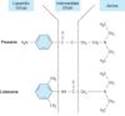
Most local anesthetic agents consist of a lipophilic group (eg, an aromatic ring) connected by an intermediate chain via an ester or amide to an ionizable group (eg, a tertiary amine) (Figure 26-1; Table 26-1). In addition to the general physical properties of the molecules, specific stereochemical configurations are associated with differences in the potency of stereoisomers (eg, bupivacaine, ropivacaine). Because ester links are more prone to hydrolysis than amide links, esters usually have a shorter duration of action.
Local anesthetics are weak bases and are usually made available clinically as salts to increase solubility and stability. In the body, they exist either as the uncharged base or as a cation. The relative proportions of these two forms is governed by their pKa and the pH of the body fluids according to the Henderson-Hasselbalch equation:
Because the pKa of most local anesthetics is in the range of 8.0-9.0, the larger percentage in body fluids at physiologic pH will be the charged, cationic form. The cationic form is the most active form at the receptor site because it cannot readily exit from closed channels. However, the uncharged form is important for rapid penetration of biologic membranes and producing a clinical effect, since the local anesthetic receptor is not readily accessible from the external side of the cell membrane. Therefore, local anesthetics are much less effective when they are injected into infected tissues because a smaller percentage of the local anesthetic is nonionized and available for diffusion across the membrane in an environment with a low extracellular pH.
|
|
Figure 26-1. Structures of two prototypical local anesthetics, the aminoester procaine and the aminoamide lidocaine. In both drugs, a lipophilic aromatic group is joined to a more hydrophilic tertiary amine base, by an intermediate amide or ester bond. |
Pharmacokinetics
The pharmacokinetic properties of the commonly used amide local anesthetics are summarized in Table 26-2. The pharmacokinetics of the ester-based local anesthetics have not been extensively studied owing to their rapid breakdown in plasma (elimination half-life < 1 minute). Local anesthetics are usually administered by injection into dermis and soft tissues located in the area of nerves. Thus, absorption and distribution are not as important in controlling the onset of effect as in determining the rate of offset of local analgesia, and the likelihood of CNS and cardiac toxicity. Topical application of local anesthetics (eg, transmucosal or transdermal) requires drug diffusion for both onset and offset of anesthetic effect.
A. ABSORPTION
Systemic absorption of injected local anesthetic from the site of administration is determined by several factors, including dosage, site of injection, drug-tissue binding, local blood flow, use of vasoconstrictors (eg, epinephrine), and the physicochemical properties of the drug itself. Application of a local anesthetic to a highly vascular area such as the tracheal mucosa or the tissue surrounding intercostal nerves results in more rapid absorption and thus higher blood levels than if the local anesthetic had been injected into a poorly perfused tissue such as tendon, dermis, or subcutaneous fat. For regional anesthesia involving block of large nerves, maximum blood levels of local anesthetic decrease according to the site of administration in the following order: intercostal (highest) > caudal > epidural > brachial plexus > sciatic nerve (lowest).
Vasoconstrictor substances such as epinephrine reduce systemic absorption of local anesthetics from the injection site by decreasing blood flow in these areas. This is important for drugs with intermediate or short durations of action such as procaine, lidocaine, and mepivacaine (but not prilocaine).
Since blood levels are lowered up to 30% when vasoconstrictors are added to local anesthetics, localized neuronal uptake is enhanced because of higher local tissue concentrations in the region of drug administration, and the systemic toxic effects are reduced. Furthermore, when used in spinal anesthesia, epinephrine acts directly on the cord to both enhance and prolong local anesthetic-induced spinal anesthesia by acting on a2 adrenoceptors, which inhibit release of substance P (neurokinin-1) and reduce sensory neuron firing. The recognition of this fact has led to the use of clonidine and dexmedetomidine to augment local anesthetic effect in the subarachnoid space and on peripheral nerves. The combination of reduced systemic absorption, enhanced local neuronal anesthetic uptake, and a2 activation by epinephrine in the CNS is responsible for prolonging the local anesthetic effect by up to 50%. Vasoconstrictors are less effective in prolonging anesthetic action of the more lipid-soluble, long-acting drugs (eg, bupivacaine and ropivacaine), possibly because these molecules are highly tissue-bound. Finally, cocaine is unique owing to its intrinsic sympathomimetic properties.
B. DISTRIBUTION
The amide local anesthetics are widely distributed after intravenous bolus administration. There is also evidence that sequestration can occur in lipophilic storage sites (eg, fat). After an initial rapid distribution phase, which consists of uptake into highly perfused organs such as the brain, liver, kidney, and heart, a slower distribution phase occurs with uptake into moderately well-perfused tissues, such as muscle and the gastrointestinal tract. As a result of the extremely short plasma half-lives of the ester type agents, their tissue distribution has not been extensively studied.
C. METABOLISM AND EXCRETION
The local anesthetics are converted in the liver (amide type) or in plasma (ester type) to more water-soluble metabolites and then excreted in the urine. Since local anesthetics in the uncharged form diffuse readily through lipid membranes, little or no urinary excretion of the neutral form occurs. Acidification of urine promotes ionization of the tertiary amine base to the more water-soluble charged form, which is more readily excreted.
Ester-type local anesthetics are hydrolyzed very rapidly in the blood by circulating butyrylcholinesterase (pseudocholinesterase) to inactive metabolites. Therefore, procaine and chloroprocaine have very short plasma half-lives (< 1 minute).
The amide linkage of amide local anesthetics is hydrolyzed by liver microsomal cytochrome P450 isozymes. There is considerable variation in the rate of liver metabolism of individual amide compounds, the approximate order being prilocaine (fastest) > lidocaine > mepivacaine > ropivacaine > bupivacaine and levobupivacaine (slowest). As a result, toxicity from amide-type local anesthetics is more likely to occur in patients with hepatic disease. For example, the average elimination half-life of lidocaine may be increased from 1.6 hours in normal patients (t1/2, Table 26-2) to more than 6 hours in patients with severe liver disease. Many other drugs used in anesthesia are metabolized by the same P450 isozymes, and concomitant use of these competing drugs may slow the hepatic metabolism of the local anesthetics.
Decreased hepatic elimination of local anesthetics would also be anticipated in patients with reduced hepatic blood flow. For example, the hepatic elimination of lidocaine in patients anesthetized with volatile anesthetics (which reduce liver blood flow) is slower than in patients anesthetized with intravenous anesthetics.
Pharmacodynamics
A. MECHANISM OF ACTION
The primary mechanism of action of local anesthetics is blockade of voltage-gated sodium channels (Figure 26-2). The excitable membrane of nerve axons, like the membrane of cardiac muscle (see Chapter 14) and neuronal cell bodies (see Chapter 21), maintains a resting transmembrane potential of -90 to -60 mV. During excitation, the sodium channels open, and a fast inward sodium current quickly depolarizes the membrane toward the sodium equilibrium potential (+40 mV). As a result of depolarization, the sodium channels close (inactivate) and potassium channels open. The outward flow of potassium repolarizes the membrane toward the potassium equilibrium potential (about -95 mV); repolarization returns the sodium channels to the rested state. The transmembrane ionic gradients are maintained by the sodium pump. These ionic fluxes are similar to those in heart muscle, and local anesthetics have similar effects in both tissues.
The function of sodium channels can be disrupted in several ways. Biologic toxins such as batrachotoxin, aconitine, veratridine, and some scorpion venoms bind to receptors within the channel and prevent inactivation. This results in prolonged influx of sodium through the channel and depolarization of the resting potential. The marine toxins tetrodotoxin (TTX) and saxitoxin block sodium channels by binding to channel receptors near the extracellular surface. Their clinical effects superficially resemble those of local anesthetics (ie, block of conduction without a change in the resting potential) even though their receptor site is quite different. Spinal neurons can be differentiated on the basis of tetrodotoxin effect into TTX-sensitive and TTX-resistant neurons. Some evidence suggests that the TTX-resistant neurons are responsible for pain transmission and are the primary targets for local anesthetics in producing spinal anesthesia. Local anesthetics bind to receptors near the intracellular end of the sodium channel and block the channel in a time- and voltage-dependent fashion (see below). The sodium channel has been cloned, its primary structure has been characterized, and mutational analysis has allowed identification of parts of the local anesthetic binding site.
When progressively increasing concentrations of a local anesthetic are applied to a nerve fiber, the threshold for excitation increases, impulse conduction slows, the rate of rise of the action potential declines, the action potential amplitude decreases, and, finally, the ability to generate an action potential is completely abolished. These progressive effects result from binding of the local anesthetic to more and more sodium channels. If the sodium current is blocked over a critical length of the nerve, propagation across the blocked area is no longer possible. In myelinated nerves, the critical length is 2 to 3 nodes of Ranvier. At the minimum dose required to block propagation, the resting potential is not significantly altered.
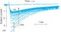
The blockade of sodium channels by most local anesthetics is both voltage- and time-dependent: Channels in the rested state, which predominate at more negative membrane potentials, have a much lower affinity for local anesthetics than activated (open state) and inactivated channels, which predominate at more positive membrane potentials (see Figure 14-8). Thus, the effect of a given drug concentration is more marked in rapidly firing axons than in resting fibers (Figure 26-3).
Between successive action potentials, a portion of the sodium channels will recover from the local anesthetic block (see Figure 14-8). The recovery from drug-induced block is 10 to 1000 times slower than the recovery of channels from normal inactivation (as shown for the cardiac membrane in Figure 14-4). As a result, the refractory period is lengthened and the nerve conducts fewer impulses.
Elevated extracellular calcium partially antagonizes the action of local anesthetics owing to the calcium-induced increase in the surface potential on the membrane (which favors the low-affinity rested state). Conversely, increases in extracellular potassium depolarize the membrane potential and favor the inactivated state, enhancing the effect of local anesthetics.
Local anesthetics can be shown to block a variety of other ion channels, including nicotinic acetylcholine channels in the spinal cord. However, there is no convincing evidence that this mechanism is important in the clinical effects of these drugs. High concentrations of local anesthetics can interfere with intra-axonal transport and calcium homeostasis in the nerve terminal, contributing to potential spinal toxicity.
B. STRUCTURE-ACTIVITY CHARACTERISTICS OF LOCAL ANESTHETICS
The smaller and more lipophilic the local anesthetic, the faster the rate of interaction with the sodium channel receptor. Potency is also positively correlated with lipid solubility as long as the agent retains sufficient water solubility to diffuse to the site of action on the neuronal membrane. Lidocaine, procaine, and mepivacaine are more water-soluble than tetracaine, bupivacaine, and ropivacaine. The latter agents are more potent and have longer durations of local anesthetic action. These long-acting local anesthetics also bind more extensively to proteins and can be displaced from these binding sites by other protein-bound drugs. In the case of optically active agents (eg, bupivacaine), the S(+)isomer can usually be shown to be moderately more potent than the R(-) isomer.
C. OTHER ACTIONS ON NERVES
Since local anesthetics are capable of blocking all nerves, their actions are not limited to the desired loss of sensation from sites of noxious (painful) stimuli. Although motor paralysis can be desirable during surgery, it may also limit the ability of the patient to cooperate during obstetric delivery or ambulate after outpatient surgery. During spinal anesthesia, motor paralysis may impair respiratory activity and autonomic nerve blockade can lead to hypotension upon ambulation. Residual autonomic blockade interferes with bladder function resulting in urinary retention and the need for catheterization.
Nerve fibers differ significantly in their susceptibility to local anesthetic blockade on the basis of differences in size and degree of myelination (Table 26-3). Upon direct application of a local anesthetic to a nerve root, the smaller B and C fibers are blocked first, followed by other sensations, and motor function is the last to be blocked.
1. Effect of fiber diameter¾ Local anesthetics preferentially block small fibers because the distance over which such fibers can passively propagate an electrical impulse is shorter. During the onset of local anesthesia, when short sections of the nerve are blocked, the small-diameter fibers are the first to fail to conduct. For myelinated nerves, at least two and preferably three successive nodes of Ranvier must be blocked by the local anesthetic to halt impulse propagation. With larger and thicker nerve fibers (eg, motor neurons), the nodes are farther apart, creating greater resistance to blockade. Myelinated nerves tend to become blocked before unmyelinated nerves of the same diameter. For this reason, the preganglionic B fibers are blocked before the smaller unmyelinated C fibers involved in pain transmission.
2. Effect of firing frequency¾ Another important reason for preferential blockade of sensory fibers follows directly from the state- and use-dependent mechanism of action of local anesthetics. Blockade by these drugs is more marked at higher frequencies of depolarization. Sensory (pain) fibers have a high firing rate and a relatively long action potential duration. Motor fibers fire at a slower rate and have a shorter action potential duration. Type A delta and C fibers are smaller-diameter fibers that participate in high-frequency pain transmission. Therefore, these fibers are blocked earlier with lower concentrations of local anesthetics than are the large A alpha fibers.
3. Effect of fiber position in the nerve bundle¾ An anatomic circumstance that sometimes creates exceptions to the above rules for differential nerve block is the location of the fibers in the peripheral nerve bundle. In large nerve trunks, motor nerves are usually located circumferentially, and for that reason they are the first to be exposed to the local anesthetic when it is administered into the tissue surrounding the nerve. Therefore, it is not uncommon for motor nerve block to occur before sensory block in large mixed nerves. In the extremities, proximal sensory fibers are located in the outer portion of the nerve trunk, whereas the distal sensory innervation is in the core of the nerve. Thus, during infiltration block of a large nerve, sensory analgesia first develops proximally and then spreads distally as the drug penetrates into the core of the nerve.
D. EFFECTS ON OTHER EXCITABLE MEMBRANES
Local anesthetics have weak neuromuscular blocking effects that are of little clinical importance. However, their effects on cardiac cell membranes are of major clinical significance, and some local anesthetics are useful antiarrhythmic agents (see Chapter 14) at concentrations lower than those required to produce nerve block. Others (eg, bupivacaine, ropivacaine) can cause lethal arrhythmias in high concentrations.
|
|
Figure 14-4. Dependence of sodium channel function on the membrane potential preceding the stimulus. Left: The fraction of sodium channels available for opening in response to a stimulus is determined by the membrane potential immediately preceding the stimulus. The decrease in the fraction available when the resting potential is depolarized in the absence of a drug (control curve) results from the voltage-dependent closure of h gates in the channels. The curve labeled Drug illustrates the effect of a typical local anesthetic antiarrhythmic drug. Most sodium channels are inactivated during the plateau of the action potential. Right: The time constant for recovery from inactivation after repolarization also depends on the resting potential. In the absence of drug, recovery occurs in less than 10 ms at normal resting potentials (-85 to -95 mV). Depolarized cells recover more slowly (note logarithmic scale). In the presence of a sodium channel-blocking drug, the time constant of recovery is increased, but the increase is far greater at depolarized potentials than at more negative ones. |
|
|
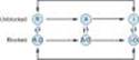
Figure 14-8. Diagram of a mechanism for the selective depressant action of antiarrhythmic drugs on sodium channels. The upper portion of the figure shows the population of channels moving through a cycle of activity during an action potential in the absence of drugs: R (rested) ® A (activated) ® I (inactivated). Recovery takes place via the I ® R pathway. Antiarrhythmic drugs (D) that act by blocking sodium channels can bind to their receptors in the channels, as shown by the vertical arrows, to form drug-channel complexes, indicated as R-D, A-D, and I-D. Binding of the drugs to the receptor varies with the state of the channel. The data available for a variety of sodium channel blockers indicate that the binding of the drugs to the active and inactivated channel receptor is much greater than the binding to the rested channel. Furthermore, recovery from the I-D state to the R-D state is much slower than from I to R. As a result, rapid activity (more activations and inactivations) and depolarization of the resting potential (more channels in the I state) will favor blockade of the channels and selectively suppress arrhythmic cells. |
|
|
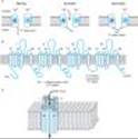
Figure 26-2. Functional and structural features of the Na+ channel that determine local anesthetic (LA) interactions. A: Cartoon of the sodium channel in an axonal membrane in the resting (h gate open), activated (h gate open), and inactivated states (h gate closed). Recovery from the inactivated, refractory state requires opening of the h gate. Local anesthetics bind to a receptor (R) within the channel and access it via the membrane phase or from the cytoplasm. B: Molecular arrangement of the 6 membrane-spanning peptides, four of which combine to form the channel around a central pore. The S4 segments (marked with "+" signs) are thought to constitute the voltage-sensing "m" gates of the channel. The linker peptide connecting the III and IV hexamers acts as the inactivation "h" gate. Ions travel through an open channel along a pore defined at its narrowest dimension by partial membrane penetration of the four extracellular loops of protein connecting S5 and S6 in each domain. Local anesthetic binding occurs on S6 segments and at other regions of the channel. C: Three-dimensional cartoon showing the configuration of the four hexamers around the central pore in the membrane. |
|
|
|
Figure 26-3. Effect of repetitive activity on the block of sodium current produced by a local anesthetic in a myelinated axon. A series of 25 pulses was applied, and the resulting sodium currents (downward deflections) are superimposed. Note that the current produced by the pulses rapidly decreased from the first to the 25th pulse. A long rest period following the train resulted in recovery from block, but the block could be reinstated by a subsequent train. (nA, nanoamperes.) (Modified and reproduced, with permission, from Courtney KR: Mechanism of frequency-dependent inhibition of sodium currents in frog myelinated nerve by the lidocaine derivative GEA. J Pharmacol Exp Ther 1975;195:225.) |
|
II. CLINICAL PHARMACOLOGY OF LOCAL ANESTHETICS
Introduction
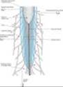
Local anesthetics can provide highly effective analgesia in well-defined regions of the body. The usual routes of administration include topical application (eg, nasal mucosa, wound margins), injection in the vicinity of peripheral nerve endings (infiltration) and major nerve trunks (blocks), and injection into the epidural or subarachnoid spaces surrounding the spinal cord (Figure 26-4). Intravenous regional anesthesia (so-called Bier block) is used for short surgical procedures (< 60 minutes) involving the upper and lower extremities. This is accomplished by intravenous injection of the anesthetic agent into a distal vein while the circulation of the limb is isolated with a proximally placed tourniquet. Finally, an infiltration block of autonomic sympathetic fibers can be used to evaluate the role of sympathetic tone in patients with peripheral vasospastic disorders.
The choice of local anesthetic for infiltration, peripheral nerve blocks, and central neuraxis blockade is usually based on the duration of action required. Procaine and chloroprocaine are short-acting; lidocaine, mepivacaine, and prilocaine have an intermediate duration of action; and tetracaine, bupivacaine, levobupivacaine, and ropivacaine are long-acting local anesthetics (Table 26-1).
The anesthetic effect of the agents with short and intermediate durations of action can be prolonged by increasing the dose or adding a vasoconstrictor agent (eg, epinephrine or phenylephrine). The vasoconstrictor slows the removal of the local anesthetic from the injection site. In addition, it decreases the blood level and the probability of CNS toxicity.
The onset of local anesthesia can be accelerated by the addition of sodium bicarbonate (1-2 mL) to the local anesthetic solution. This maximizes the amount of drug in the more lipid-soluble form.
Repeated injections of local anesthetics can result in loss of effectiveness (ie, tachyphylaxis) due to extracellular acidosis. Local anesthetics are commonly marketed as hydrochloride salts (pH 4.0-6.0). After injection, the salts are buffered in the tissue to physiologic pH, thereby providing sufficient free base concentration for diffusion through the axonal membrane. However, repeated injections of the local anesthetic can deplete the buffering capacity of the local tissues. The ensuing acidosis increases the extracellular cationic form, which diffuses poorly and results in tachyphylaxis. Tachyphylaxis to local anesthetics is common in areas with a limited buffer capacity (eg, the cerebrospinal fluid).
Pregnancy appears to increase susceptibility to local anesthetic toxicity, with reductions in the median doses required for neural blockade and to induce toxicity. Cardiac arrest leading to death following the epidural administration of 0.75% bupivacaine to women in labor resulted in the temporary withdrawal of the high concentration of this widely used long-acting local anesthetic. The subsequent introduction of purportedly less cardiotoxic alternatives to bupivacaine (ie, ropivacaine and levobupivacaine) has led to controversy because the evidence supporting enhanced safety is based on animal models. It is not clear whether the increased sensitivity to bupivacaine during pregnancy is due to elevated levels of estrogen, progesterone, or some other unrecognized factor.
Topical local anesthesia is often used for eye, ear, nose, and throat procedures and for cosmetic surgery. Satisfactory topical local anesthesia requires an agent capable of rapid penetration across the skin or mucosa, and with limited tendency to diffuse away from the site of application. Cocaine, because of its excellent penetration and local vasoconstrictor effects, has been used extensively for ear, nose and throat (ENT) procedures. Cocaine is somewhat irritating and therefore is less popular for ophthalmic procedures. Recent concern about its potential cardiotoxicity when combined with epinephrine has led most otolaryngologists and plastic surgeons to switch to a combination containing lidocaine and epinephrine. Other drugs used for topical anesthesia include tetracaine, pramoxine, dibucaine, benzocaine, and dyclonine.
Since local anesthetics have membrane-stabilizing effects, both parenteral (eg, intravenous lidocaine) and oral (eg, mexiletine, tocainide) formulations of local anesthetics have been used to treat patients with neuropathic pain syndromes because these syndromes are thought to involve uncontrolled, rapid, sensory fiber firing. Systemic local anesthetic drugs are commonly used as adjuvants to the combination of a tricyclic antidepressant (eg, amitriptyline) and an anticonvulsant (eg, carbamazepine) in patients who fail to respond to the combination of antidepressant and anticonvulsant. A period of 1-3 weeks may be required to observe a therapeutic effect after introduction of the local anesthetic in patients with neuropathic pain.
|
|
Figure 26-4. Schematic diagram of sites of injection of local anesthetics in and near the spinal canal. |
Toxicity
The two major forms of local anesthetic toxicity are: (1) systemic effects following absorption of the local anesthetic from their site of administration and (2) direct neurotoxicity from the local effects of these drugs when administered in close proximity to the spinal cord and other major nerve trunks. When blood levels of local anesthetics rise rapidly, adverse effects on several major organ systems may be observed.
A. CENTRAL NERVOUS SYSTEM
1. All local anesthetics¾ At low concentrations, all local anesthetics have the ability to produce sleepiness, light-headedness, visual and auditory disturbances, and restlessness. An early symptom of local anesthetic toxicity is circumoral and tongue numbness and a metallic taste. At higher concentrations, nystagmus and muscular twitching occur, followed by overt tonic-clonic convulsions. Local anesthetics apparently cause depression of cortical inhibitory pathways, thereby allowing unopposed activity of excitatory neuronal pathways. This transitional stage of unbalanced excitation (ie, seizure activity) is then followed by generalized CNS depression.
Convulsions due to excessive blood levels can usually be prevented by administering the smallest effective dose of the local anesthetic required for adequate surgical analgesia and by avoiding inadvertent intravascular injection, or injection into highly perfused tissues. When large doses of a local anesthetic are required (eg, for major peripheral nerve block), premedication with a parenteral benzodiazepine (eg, diazepam or midazolam) provides significant prophylaxis against local anesthetic-induced CNS toxicity by raising the seizure threshold.
If seizures do occur, it is important to prevent hypoxemia and acidosis. Although administration of oxygen does not prevent seizure activity, hyperoxemia may be beneficial after onset of seizures. Hypercapnia and acidosis may lower the seizure threshold, and so hyperventilation is recommended during treatment of seizures. In addition, hyperventilation increases blood pH, which in turn lowers extracellular potassium. This action hyperpolarizes the transmembrane potential of axons, which favors the rested (or low-affinity) state of the sodium channels, resulting in decreased local anesthetic toxicity.
Seizures induced by local anesthetics can also be treated with intravenous anesthetic drugs (eg, thiopental 1-2 mg/kg, propofol 0.5-1 mg/kg, midazolam 0.03-0.06 mg/kg, or diazepam 0.1-0.2 mg/kg). The muscular manifestations of a seizure can be blocked using a short-acting neuromuscular relaxant drug (eg, succinylcholine, 0.5-1 mg/kg IV). It should be emphasized that succinylcholine does not obliterate CNS manifestations of seizure activity. Rapid tracheal intubation and mechanical ventilation can prevent pulmonary aspiration of gastric contents and facilitate hyperventilation.
2. Cocaine¾ Since prehistoric times, the natives of Peru and Bolivia have chewed the leaves of the indigenous plant Erythroxylon coca, the source of cocaine, to obtain a feeling of well-being and reduce fatigue. Intense CNS effects can be achieved by sniffing cocaine powder and smoking cocaine base ("free basing"). Cocaine has become one of the most widely abused drugs (see Chapter 32). High doses of inhaled and injected cocaine have all of the toxicities described for other local anesthetics. In addition, cocaine can produce severe cardiovascular toxicity, including hypertension, arrhythmias, and myocardial failure.
B. NEUROTOXICITY
When applied at excessively high concentrations, all local anesthetics can produce direct neural toxicity. Chloroprocaine and lidocaine appear to be more neurotoxic than other local anesthetics when used for spinal anesthesia, with high local concentrations producing so-called transient radicular irritation (or transient neuropathic symptoms). It has been suggested that this toxicity results from pooling of high concentrations of the local anesthetic in the cauda equina (Figure 26-4). Although the precise mechanism of this neurotoxic action has not been established, both interference with axonal transport and disruption of calcium homeostasis have been implicated. Spinal neurotoxicity does not result from excessive sodium channel blockade.
C. CARDIOVASCULAR SYSTEM
The cardiovascular effects of local anesthetics result partly from direct effects on the cardiac and smooth muscle membranes and partly from indirect effects on the autonomic nervous system. As described in Chapter 14, local anesthetics block cardiac sodium channels and thus depress abnormal cardiac pacemaker activity, excitability, and conduction. At extremely high concentrations, local anesthetics can also block calcium channels. With the notable exception of cocaine, local anesthetics also depress the strength of cardiac contraction and cause arteriolar dilation, leading to systemic hypotension. Cardiovascular collapse is rare, but has been reported after large doses of bupivacaine and ropivacaine.
Cocaine differs from the other local anesthetics with respect to its cardiovascular effects. Cocaine's blockade of norepinephrine reuptake results in vasoconstriction and hypertension, as well as cardiac arrhythmias. The vasoconstriction produced by cocaine can lead to local ischemia and, in chronic abusers who use the nasal route, ulceration of the mucous membrane and damage to the nasal septum have been reported. The vasoconstrictor properties of cocaine can be used clinically to decrease bleeding from mucosal damage in the nasopharynx.
It has been suggested that bupivacaine may be more cardiotoxic than other long-acting local anesthetics. This reflects the fact that bupivacaine-induced blockade of sodium channels is potentiated by the long action potential duration of cardiac cells compared with nerve fibers. The most common electrocardiographic finding in patients with bupivacaine intoxication is a slow idioventricular rhythm with broad QRS complexes and eventually electromechanical dissociation.
Resuscitation from bupivacaine cardiovascular toxicity is extremely difficult even for experienced clinicians. The (S)-isomer, levobupivacaine, appears to have a lower propensity for cardiovascular toxicity than the racemic mixture or the (R)-isomer and has been approved for clinical use. Ropivacaine has clinical (pharmacodynamic) effects similar to those of bupivacaine, but is allegedly associated with a lower potential for cardiovascular toxicity. Ropivacaine is available only as the (S)-stereoisomer, which has inherently less affinity for the cardiac sodium channel. However, both cardiac toxicity and CNS toxicity have been reported when ropivacaine was used for peripheral nerve blocks.
D. HEMATOLOGIC EFFECTS
The administration of large doses (> 10 mg/kg) of prilocaine during regional anesthesia may lead to accumulation of the metabolite o-toluidine, an oxidizing agent capable of converting hemoglobin to methemoglobin. When sufficient methemoglobin is present (3-5 mg/dL), the patient may appear cyanotic and the blood "chocolate-colored." Although moderate levels of methemoglobinemia are well tolerated by healthy individuals, elevated methemoglobinemia may cause decompensation in patients with preexisting cardiac or pulmonary disease. The treatment of methemoglobinemia involves the intravenous administration of a reducing agent (eg, methylene blue or ascorbic acid), which rapidly converts methemoglobin to hemoglobin.
E. ALLERGIC REACTIONS
The ester-type local anesthetics are metabolized to p-aminobenzoic acid derivatives. These metabolites are responsible for allergic reactions in a small percentage of the patient population. Amides are not metabolized to p-aminobenzoic acid, and allergic reactions to amide local anesthetics are extremely rare.
PREPARATIONS AVAILABLE
Articaine (Septocaine)
Parenteral: 4% with 1:100,000 epinephrine
Benzocaine (generic)
Topical: 5, 6% creams; 15, 20% gels; 5, 20% ointments; 0.8% lotion; 20% liquid; 20% spray
Bupivacaine (generic, Marcaine, Sensorcaine)
Parenteral: 0.25, 0.5, 0.75% for injection; 0.25, 0.5, 0.75% with 1:200,000 epinephrine
Butamben picrate (Butesin Picrate)
Topical: 1% ointment
Chloroprocaine (generic, Nesacaine)
Parenteral: 1, 2, 3% for injection
Cocaine (generic)
Topical: 40, 100 mg/mL regular and viscous solutions; 5, 25 g powder
Dibucaine (generic, Nupercainal)
Topical: 0.5% cream; 1% ointment
Dyclonine (Dyclone)
Topical: 0.5, 1% solution
Levobupivacaine (Chirocaine)
Parenteral: 2.5, 5, 7.5 mg/mL
Lidocaine (generic, Xylocaine)
Parenteral: 0.5, 1, 1.5, 2, 4% for injection; 0.5, 1, 1.5, 2% with 1:200,000 epinephrine; 1, 2% with 1:100,000 epinephrine, 2% with 1:50,000 epinephrine
Topical: 2.5, 5% ointments; 0.5, 4% cream; 0.5, 2.5% gel; 2, 2.5, 4% solutions; 23, 46 mg/2 cm2 patch
Lidocaine and bupivacaine mixture (Duocaine)
Parenteral: 10 mg/mL lidocaine plus 3.75 mg/mL bupivacaine for injection
Lidocaine and prilocaine eutectic mixture (EMLA cream)
Topical: lidocaine 2.5% plus prilocaine 2.5%
Mepivacaine (generic, Carbocaine)
Parenteral: 1, 1.5, 2, 3% for injection; 2% with 1:20,000 levonordefrin
Pramoxine (generic, Tronothane)
Topical: 1% cream, lotion, spray, and gel
Procaine (generic, Novocain)
Parenteral: 1, 2, 10% for injection
Proparacaine (generic, Alcain, others)
0.5% solution for ophthalmic use
Ropivacaine (Naropin)
Parenteral: 0.2, 0.5, 0.75, 1.0 % solution for injection
Tetracaine (generic, Pontocaine)
Parenteral: 1% for injection; 0.2, 0.3% with 6% dextrose for spinal anesthesia
Topical: 1% ointment; 0.5% solution (ophthalmic); 1, 2% cream; 2% solution for nose and throat; 2% gel
REFERENCES
Brau ME et al: Effect of drugs used for neuropathic pain management on tetrodotoxin-resistant Na+ currents in rat sensory neurons. Anesthesiology 2001;94:137.
Davies PS, Galer BS: Review of lidocaine patch 5% studies in the treatment of postherpetic neuralgia. Drugs 2004;64:937.
Ferreira S et al: Effects of cocaine and its major metabolites on the HERG-encoded potassium channel. J Pharmacol Exp Ther 2001;299:220.
Hille B: Local anesthetics: Hydrophilic and hydrophobic pathways for the drug-receptor reactions. J Gen Physiol 1977;69:497.
Johnson ME et al: Effect of local anesthetic on neuronal cytoplasmic calcium and plasma membrane lysis (necrosis) in a cell culture model. Anesthesiology 2002;97:1466.
Kanai Y, Katsuki H, Takasaki M: Comparisons of the anesthetic potency and intracellular concentrations of S(-) and R(+) bupivacaine and ropivacaine in crayfish giant axon in vitro.Anesth Analg 2000;90:415.
Kanai Y, Katsuki H, Takasaki M: Lidocaine disrupts axonal membrane of rat sciatic nerve in vitro. Anesth Analg 2000;91:944.
Miyamoto Y et al: Direct inhibition of microtubule-based kinesin motility by local anesthetics. Biophys J 2000;78:940.
Oda A et al: Characteristics of ropivacaine block of Na+ channels in rat dorsal root ganglion neurons. Anesth Analg 2000;91:1213.
Scholtz A: Mechanisms of (local) anaesthetics on voltage-gated sodium and other ion channels. Br J Anaesth 2002;89:52.
Sinnott CJ et al: On the mechanism by which epinephrine potentiates lidocaine's peripheral nerve block. Anesthesiology 2003;98:181.
White JL, Durieux ME: Clinical pharmacology of local anesthetics. Anesthesiol Clin North Am 2005;23:73.
White PF: The changing role of non-opioid analgesic techniques in the management of postoperative pain. Anesth Analg 2005;101:S5.
Zapata-Sudo G et al: Is comparative cardiotoxicity of S(-) and R(+) bupivacaine related to enantiomer-selective inhibition of L-type Ca2+ channels? Anesth Analg 2001;92:496.
Zhou W et al: Mechanism underlying bupivacaine inhibition of G protein-gated inwardly rectifying K+ channels. Proc Natl Acad Sci USA 2001;98:6482.