INTRODUCTION
Hemostasis refers to the finely regulated dynamic process of maintaining fluidity of the blood, repairing vascular injury, and limiting blood loss while avoiding vessel occlusion (thrombosis) and inadequate perfusion of vital organs. Either extreme¾excessive bleeding or thrombosis¾represents a breakdown of the hemostatic mechanism. Common causes of dysregulated hemostasis include hereditary or acquired defects in the clotting mechanism and secondary effects of infection or cancer. The drugs used to limit abnormal bleeding and to inhibit thrombosis are the subjects of this chapter.
MECHANISMS OF BLOOD COAGULATION
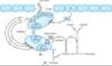
The vascular endothelial cell layer lining blood vessels has an anticoagulant phenotype, and circulating blood platelets and clotting factors do not normally adhere to it to an appreciable extent. In the setting of vascular injury, the endothelial cell layer rapidly undergoes a series of changes resulting in a more procoagulant phenotype. Injury exposes reactive subendothelial matrix proteins such as collagen and von Willebrand factor, which results in platelet adherence and activation, and secretion and synthesis of vasoconstrictors and platelet-recruiting and activating molecules. Thus, thromboxane A2 (TXA2) is synthesized from arachadonic acid within platelets and is a platelet activator and potent vasoconstrictor. Products secreted from platelet granules include adenosine diphosphate (ADP), a powerful inducer of platelet aggregation, and serotonin (5-HT), which stimulates aggregation and vasoconstriction. Activation of platelets results in a conformational change in the aIIbbIII integrin (IIb/IIIa) receptor, enabling it to bind fibrinogen, which cross-links adjacent platelets, resulting in aggregation and formation of a platelet plug (Figure 34-1). Simultaneously, the coagulation system cascade is activated, resulting in thrombin generation and a fibrin clot, which stabilizes the platelet plug (see below). Knowledge of the hemostatic mechanism is important for diagnosis of bleeding disorders. Patients with defects in the formation of the primary platelet plug (defects in primary hemostasis, eg, platelet function defects, von Willebrand disease) typically bleed from mucosal sites (gingiva, skin, heavy menses) with injury. In contrast, patients with defects in the clotting mechanism (secondary hemostasis, eg, hemophilia A) tend to bleed into deep tissues (joints, muscle, retroperitoneum), often with no apparent inciting event, and bleeding may recur unpredictably.
The platelet is central to normal hemostasis and thromboembolic disease, and is the target of many therapies discussed in this chapter. Platelet-rich thrombi (white thrombi) form in the high flow rate and high shear force environment of arteries. Occlusive arterial thrombi cause serious disease by producing downstream ischemia of extremities or vital organs, and can result in limb amputation or organ failure. Venous clots tend to be more fibrin-rich, contain large numbers of trapped red blood cells, and are recognized pathologically as red thrombi. Venous thrombi can cause severe swelling and pain of the affected extremity, but the most feared consequence is pulmonary embolism. This occurs when part or all of the clot breaks off from its location in the deep venous system and travels as an embolus through the right side of the heart and into the pulmonary arterial circulation. Sudden occlusion of a large pulmonary artery can cause acute right heart failure and sudden death. In addition lung ischemia or infarction will occur distal to the occluded pulmonary arterial segment. Such emboli usually arise from the deep venous system of the proximal lower extremities or pelvis. Although all thrombi are mixed, the platelet nidus dominates the arterial thrombus and the fibrin tail dominates the venous thrombus.
|
|
Figure 34-1. Thrombus formation at the site of the damaged vascular wall (EC, endothelial cell) and the role of platelets and clotting factors. Platelet membrane receptors include the glycoprotein (GP) Ia receptor, binding to collagen (C); GP Ib receptor binding von Willebrand factor (vWF), and GP IIb/IIIa, which binds fibrinogen and other macromolecules. Antiplatelet prostacyclin (PGI2) is released from the endothelium. Aggregating substances released from the degranulating platelet include adenosine diphosphate (ADP), thromboxane A2 (TXA2), and serotonin (5-HT). Production of factor Xa is detailed in Figure 34-2. (Redrawn and reproduced, with permission, from Simoons ML, Decker JW: New directions in anticoagulant and antiplatelet treatment. [Editorial.] Br Heart J 1995;74:337.) |
BLOOD COAGULATION CASCADE
Introduction
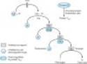
Blood coagulates by the transformation of soluble fibrinogen into insoluble fibrin. Several circulating proteins interact in a cascading series of limited proteolytic reactions (Figure 34-2). At each step, a clotting factor zymogen undergoes limited proteolysis and becomes an active protease (eg, factor VII is converted to factor VIIa). Each protease factor activates the next clotting factor in the sequence, culminating in the formation of thrombin (factor IIa). Several of these factors are targets for drug therapy (Table 34-1).
Thrombin has a central role in hemostasis and has many functions. In clotting, thrombin proteolytically cleaves small peptides from fibrinogen, allowing fibrinogen to polymerize and form a fibrin clot. Thrombin also activates many upstream clotting factors, leading to more thrombin generation, and activates factor XIII, a transaminase that cross-links the fibrin polymer and stabilizes the clot. Thrombin is a potent platelet activator and mitogen. Thrombin also exerts anticoagulant effects by activating the protein C pathway, which attenuates the clotting response (Figure 34-2). It should therefore be apparent that the response to vascular injury is a complex and precisely modulated process that ensures that under normal circumstances, repair of vascular injury occurs without thrombosis and downstream ischemia; that is, the response is proportionate and reversible. Eventually vascular remodeling and repair occur with reversion to the quiescent resting anticoagulant endothelial cell phenotype.
|
|
Figure 34-2. A model of blood coagulation. With tissue factor (TF), factor VII forms an activated complex (VIIa-TF) that catalyzes the activation of factor IX to factor IXa. Activated factor XIa also catalyzes this reaction. Tissue factor pathway inhibitor (TFPI) inhibits the catalytic action of the VIIa-TF complex. The cascade proceeds as shown, resulting ultimately in the conversion of fibrinogen to fibrin, an essential component of a functional clot. The two major anticoagulant drugs, heparin and warfarin (an oral anticoagulant), have very different actions. Heparin, acting in the blood, directly activates anticlotting factors, specifically antithrombin, which inactivates the factors enclosed in rectangles. Warfarin, acting in the liver, inhibits the synthesis of the factors enclosed in circles. Proteins C and S exert anticlotting effects by inactivating activated factors Va and VIIIa. |
Initiation of Clotting: The Tissue Factor-VIIa Complex
The main initiator of blood coagulation in vivo is the tissue factor (TF)-factor VIIa pathway (Figure 34-2). Tissue factor is a transmembrane protein ubiquitously expressed outside the vasculature, but not normally expressed in an active form within vessels. The exposure of TF on damaged endothelium or to blood that has extravasated into tissue binds TF to factor VIIa. This complex, in turn, activates factors X and IX. Factor Xa along with factor Va forms the prothrombinase complex on activated cell surfaces, which catalyzes the conversion of prothrombin (factor II) to thrombin (factor IIa). Thrombin, in turn, activates upstream clotting factors, primarily factors V, VIII, and XI, resulting in amplification of thrombin generation. The TF-factor VIIa-catalyzed activation of factor Xa is regulated by tissue factor pathway inhibitor (TFPI). Thus after initial activation of factor X to Xa by TF-VIIa, further propagation of the clot is by feedback amplification of thrombin through the intrinsic pathway factors VIII and IX (this provides an explanation of why patients with deficiency of factor VIII or IX¾hemophilia A and hemophilia B, respectively¾have a severe bleeding disorder).
It is also important to note that the coagulation mechanism in vivo does not occur in solution, but is localized to activated cell surfaces expressing anionic phospholipids such as phosphatidylserine, and is mediated by Ca2+ bridging between the anionic phospholipids and g-carboxyglutamic acid residues of the clotting factors. This is the basis for using calcium chelators such as ethylenediamine tetraacetic acid (EDTA) or citrate to prevent blood from clotting in a test tube.
Antithrombin (AT) is an endogenous anticoagulant and a member of the serine protease inhibitor (serpin) family; it inactivates the serine proteases IIa, IXa, Xa, XIa, and XIIa. The endogenous anticoagulants protein C and protein S attenuate the blood clotting cascade by proteolysis of the two cofactors Va and VIIIa. From an evolutionary standpoint, it is of interest that factors V and VIII have an identical overall domain structure and considerable homology, consistent with a common ancestor gene; likewise the serine proteases are descendents of a trypsin-like common ancestor. Thus, the TF-VIIa initiating complex, serine proteases, and cofactors each have their own lineage-specific attenuation mechanism (see Figure 34-2). Defects in natural anticoagulants result in an increased risk of venous thrombosis. The most common defect in the natural anticoagulant system is a mutation in factor V (factor V Leiden), which results in resistance to inactivation by the protein C, protein S mechanism.
Fibrinolysis
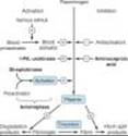
Fibrinolysis refers to the process of fibrin digestion by the fibrin-specific protease, plasmin. The fibrinolytic system is similar to the coagulation system in that the precursor form of the serine protease plasmin circulates in an inactive form as plasminogen. In response to injury, endothelial cells synthesize and release tissue plasminogen activator (t-PA), which converts plasminogen to plasmin (Figure 34-3). Plasmin remodels the thrombus and limits its extension by proteolytic digestion of fibrin.
Both plasminogen and plasmin have specialized protein domains (kringles) that bind to exposed lysines on the fibrin clot and impart clot specificity to the fibrinolytic process. It should be noted that this clot specificity is only observed at physiologic levels of t-PA. At the pharmacologic levels of t-PA used in thrombolytic therapy, clot specificity is lost and a systemic lytic state is created, with attendant increase in bleeding risk. As in the coagulation cascade, there are negative regulators of fibrinolysis: endothelial cells synthesize and release plasminogen activator inhibitor (PAI), which inhibits t-PA; in addition a2 antiplasmin circulates in the blood at high concentrations and under physiologic conditions will rapidly inactivate any plasmin that is not clot-bound. However, this regulatory system is overwhelmed by therapeutic doses of plasminogen activators.
If the coagulation and fibrinolytic systems are pathologically activated, the hemostatic system may careen out of control, leading to generalized intravascular clotting and bleeding. This process is called disseminated intravascular coagulation (DIC) and may follow massive tissue injury, advanced cancers, obstetric emergencies such as abruptio placentae or retained products of conception, or bacterial sepsis. The treatment of DIC is to control the underlying disease process; if this is not possible, DIC is often fatal.
Regulation of the fibrinolytic system is useful in therapeutics. Increased fibrinolysis is effective therapy for thrombotic disease. Tissue plasminogen activator, urokinase, and streptokinase all activate the fibrinolytic system (Figure 34-3). Conversely, decreased fibrinolysis protects clots from lysis and reduces the bleeding of hemostatic failure. Aminocaproic acid is a clinically useful inhibitor of fibrinolysis. Heparin and the oral anticoagulant drugs do not affect the fibrinolytic mechanism.
|
|
Figure 34-3. Schematic representation of the fibrinolytic system. Plasmin is the active fibrinolytic enzyme. Several clinically useful activators are shown on the left in bold. Anistreplase is a combination of streptokinase and the proactivator plasminogen. Aminocaproic acid (right) inhibits the activation of plasminogen to plasmin and is useful in some bleeding disorders. (t-PA, tissue plasminogen activator.) |
I. BASIC PHARMACOLOGY OF THE ANTICOAGULANT DRUGS
INTRODUCTION
The ideal anticoagulant drug would prevent pathologic thrombosis and limit reperfusion injury, yet allow a normal response to vascular injury and limit bleeding. Theoretically this could be accomplished by preservation of the TF-VIIa initiation phase of the clotting mechanism with attenuation of the secondary intrinsic pathway propagation phase of clot development. At this time such a drug does not exist; all anticoagulants and fibrinolytic drugs have an increased bleeding risk as their principle toxicity.
INDIRECT THROMBIN INHIBITORS
INTRODUCTION
The indirect thrombin inhibitors are so-named because their antithrombotic effect is exerted by their interaction with a separate protein, antithrombin. Unfractionated heparin (UFH), low-molecular-weight heparin (LMWH), and the synthetic pentasaccharide fondaparinux bind to antithrombin and enhance its inactivation of factor Xa. Unfractionated heparin and to a lesser extent LMWH also enhance antithrombin's inactivation of thrombin.
HEPARIN
Chemistry & Mechanism of Action
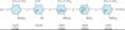
Heparin is a heterogeneous mixture of sulfated mucopolysaccharides. It binds to endothelial cell surfaces and a variety of plasma proteins. Its biologic activity is dependent upon the endogenous anticoagulant antithrombin. Antithrombin inhibits clotting factor proteases, especially thrombin (IIa), IXa, and Xa, by forming equimolar stable complexes with them. In the absence of heparin, these reactions are slow; in the presence of heparin, they are accelerated 1000-fold. Only about a third of the molecules in commercial heparin preparations have an accelerating effect because the remainder lack the unique pentasaccharide sequence needed for high-affinity binding to antithrombin (Figure 34-4). The active heparin molecules bind tightly to antithrombin and cause a conformational change in this inhibitor. The conformational change of antithrombin exposes its active site for more rapid interaction with the proteases (the activated clotting factors). Heparin functions as a cofactor for the antithrombin-protease reaction without being consumed. Once the antithrombin-protease complex is formed, heparin is released intact for renewed binding to more antithrombin.
The antithrombin binding region of commercial unfractionated heparin consists of repeating sulfated disaccharide units composed of D-glucosamine-L-iduronic acid and D-glucosamine-D-glucuronic acid (Figure 34-4). High-molecular-weight fractions of heparin with high affinity for antithrombin markedly inhibit blood coagulation by inhibiting all three factors, especially thrombin and factor Xa. Unfractionated heparin has a molecular weight range of 5000-30,000. In contrast, the shorter-chain low-molecular-weight (LMW) fractions of heparin inhibit activated factor X but have less effect on thrombin than the HMW species. Nevertheless, numerous studies have demonstrated that LMW heparins such as enoxaparin, dalteparin, and tinzaparin are effective in several thromboembolic conditions. In fact, these LMW heparins¾in comparison with UFH¾have equal efficacy, increased bioavailability from the subcutaneous site of injection, and less frequent dosing requirements (once or twice daily is sufficient).
Because commercial heparin consists of a family of molecules of different molecular weights, the correlation between the concentration of a given heparin preparation and its effect on coagulation often is poor. Therefore, UFH is standardized by bioassay. Heparin sodium USP must contain at least 120 USP units per milligram. Heparin is generally used as the sodium salt, but calcium heparin is equally effective. Lithium heparin is used in vitro as an anticoagulant for blood samples. Commercial heparin is extracted from porcine intestinal mucosa and bovine lung. Enoxaparin is obtained from the same sources as regular heparin, but doses are specified in milligrams. Dalteparin, tinzaparin and danaparoid (an LMW heparanoid containing heparan sulfate, dermatan sulfate, and chondroitin sulfate), on the other hand, are specified in anti-factor Xa units.
|
|
Figure 34-4. Subunit structure of heparin. The small polymer section shown illustrates the repeating disaccharide units typical of heparin. The sequence shows the critical pentasaccharide portion required for binding to antithrombin. In addition to those shown, other saccharides occur. Heparin is a strongly acidic molecule because of its high content of anionic sulfate and carboxylic acid groups. (GlcN, glucosamine; IdUA, iduronic acid; GlcUA, glucuronic acid. The same five residues with the terminal groups shown in parentheses, constitute fondaparinux.) |
Monitoring of Heparin Effect
Close monitoring of the activated partial thromboplastin time (aPTT) is necessary in patients receiving UFH. Levels of UFH may also be determined by protamine titration (therapeutic levels 0.2-0.4 unit/mL) or anti-Xa units (therapeutic levels 0.3-0.7 unit/mL). Weight-based dosing of the LMW heparins results in predictable pharmacokinetics and plasma levels in patients with normal renal function. Therefore, LMW heparin levels are not generally measured except in the setting of renal insufficiency, obesity, and pregnancy. LMW heparin levels can be determined by anti-Xa units. Peak therapeutic levels should be 0.5-1 unit/mL for twice-daily dosing, determined 4 hours after administration, and approximately 1.5 units/mL for once-daily dosing.
Toxicity
A. BLEEDING
The major adverse effect of heparin is bleeding. This risk can be decreased by scrupulous patient selection, careful control of dosage, and close monitoring. Elderly women and patients with renal failure are more prone to hemorrhage. Heparin is of animal origin and should be used cautiously in patients with allergy. Increased loss of hair and reversible alopecia have been reported. Long-term heparin therapy is associated with osteoporosis and spontaneous fractures. Heparin accelerates the clearing of postprandial lipemia by causing the release of lipoprotein lipase from tissues, and long-term use is associated with mineralocorticoid deficiency.
B. HEPARIN-INDUCED THROMBOCYTOPENIA
Heparin-induced thrombocytopenia (HIT) is a systemic hypercoagulable state that occurs in 1-4% of individuals treated with UFH for a minimum of 7 days. Surgical patients are at greatest risk. The reported incidence of HIT is lower in pediatric populations outside the critical care setting and is relatively rare in pregnant women. The risk of HIT may be higher in individuals treated with UFH of bovine origin compared with porcine heparin and is lower in those treated exclusively with LMWH.
Morbidity and mortality in HIT are related to thrombotic events. Venous thrombosis occurs most commonly, but occlusion of peripheral or central arteries is not infrequent. If an indwelling catheter is present, the risk of thrombosis is increased in that extremity. Skin necrosis has been described, particularly in individuals treated with warfarin in the absence of a direct thrombin inhibitor, presumably due to acute depletion of the vitamin K-dependent anticoagulant protein C occurring in the presence of high levels of procoagulant proteins and an active hypercoagulable state.
The following points should be considered in all patients receiving heparin: Platelet counts should be performed frequently; thrombocytopenia appearing in a time frame consistent with an immune response to heparin should be considered suspicious for HIT; and any new thrombus occuring in a patient receiving heparin therapy should raise suspicion of HIT. Patients who develop HIT are treated by discontinuance of heparin and administration of a direct thrombin inhibitor or fondaparinux (see below).
Contraindications
Heparin is contraindicated in patients with HIT, hypersensitivity to the drug, active bleeding, hemophilia, significant thrombocytopenia, purpura, severe hypertension, intracranial hemorrhage, infective endocarditis, active tuberculosis, ulcerative lesions of the gastrointestinal tract, threatened abortion, visceral carcinoma, or advanced hepatic or renal disease. Heparin should be avoided in patients who have recently had surgery of the brain, spinal cord, or eye, and in patients who are undergoing lumbar puncture or regional anesthetic block. Despite the apparent lack of placental transfer, heparin should be used in pregnant women only when clearly indicated.
Administration & Dosage
The indications for the use of heparin are described in the section on clinical pharmacology. A plasma concentration of heparin of 0.2-0.4 unit/mL (by protamine titration) or 0.3-0.7 unit/mL (anti-Xa units) usually prevents pulmonary emboli in patients with established venous thrombosis. This concentration of heparin will prolong the aPTT to 2-2.5 times that of the control value. This degree of anticoagulant effect should be maintained throughout the course of continuous intravenous heparin therapy. When intermittent heparin administration is used, the aPTT should be measured 6 hours after the administered dose to maintain prolongation of the aPTT to 2-2.5 times that of the control value.
Continuous intravenous administration of heparin is accomplished via an infusion pump. After an initial bolus injection of 80-100 units/kg, a continuous infusion of about 15-22 units/kg/h is required to maintain the aPTT at 2-2.5 times control. Patients with acute pulmonary emboli often require larger doses during the first few days because of binding to a variety of acute phase proteins, such as factor VIII and von Willebrand factor, and increased heparin clearance. Low-dose prophylaxis is achieved with subcutaneous administration of heparin, 5000 units every 8-12 hours. Because of the danger of hematoma formation at the injection site, heparin must never be administered intramuscularly.
Prophylactic enoxaparin is given subcutaneously in a dosage of 30 mg twice daily or 40 mg once daily. Full-dose enoxaparin therapy is 1 mg/kg subcutaneously every 12 hours. This corresponds to a therapeutic anti-factor Xa level of 0.5-1 unit/mL. Selected patients may be treated with enoxaparin 1.5 mg/kg once a day, with a target anti-Xa level of 1.5 units/mL. The prophylactic dose of dalteparin is 5000 units subcutaneously once a day; therapeutic dosing is 200 units/kg once a day for venous disease or 120 units/kg every 12 hours for acute coronary syndrome. Low-molecular-weight heparins should be used with caution in patients with renal insufficiency or body weight greater than 150 kg. Measurement of the anti-Xa level is useful to guide dosing in these individuals.
The synthetic pentasaccharide molecule fondaparinux (Figure 34-4) avidly binds antithrombin with high specific activity, resulting in efficient inactivation of factor Xa. Fondaparinux has a long half-life of 15 hours, allowing for once-daily dosing by subcutaneous administration. Fondaparinux is effective in the prevention and treatment of venous thromboembolism, and appears to not cross-react with pathologic HIT antibodies in most individuals. The use of fondaparinux as an alternative anticoagulant in HIT is currently being tested in clinical trials. Pharmaceutical companies have put much effort into developing an orally bioavailable anti-Xa inhibitor. The first of these have recently completed phase I clinical trials with promising results and are currently being evaluated for efficacy in prophylaxis and treatment of venous thromboembolism.
Reversal of Heparin Action
Excessive anticoagulant action of heparin is treated by discontinuance of the drug. If bleeding occurs, administration of a specific antagonist such as protamine sulfate is indicated. Protamine is a highly basic peptide that combines with heparin as an ion pair to form a stable complex devoid of anticoagulant activity. For every 100 units of heparin remaining in the patient, 1 mg of protamine sulfate is given intravenously; the rate of infusion should not exceed 50 mg in any 10-minute period. Excess protamine must be avoided; it also has an anticoagulant effect. Neutralization of LMW heparin by protamine is incomplete. Limited experience suggests that 1 mg of protamine sulfate may be used to partially neutralize 1 mg of enoxaparin. Protamine will not reverse the activity of fondaparinux. Excess danaparoid can be removed by plasmapheresis.
DIRECT THROMBIN INHIBITORS
The direct thrombin inhibitors (DTIs) exert their anticoagulant effect by directly binding to the active site of thrombin, thereby inhibiting thrombin's downstream effects. This is in contrast to indirect thrombin inhibitors such as heparin and LMWH (see above), which act through antithrombin. Hirudin and bivalirudin are bivalent DTIs in that they bind at both the catalytic or active site of thrombin as well as at a substrate recognition site. Argatroban and melagatran are small molecules that bind only at the thrombin active site.
Leeches have been used for bloodletting since the age of Hippocrates. More recently, surgeons have used medicinal leeches (Hirudo medicinalis) to prevent thrombosis in the fine vessels of reattached digits. Hirudin is a specific, irreversible thrombin inhibitor from leech saliva that is now available in recombinant form as lepirudin. Its action is independent of antithrombin, which means it can reach and inactivate fibrin-bound thrombin in thrombi. Lepirudin has little effect on platelets or the bleeding time. Like heparin, it must be administered parenterally and is monitored by the aPTT. Lepirudin is approved by the Food and Drug Administration for use in patients with thrombosis related to heparin-induced thrombocytopenia. Lepirudin is excreted by the kidney and should be used with great caution in patients with renal insufficiency as no antidote exists. Up to 40% of patients who receive long-term infusions develop an antibody directed against the thrombin-lepirudin complex. These antigen-antibody complexes are not cleared by the kidney and may result in an enhanced anticoagulant effect. Some patients re-exposed to the drug have developed life-threatening anaphylactic reactions.
Bivalirudin, another bivalent inhibitor of thrombin, is administered intravenously, with a rapid onset and offset of action. The drug has a short half-life with clearance that is 20% renal and the remainder metabolic. Bivalirudin also inhibits platelet activation and has been FDA-approved for use in percutaneous coronary angioplasty.
Argatroban is a small molecule thrombin inhibitor that is FDA-approved for use in patients with HIT with or without thrombosis and coronary angioplasty in patients with HIT. It, too, has a short half-life, is given by continuous intravenous infusion, and is monitored by aPTT. Its clearance is not affected by renal disease but is dependent on liver function; dose reduction is required in patients with liver disease. Patients on argatroban will demonstrate elevated international normalized ratios (INRs), rendering the transition to warfarin difficult (ie, the INR will reflect contributions from both warfarin and argatroban). (INR is discussed in detail in the discussion of warfarin administration.) A nomogram is supplied by the manufacturer to assist in this transition. No properly designed head-to-head trials have been performed to determine whether argatroban or lepirudin is superior in the treatment of HIT. However in practice, the choice of which DTI to use in a patient with HIT is usually dictated by the condition of the clearing organ. If the patient has severe renal insufficiency, then argatroban would be preferred. If there is severe hepatic insufficiency, then lepirudin would be a better choice.
Ximelagatran is an oral prodrug that is metabolized to the DTI melagatran. Potential advantages of ximelagatran include predictable pharmacokinetics and bioavailability. This allows for fixed dosing and predictable anticoagulant response; no need for routine coagulation monitoring; lack of interaction with P450-interacting drugs; and rapid onset and offset of action, which allow for immediate anticoagulation and thus no need for overlap with additional anticoagulant drugs. While clinical trials found that ximelagatran was as effective as other anticoagulants in venous thromboembolism and atrial fibrillation, hepatic toxicity was observed in 5-10% of individuals treated for more than one month. Thus while there is much enthusiasm for a nontoxic oral anticoagulant that might replace warfarin and not require routine monitoring, no such drug is currently FDA-approved.
WARFARIN & THE COUMARIN ANTICOAGULANTS
Chemistry & Pharmacokinetics
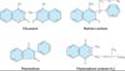
The clinical use of the coumarin anticoagulants began with the discovery of an anticoagulant substance formed in spoiled sweet clover silage which caused hemorrhagic disease in cattle. At the behest of local farmers, a chemist at the University of Wisconsin identified the toxic agent as bishydroxycoumarin. A synthesized derivative, dicumarol and its congeners, most notably warfarin (Wisconsin Alumni Research Foundation, with "arin" from coumarin added; Figure 34-5), were initially used as rodenticides. In the 1950s warfarin (under the brand name Coumadin) was introduced as an antithrombotic agent in humans. Warfarin is one of the most commonly prescribed drugs, used by approximately 1.5 million individuals, and several studies have indicated that the drug is significantly underused in clinical situations where it has proven benefit.
Warfarin is generally administered as the sodium salt and has 100% bioavailability. Over 99% of racemic warfarin is bound to plasma albumin, which may contribute to its small volume of distribution (the albumin space), its long half-life in plasma (36 hours), and the lack of urinary excretion of unchanged drug. Warfarin used clinically is a racemic mixture composed of equal amounts of two enantiomorphs. The levorotatory S-warfarin is four times more potent than the dextrorotatory R-warfarin. This observation is useful in understanding the stereoselective nature of several drug interactions involving warfarin.
|
|
Figure 34-5. Structural formulas of several oral anticoagulant drugs and of vitamin K. The carbon atom of warfarin shown at the asterisk is an asymmetric center. |
Mechanism of Action
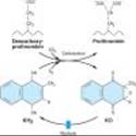
Coumarin anticoagulants block the g-carboxylation of several glutamate residues in prothrombin and factors VII, IX, and X as well as the endogenous anticoagulant proteins C and S (See Figure 34-2, Table 34-1). The blockade results in incomplete coagulation factor molecules that are biologically inactive. The protein carboxylation reaction is coupled to the oxidation of vitamin K. The vitamin must then be reduced to reactivate it. Warfarin prevents reductive metabolism of the inactive vitamin K epoxide back to its active hydroquinone form (Figure 34-6). Mutational change of the responsible enzyme, vitamin K epoxide reductase, can give rise to genetic resistance to warfarin in humans and especially in rats.
There is an 8- to 12-hour delay in the action of warfarin. Its anticoagulant effect results from a balance between partially inhibited synthesis and unaltered degradation of the four vitamin K-dependent clotting factors. The resulting inhibition of coagulation is dependent on their degradation half-lives in the circulation. These half-lives are 6, 24, 40, and 60 hours for factors VII, IX, X, and II, respectively. Larger initial doses of warfarin¾up to about 0.75 mg/kg¾hasten the onset of the anticoagulant effect. Beyond this dosage, the speed of onset is independent of the dose size. The only effect of a larger loading dose is to prolong the time that the plasma concentration of drug remains above that required for suppression of clotting factor synthesis. The only difference among oral anticoagulants in producing and maintaining hypoprothrombinemia is the half-life of each drug.
|
|
Figure 34-6. Vitamin K cycle¾metabolic interconversions of vitamin K associated with the synthesis of vitamin K-dependent clotting factors. Vitamin K1 or K2 is activated by reduction to the hydroquinone form (KH2). Stepwise oxidation to vitamin K epoxide (KO) is coupled to prothrombin carboxylation by the enzyme carboxylase. The reactivation of vitamin K epoxide is the warfarin-sensitive step (warfarin). The R on the vitamin K molecule represents a 20-carbon phytyl side chain in vitamin K1 and a 30- to 65-carbon polyprenyl side chain in vitamin K2. |
Toxicity
Warfarin crosses the placenta readily and can cause a hemorrhagic disorder in the fetus. Furthermore, fetal proteins with g-carboxyglutamate residues found in bone and blood may be affected by warfarin; the drug can cause a serious birth defect characterized by abnormal bone formation. Thus, warfarin should never be administered during pregnancy. Cutaneous necrosis with reduced activity of protein C sometimes occurs during the first weeks of therapy. Rarely, the same process causes frank infarction of the breast, fatty tissues, intestine, and extremities. The pathologic lesion associated with the hemorrhagic infarction is venous thrombosis, suggesting that it is caused by warfarin-induced depression of protein C synthesis.
Administration & Dosage
Treatment with warfarin should be initiated with standard doses of 5-10 mg rather than the large loading doses formerly used. The initial adjustment of the prothrombin time takes about 1 week, which usually results in a maintenance dose of 5-7 mg/d. The prothrombin time (PT) should be increased to a level representing a reduction of prothrombin activity to 25% of normal and maintained there for long-term therapy. When the activity is less than 20%, the warfarin dosage should be reduced or omitted until the activity rises above 20%.
The therapeutic range for oral anticoagulant therapy is defined in terms of an international normalized ratio (INR). The INR is the prothrombin time ratio (patient prothrombin time/mean of normal prothrombin time for lab)ISI, where the ISI exponent refers to the International Sensitivity Index, and is dependent on the specific reagents and instruments used for the determination. The ISI serves to relate measured prothrombin times to a World Health Organization reference standard thromboplastin; thus the prothrombin times performed on different properly calibrated instruments with a variety of thromboplastin reagents should give the same INR results for a given sample. For most reagent and instrument combinations in current use, the ISI is close to 1, making the INR roughly the ratio of the patient prothrombin time to the mean normal prothrombin time. The recommended INR for prophylaxis and treatment of thrombotic disease is 2-3. Patients with some types of artificial heart valves (eg, tilting disk) or other medical conditions increasing thrombotic risk have a recommended range of 2.5-3.5.
Occasionally patients exhibit warfarin resistance, defined as progression or recurrence of a thrombotic event while in therapeutic range. These individuals may have their INR target raised (which is accompanied by an increase in bleeding risk) or be changed to an alternative form of anticoagulation (eg, daily injections of LMWH). Warfarin resistance is most commonly seen in patients with advanced cancers, typically of gastrointestinal origin (Trousseau's syndrome). A recent study has demonstrated the superiority of LMWH over warfarin in preventing recurrent venous thromboembolism in patients with cancer.
Drug Interactions
The oral anticoagulants often interact with other drugs and with disease states. These interactions can be broadly divided into pharmacokinetic and pharmacodynamic effects (Table 34-2). Pharmacokinetic mechanisms for drug interaction with oral anticoagulants are mainly enzyme induction, enzyme inhibition, and reduced plasma protein binding. Pharmacodynamic mechanisms for interactions with warfarin are synergism (impaired hemostasis, reduced clotting factor synthesis, as in hepatic disease), competitive antagonism (vitamin K), and an altered physiologic control loop for vitamin K (hereditary resistance to oral anticoagulants).
The most serious interactions with warfarin are those that increase the anticoagulant effect and the risk of bleeding. The most dangerous of these interactions are the pharmacokinetic interactions with the pyrazolones phenylbutazone and sulfinpyrazone. These drugs not only augment the hypoprothrombinemia but also inhibit platelet function and may induce peptic ulcer disease (see Chapter 36). The mechanisms for their hypoprothrombinemic interaction are a stereoselective inhibition of oxidative metabolic transformation of S-warfarin (the more potent isomer) and displacement of albumin-bound warfarin, increasing the free fraction. For this and other reasons, neither phenylbutazone nor sulfinpyrazone is in common use in the USA. Metronidazole, fluconazole, and trimethoprim-sulfamethoxazole also stereoselectively inhibit the metabolic transformation of S-warfarin, whereas amiodarone, disulfiram, and cimetidine inhibit metabolism of both enantiomorphs of warfarin. Aspirin, hepatic disease, and hyperthyroidism augment warfarin pharmacodynamically¾aspirin by its effect on platelet function and the latter two by increasing the turnover rate of clotting factors. The third-generation cephalosporins eliminate the bacteria in the intestinal tract that produce vitamin K and, like warfarin, also directly inhibit vitamin K epoxide reductase.
Barbiturates and rifampin cause a marked decrease of the anticoagulant effect by induction of the hepatic enzymes that transform racemic warfarin. Cholestyramine binds warfarin in the intestine and reduces its absorption and bioavailability.
Pharmacodynamic reductions of anticoagulant effect occur with vitamin K (increased synthesis of clotting factors), the diuretics chlorthalidone and spironolactone (clotting factor concentration), hereditary resistance (mutation of vitamin K reactivation cycle molecules), and hypothyroidism (decreased turnover rate of clotting factors).
Drugs with no significant effect on anticoagulant therapy include ethanol, phenothiazines, benzodiazepines, acetaminophen, opioids, indomethacin, and most antibiotics.
Reversal of Warfarin Action
Excessive anticoagulant effect and bleeding from warfarin can be reversed by stopping the drug and administering oral or parenteral vitamin K1 (phytonadione), fresh-frozen plasma, prothrombin complex concentrates such as Bebulin and Proplex T, and recombinant factor VIIa (rFVIIa). The disappearance of excessive effect is not correlated with plasma warfarin concentrations but rather with reestablishment of normal activity of the clotting factors. A modest excess of anticoagulant effect without bleeding may require no more than cessation of the drug. The warfarin effect can be rapidly reversed in the setting of severe bleeding with the administration of prothrombin complex or rFVIIa coupled with intravenous vitamin K. It is important to note that due to the long half life of warfarin, a single dose of vitamin K or rFVIIa may not be sufficient.
II. BASIC PHARMACOLOGY OF THE FIBRINOLYTIC DRUGS
Introduction
Fibrinolytic drugs rapidly lyse thrombi by catalyzing the formation of the serine protease plasmin from its precursor zymogen, plasminogen (Figure 34-3). These drugs create a generalized lytic state when administered intravenously. Thus, both protective hemostatic thrombi and target thromboemboli are broken down. The Box: Thrombolytic Drugs for Acute Myocardial Infarction, describes the use of these drugs in one major application.
Pharmacology
Streptokinase is a protein (but not an enzyme in itself) synthesized by streptococci that combines with the proactivator plasminogen. This enzymatic complex catalyzes the conversion of inactive plasminogen to active plasmin. Urokinase is a human enzyme synthesized by the kidney that directly converts plasminogen to active plasmin. Plasmin itself cannot be used because naturally occurring inhibitors in plasma prevent its effects. However, the absence of inhibitors for urokinase and the streptokinase-proactivator complex permit their use clinically. Plasmin formed inside a thrombus by these activators is protected from plasma antiplasmins, which allows it to lyse the thrombus from within.
Anistreplase (anisoylated plasminogen streptokinase activator complex; APSAC) consists of a complex of purified human plasminogen and bacterial streptokinase that has been acylated to protect the enzyme's active site. When administered, the acyl group spontaneously hydrolyzes, freeing the activated streptokinase-proactivator complex. This product (recently discontinued in the USA) allows for rapid intravenous injection, greater clot selectivity (ie, more activity on plasminogen associated with clots than on free plasminogen in the blood), and more thrombolytic activity.
Plasminogen can also be activated endogenously by tissue plasminogen activators (t-PAs). These activators preferentially activate plasminogen that is bound to fibrin, which (in theory) confines fibrinolysis to the formed thrombus and avoids systemic activation. Human t-PA is manufactured as alteplase by means of recombinant DNA technology.
Reteplase is another recombinant human t-PA from which several amino acid sequences have been deleted. Reteplase is less expensive to produce than t-PA. Because it lacks the major fibrin-binding domain, reteplase is less fibrin-specific than t-PA. Tenecteplase is a mutant form of t-PA that has a longer half-life, and it can be given as an intravenous bolus. Tenecteplase is slightly more fibrin-specific than t-PA.
THROMBOLYTIC DRUGS FOR ACUTE MYOCARDIAL INFARCTION
The paradigm shift in 1980 on the causation of acute myocardial infarction to acute coronary occlusion by a thrombus created the rationale for thrombolytic therapy of this common lethal disease. At that time¾and for the first time¾intravenous thrombolytic therapy for acute myocardial infarction in the European Cooperative Study Group trial was found to reduce mortality significantly. Later studies, with thousands of patients in each trial, provided enough statistical power for the 20% reduction in mortality to be considered statistically significant. Although the standard of care in areas with adequate facilities and experience in percutaneous coronary intervention (PCI) now favors catheterization and placement of a stent, thrombolytic therapy is still very important where PCI is not readily available.
The proper selection of patients for thrombolytic therapy is critical. The diagnosis of acute myocardial infarction is made clinically and is confirmed by electrocardiography. Patients with ST-segment elevation and bundle branch block on electrocardiography have the best outcomes. All trials to date show the greatest benefit for thrombolytic therapy when it is given early, within 6 hours after symptomatic onset of acute myocardial infarction.
Thrombolytic drugs reduce the mortality of acute myocardial infarction. The early and appropriate use of any thrombolytic drug probably transcends possible advantages of a particular drug. Adjunctive drugs such as aspirin, heparin, b blockers, and angiotensin-converting enzyme (ACE) inhibitors reduce mortality even further. The principles of management are outlined in part 7 of the American Heart Association Guidelines, 2000.
Indications & Dosage
Administration of fibrinolytic drugs by the intravenous route is indicated in cases of pulmonary embolism with hemodynamic instability, severe deep venous thrombosis such as the superior vena caval syndrome, and ascending thrombophlebitis of the iliofemoral vein with severe lower extremity edema. These drugs are also given intra-arterially, especially for peripheral vascular disease.
Thrombolytic therapy in the management of acute myocardial infarction requires careful patient selection, the use of a specific thrombolytic agent, and the benefit of adjuvant therapy. Streptokinase is administered by intravenous infusion of a loading dose of 250,000 units, followed by 100,000 units/h for 24-72 hours. Patients with antistreptococcal antibodies can develop fever, allergic reactions, and therapeutic resistance. Urokinase requires a loading dose of 300,000 units given over 10 minutes and a maintenance dose of 300,000 units/h for 12 hours. Alteplase (t-PA) is given by intravenous infusion of 60 mg over the first hour and then 40 mg at a rate of 20 mg/h. Reteplase is given as two intravenous bolus injections of 10 units each, separated by 30 minutes. Tenecteplase is given as a single intravenous bolus of 0.5 mg/kg. Anistreplase (where available) is given as a single intravenous injection of 30 units over 3-5 minutes. A single course of fibrinolytic drugs is expensive: hundreds of dollars for streptokinase and thousands for urokinase and t-PA.
Recombinant t-PA has also been approved for use in acute ischmic stroke within 3 hours of symptom onset. In patients without hemorrhagic infarct or other contraindications, this therapy has been demonstrated to provide better outcomes in several randomized clinical trials. The recommended dose is 0.9 mg/kg, not to exceed 90 mg, with 10% given as a bolus and the remainder during a 1 hour infusion. Streptokinase has been associated with increased bleeding risk in acute ischemic stroke when given at a dose of 1.5 million units, and its use is not recommended in this setting.
III. BASIC PHARMACOLOGY OF ANTIPLATELET AGENTS
INTRODUCTION
Platelet function is regulated by three categories of substances. The first group consists of agents generated outside the platelet that interact with platelet membrane receptors, eg, catecholamines, collagen, thrombin, and prostacyclin. The second category contains agents generated within the platelet that interact with membrane receptors, eg, ADP, prostaglandin D2, prostaglandin E2, and serotonin. The third group comprises agents generated within the platelet that act within the platelet, eg, prostaglandin endoperoxides and thromboxane A2, the cyclic nucleotides cAMP and cGMP, and calcium ion. From this list of agents, several targets for platelet inhibitory drugs have been identified (see Figure 34-1): inhibition of prostaglandin synthesis (aspirin), inhibition of ADP-induced platelet aggregation (clopidogrel, ticlopidine), and blockade of glycoprotein IIb/IIIa receptors on platelets (abciximab, tirofiban, and eptifibatide). Dipyridamole and cilostazol are additional antiplatelet drugs.
ASPIRIN
The prostaglandin thromboxane A2 is an arachidonate product that causes platelets to change shape, release their granules, and aggregate (see Chapter 18). Drugs that antagonize this pathway interfere with platelet aggregation in vitro and prolong the bleeding time in vivo. Aspirin is the prototype of this class of drugs.
As described in Chapter 18, aspirin inhibits the synthesis of thromboxane A2 by irreversible acetylation of the enzyme cyclooxygenase. Other salicylates and nonsteroidal anti-inflammatory drugs also inhibit cyclooxygenase but have a shorter duration of inhibitory action because they cannot acetylate cyclooxygenase; that is, their action is reversible.
The FDA has approved the use of 325 mg/d for primary prophylaxis of myocardial infarction but urges caution in this use of aspirin by the general population except when prescribed as an adjunct to risk factor management by smoking cessation and lowering of blood cholesterol and blood pressure. Meta-analysis of many published trials of aspirin and other antiplatelet agents confirms the value of this intervention in the secondary prevention of vascular events among patients with a history of vascular events.
CLOPIDOGREL & TICLOPIDINE
Introduction
Clopidogrel and ticlopidine reduce platelet aggregation by inhibiting the ADP pathway of platelets. These drugs are thienopyridine derivatives that achieve their antiplatelet effects by irreversibly blocking the ADP receptor on platelets. Unlike aspirin, these drugs have no effect on prostaglandin metabolism. Randomized clinical trials with both drugs report efficacy in the prevention of vascular events among patients with transient ischemic attacks, completed strokes, and unstable angina pectoris. Use of clopidogrel or ticlopidine to prevent thrombosis is now considered standard practice in patients undergoing placement of a coronary stent.
Adverse effects of ticlopidine include nausea, dyspepsia, and diarrhea in up to 20% of patients, hemorrhage in 5%, and, most seriously, leukopenia in 1%. The leukopenia is detected by regular monitoring of the white blood cell count during the first 3 months of treatment. Development of thrombotic thrombocytopenic purpura has also been associated with the ingestion of ticlopidine. The dosage of ticlopidine is 250 mg twice daily. It is particularly useful in patients who cannot tolerate aspirin. Doses of ticlopidine less than 500 mg/d may be efficacious with fewer adverse effects.
Clopidogrel has fewer adverse effects than ticlopidine and is rarely associated with neutropenia. Thrombotic thrombocytopenic purpura associated with clopidogrel has been reported. Because of its superior side effect profile and dosing requirements, clopidogrel is preferred over ticlopidine. The antithrombotic effects of clopidogrel are dose-dependent; within 5 hours after an oral loading dose of 300 mg, 80% of platelet activity will be inhibited. The maintenance dose of clopidogrel is 75 mg/d, which achieves maximum platelet inhibition. The duration of the antiplatelet effect is 7-10 days.
Aspirin & Clopidogrel Resistance
The reported incidence of resistance to these drugs varies greatly, from less than 5% to 75%. In part this tremendous variation in incidence reflects the definition of resistance (recurrent thrombosis while on antiplatelet therapy vs in vitro testing), methods by which drug response is measured, and patient compliance. Several methods for testing aspirin and clopidogrel resistance in vitro are now FDA-approved; however, their utility outside of clinical trials remains controversial.
BLOCKADE OF PLATELET GLYCOPROTEIN IIB/IIIA RECEPTORS
The glycoprotein IIb/IIIa inhibitors are used in patients with acute coronary syndromes. These drugs target the platelet IIb/IIIa receptor complex (Figure 34-1). The IIb/IIIa complex functions as a receptor mainly for fibrinogen and vitronectin but also for fibronectin and von Willebrand factor. Activation of this receptor complex is the "final common pathway" for platelet aggregation. There are approximately 50,000 copies of this complex on the surface of each platelet. Persons lacking this receptor have a bleeding disorder called Glanzmann's thrombasthenia.
Abciximab, a chimeric monoclonal antibody directed against the IIb/IIIa complex including the vibronectin receptor, was the first agent approved in this class of drugs. It has been approved for use in percutaneous coronary intervention and in acute coronary syndromes. Eptifibatide is an analog of the sequence at the extreme carboxyl terminal of the delta chain of fibrinogen, which mediates the binding of fibrinogen to the receptor. Tirofiban is a smaller molecule with similar properties. Eptifibatide and tirofiban inhibit ligand binding to the IIb/IIIa receptor by their occupancy of the receptor but do not block the vibronectin receptor.
The three agents described above are administered parenterally. Oral formulations of IIb/IIIa antagonists have been developed and are in various stages of development.
ADDITIONAL ANTIPLATELET-DIRECTED DRUGS
Dipyridamole is a vasodilator that inhibits platelet function by inhibiting adenosine uptake and cyclic GMP phosphodiesterase activity. Dipyridamole by itself has little or no beneficial effect. Therefore, therapeutic use of this agent is primarily in combination with aspirin to prevent cerebrovascular ischemia. It may also be used in combination with warfarin for primary prophylaxis of thromboemboli in patients with prosthetic heart valves. A combination of dipyridamole complexed with 25 mg of aspirin is now available for secondary prophylaxis of cerebrovascular disease.
Cilostazol is a newer phosphodiesterase inhibitor that promotes vasodilation and inhibition of platelet aggregation. Cilostazol is used primarily to treat intermittent claudication.
IV. CLINICAL PHARMACOLOGY OF DRUGS USED TO PREVENT CLOTTING
VENOUS THROMBOSIS
Risk Factors
A. INHERITED DISORDERS
The inherited disorders characterized by a tendency to form thrombi (thrombophilia) derive from either quantitative or qualitative abnormalities of the natural anticoagulant system. Deficiencies (loss of function mutations) in the natural anticoagulants antithrombin, protein C, and protein S account for approximately 15% of selected patients with juvenile or recurrent thrombosis and 5-10% of unselected cases of acute venous thrombosis. Additional causes of thrombophilia include gain of function mutations such as the factor V Leiden mutation and the prothrombin 20210 mutation, elevated clotting factor and cofactor levels, and hyperhomocysteinemia that together account for the greater number of hypercoagulable patients. Although the loss of function mutations are less common, they are associated with the greatest thrombosis risk. Some patients have multiple inherited risk factors or combinations of inherited and acquired risk factors as discussed below. These individuals are at higher risk for recurrent thrombotic events and are often considered candidates for lifelong therapy.
B. ACQUIRED DISEASE
The increased risk of thromboembolism associated with atrial fibrillation and with the placement of mechanical heart valves has long been recognized. Similarly, prolonged bed rest, high-risk surgical procedures, and the presence of cancer are clearly associated with an increased incidence of deep venous thrombosis and embolism. Antiphospholipic antibody syndrome is another important acquired risk factor. Drugs may function as synergistic risk factors in concert with inherited risk factors. For example, women who have the factor V Leiden mutation and take oral contraceptives have a synergistic increase in risk.
Antithrombotic Management
A. PREVENTION
Primary prevention of venous thrombosis reduces the incidence of and mortality rate from pulmonary emboli. Heparin and warfarin may be used to prevent venous thrombosis. Subcutaneous administration of low-dose unfractionated heparin, low-molecular-weight heparin, or fondaparinux provides effective prophylaxis. Warfarin is also effective but requires laboratory monitoring of the prothrombin time.
B. TREATMENT OF ESTABLISHED DISEASE
Treatment for established venous thrombosis is initiated with unfractionated or low-molecular-weight heparin for the first 5-7 days, with an overlap with warfarin. Once therapeutic effects of warfarin have been established, therapy with warfarin is continued for a minimum of 3-6 months. Patients with recurrent disease or identifiable, nonreversible risk factors may be treated indefinitely. Small thrombi confined to the calf veins may be managed without anticoagulants if there is documentation over time that the thrombus is not extending.
Warfarin readily crosses the placenta. It can cause hemorrhage at any time during pregnancy as well as developmental defects when administered during the first trimester. Therefore, venous thromboembolic disease in pregnant women is generally treated with heparin, best administered by subcutaneous injection.
ARTERIAL THROMBOSIS
Activation of platelets is considered an essential process for arterial thrombosis. Thus, treatment with platelet-inhibiting drugs such as aspirin and clopidogrel or ticlopidine is indicated in patients with transient ischemic attacks and strokes or unstable angina and acute myocardial infarction. In angina and infarction, these drugs are often used in conjunction with b blockers, calcium channel blockers, and fibrinolytic drugs.
V. DRUGS USED IN BLEEDING DISORDERS
VITAMIN K
Vitamin K confers biologic activity upon prothrombin and factors VII, IX, and X by participating in their postribosomal modification. Vitamin K is a fat-soluble substance found primarily in leafy green vegetables. The dietary requirement is low, because the vitamin is additionally synthesized by bacteria that colonize the human intestine. Two natural forms exist: vitamins K1 and K2. Vitamin K1 (phytonadione; Figure 34-5) is found in food. Vitamin K2 (menaquinone) is found in human tissues and is synthesized by intestinal bacteria.
Vitamins K1 and K2 require bile salts for absorption from the intestinal tract. Vitamin K1 is available clinically in oral and parenteral forms. Onset of effect is delayed for 6 hours but the effect is complete by 24 hours when treating depression of prothrombin activity by excess warfarin or vitamin K deficiency. Intravenous administration of vitamin K1 should be slow, because rapid infusion can produce dyspnea, chest and back pain, and even death. Vitamin K repletion is best achieved with intravenous or oral administration, because its bioavailability after subcutaneous administration is erratic. Vitamin K1 is currently administered to all newborns to prevent the hemorrhagic disease of vitamin K deficiency, which is especially common in premature infants. The water-soluble salt of vitamin K3 (menadione) should never be used in therapeutics. It is particularly ineffective in the treatment of warfarin overdosage. Vitamin K deficiency frequently occurs in hospitalized patients in intensive care units because of poor diet, parenteral nutrition, recent surgery, multiple antibiotic therapy, and uremia. Severe hepatic failure results in diminished protein synthesis and a hemorrhagic diathesis that is unresponsive to vitamin K.
PLASMA FRACTIONS
Sources & Preparations
Deficiencies in plasma coagulation factors can cause bleeding (Table 34-3). Spontaneous bleeding occurs when factor activity is less than 5-10% of normal. Factor VIII deficiency (classic hemophilia, or hemophilia A) and factor IX deficiency (Christmas disease, or hemophilia B) account for most of the heritable coagulation defects. Concentrated plasma fractions are available for the treatment of these deficiencies. Administration of plasma-derived, heat- or detergent-treated factor concentrates and recombinant factor concentrates are the standard treatments for bleeding associated with hemophilia. Lyophilized factor VIII concentrates are prepared from large pools of plasma. Transmission of viral diseases such as hepatitis B and C and HIV is reduced or eliminated by pasteurization and by extraction of plasma with solvents and detergents. However, this treatment does not remove other potential causes of transmissable diseases such as prions. For this reason, recombinant clotting factor preparations are recommended whenever possible for factor replacement. The best use of these therapeutic materials requires diagnostic specificity of the deficient factor and quantitation of its activity in plasma. Intermediate purity factor VIII concentrates (as opposed to recombinant or high purity concentrates) contain significant amounts of von Willebrand factor. Humate-P is a factor VIII concentrate that is approved by the FDA for the treatment of bleeding associated with von Willebrand disease.
Clinical Uses
An uncomplicated hemorrhage into a joint should be treated with sufficient factor VIII or factor IX replacement to maintain a level of at least 30-50% of the normal concentration for 24 hours. Soft tissue hematomas require a minimum of 100% activity for 7 days. Hematuria requires at least 10% activity for 3 days. Surgery and major trauma require a minimum of 100% activity for 10 days. The initial loading dose for factor VIII is 50 units/kg of body weight to achieve 100% activity of factor VIII from a baseline of £1%, assuming a normal hemoglobin. Each unit of factor VIII per kilogram of body weight raises its activity in plasma 2%. Replacement should be administered every 12 hours. Factor IX therapy requires twice the dose of factor VIII, but with an administration of about every 24 hours because of its longer half-life. Recombinant factor IX has only 80% recovery compared with plasma-derived factor IX products. Therefore, dosing with recombinant factor IX requires 120% of the dose used with the plasma-derived product.
Desmopressin acetate increases the factor VIII activity of patients with mild hemophilia A or von Willebrand disease. It can be used in preparation for minor surgery such as tooth extraction without any requirement for infusion of clotting factors if the patient has a documented adequate response. High-dose intranasal desmopressin (see Chapter 17) is available and has been shown to be efficacious and well tolerated by patients.
Freeze-dried concentrates of plasma containing prothrombin, factors IX and X, and varied amounts of factor VII (Proplex, etc) are commercially available for treating deficiencies of these factors (Table 34-3). Each unit of factor IX per kilogram of body weight raises its activity in plasma 1.5%. Heparin is often added to inhibit coagulation factors activated by the manufacturing process. However, addition of heparin does not eliminate all thromboembolic risk.
Some preparations of factor IX concentrate contain activated clotting factors, which has led to their use in treating patients with inhibitors or antibodies to factor VIII or factor IX. Two products are available expressly for this purpose: Autoplex (with factor VIII correctional activity) and FEIBA (Factor Eight Inhibitor Bypassing Activity). These products are not uniformly successful in arresting hemorrhage, and the factor IX inhibitor titers often rise after treatment with them. Acquired inhibitors of coagulation factors may also be treated with porcine factor VIII (for factor VIII inhibitors) and recombinant activated factor VII. Recombinant activated factor VII (NovoSeven) is being increasingly used to treat coagulopathy associated with liver disease and major blood loss in trauma and surgery. These recombinant and plasma-derived factor concentrates are very expensive, and the indications for them are very precise. Therefore, close consultation with a hematologist knowledgeable in this area is essential.
Cryoprecipitate is a plasma protein fraction obtainable from whole blood. It is used to treat deficiencies or qualitative abnormalities of fibrinogen, such as that which occurs with disseminated intravascular coagulation and liver disease. A single unit of cryoprecipitate contains 300 mg of fibrinogen.
Cryoprecipitate may also be used for patients with factor VIII deficiency and von Willebrand disease if desmopressin is not indicated and a pathogen-inactivated, recombinant, or plasma-derived product is not available. The concentration of factor VIII and von Willebrand factor in cryoprecipitate is not as great as that found in the concentrated plasma fractions. Moreover, cryoprecipitate is not treated in any manner to decrease the risk of viral exposure. For infusion, the frozen cryoprecipitate unit is thawed and dissolved in a small volume of sterile citrate-saline solution and pooled with other units. Rh-negative women with potential for childbearing should receive only Rh-negative cryoprecipitate because of possible contamination of the product with Rh-positive blood cells.
FIBRINOLYTIC INHIBITORS: AMINOCAPROIC ACID
Aminocaproic acid (EACA), which is chemically similar to the amino acid lysine, is a synthetic inhibitor of fibrinolysis. It competitively inhibits plasminogen activation (Figure 34-3). It is rapidly absorbed orally and is cleared from the body by the kidney. The usual oral dosage of EACA is 6 g four times a day. When the drug is administered intravenously, a 5 g loading dose should be infused over 30 minutes to avoid hypotension. Tranexamic acid is an analog of aminocaproic acid and has the same properties. It is administered orally with a 15 mg/kg loading dose followed by 30 mg/kg every 6 hours, but the drug is not currently available in the USA.
Clinical uses of aminocaproic acid are as adjunctive therapy in hemophilia, as therapy for bleeding from fibrinolytic therapy, and as prophylaxis for rebleeding from intracranial aneurysms. Treatment success has also been reported in patients with postsurgical gastrointestinal bleeding and postprostatectomy bleeding and bladder hemorrhage secondary to radiation- and drug-induced cystitis. Adverse effects of the drug include intravascular thrombosis from inhibition of plasminogen activator, hypotension, myopathy, abdominal discomfort, diarrhea, and nasal stuffiness. The drug should not be used in patients with disseminated intravascular coagulation or genitourinary bleeding of the upper tract, eg, kidney and ureters, because of the potential for excessive clotting.
SERINE PROTEASE INHIBITORS: APROTININ
Aprotinin is a serine protease inhibitor ("serpin") that inhibits fibrinolysis by free plasmin and may have other antihemorrhagic effects as well. It also inhibits the plasmin-streptokinase complex in patients who have received that thrombolytic agent. Aprotinin will reduce bleeding¾by as much as 50%¾from many types of surgery, especially that involving extracorporeal circulation for open heart procedures and liver transplantation. It is currently approved for use in patients undergoing coronary artery bypass grafting who are at high risk of excessive blood loss. In earlier placebo-controlled trials, adverse effects of aprotinin were little different from those reported in patients in the placebo group. A more recent study indicated an increased risk of myocardial infarction, stroke, and renal damage in aprotinin-treated patients. In larger studies, a possible association with anaphylaxis has been reported in < 0.5% of cases. Therefore, a small test dose is recommended before the full therapeutic dose is given.
PREPARATIONS AVAILABLE
Abciximab (ReoPro)
Parenteral: 2 mg/mL for IV injection
Alteplase recombinant [t-PA] (Activase*)
Parenteral: 50, 100 mg lyophilized powder to reconstitute for IV injection
Aminocaproic acid (generic, Amicar)
Oral: 500 mg tablets; 250 mg/mL syrup
Parenteral: 250 mg/mL for IV injection
Anisindione (Miradon)
Oral: 50 mg tablets
Antihemophilic factor [factor VIII, AHF] (Alphanate, Bioclate*, Helixate*, Hemofil M, Koate-HP, Kogenate*, Monoclate, Recombinate,* others)
Parenteral: in vials
Anti-inhibitor coagulant complex (Autoplex T, Feiba VH Immuno)
Parenteral: in vials
Antithrombin III (Thrombate III)
Parenteral: 500, 1000 IU powder to reconstitute for IV injection
Aprotinin (Trasylol)
Parenteral: 10,000 units/mL in 100 and 200 mL vials
Argatroban
Parenteral: 100 mg/mL in 2.5 mL vials
Bivalirudin (Angiomax)
Parenteral: 250 mg per vial
Cilostazol (Pletal)
Oral: 50, 100 mg tablets
Clopidogrel (Plavix)
Oral: 75 mg tablets
Coagulation factor VIIa recombinant (NovoSeven*)
Parenteral: 1.2, 4.8 mg powder/vial for IV injection
Dalteparin (Fragmin)
Parenteral: 2500, 5000, 10,000 anti-factor Xa units/0.2 mL for SC injection only
Danaparoid (Orgaran)
Parenteral: 750 anti-Xa units/vial
Dipyridamole (Persantine)
Oral: 25, 50, 75 mg tablets
Oral combination product (Aggrenox): 200 mg extended-release dipyridamole plus 25 mg aspirin
Enoxaparin (low-molecular-weight heparin, Lovenox)
Parenteral: pre-filled, multiple-dose syringes for SC injection only
Eptifibatide (Integrilin)
Parenteral: 0.75, 2 mg/mL for IV infusion
Factor VIIa: see Coagulation factor VIIa recombinant
Factor VIII: see Antihemophilic factor
Factor IX complex, human (AlphaNine SD, Bebulin VH, BeneFix*, Konyne 80, Mononine, Profilnine SD, Proplex T, Proplex SX-T)
Parenteral: in vials
Fondaparinux (Arixtra)
Parenteral: 2.5 mg in 0.5 mL single-dose pre-filled syringes
Heparin sodium (generic, Liquaemin)
Parenteral: 1000, 2000, 2500, 5000, 10,000, 20,000, 40,000 units/mL for injection
Lepirudin (Refludan*)
Parenteral: 50 mg powder for IV injection
Phytonadione [K1] (generic, Mephyton, Aqua-Mephyton)
Oral: 5 mg tablets
Parenteral: 2, 10 mg/mL aqueous colloidal solution or suspension for injection
Protamine (generic)
Parenteral: 10 mg/mL for injection
Reteplase (Retavase*)
Parenteral: 10.8 IU powder for injection
Streptokinase (Streptase)
Parenteral: 250,000, 750,000, 1,500,000 IU per vial powders to reconstitute for injection
Tenecteplase (TNKase*)
Parenteral: 50 mg powder for injection
Ticlopidine (Ticlid)
Oral: 250 mg tablets
Tinzaparin (Innohep)
Parenteral: 20,000 anti-Xa units/mL for subcutaneous injection only
Tirofiban (Aggrastat)
Parenteral: 50, 250 mcg/mL for IV infusion
Tranexamic acid (Cyklokapron)
Oral: 500 mg tablets
Parenteral: 100 mg/mL for IV infusion
Urokinase (Abbokinase)
Parenteral: 250,000 IU per vial for systemic use
Warfarin (generic, Coumadin)
Oral: 1, 2, 2.5, 3, 4, 5, 6, 7.5, 10 mg tablets
*Recombinant product.
REFERENCES
Blood Coagulation & Bleeding Disorders
Dahlback B: Blood coagulation. Lancet 2000;355:1627.
Ginsburg D: Genetic risk factors for arterial thrombosis and inflammation. Hematology (Amer Soc Hematol Education Program) 2005;442.
Hayward CP: Diagnosis and management of mild bleeding disorders. Hematology (Amer Soc Hematol Educ Program) 2005;423.
Kessler CM: New perspectives in hemophilia treatment. Hematology (Amer Soc Hematol Educ Program) 2005;429.
Manno CS: Management of bleeding disorders in children. Hematology (Amer Soc Hematol Educ Program) 2005;416.
Rosendaal F: Venous thrombosis: The role of genes, environment and behavior. Hematology (Amer Soc Hematol Education Program) 2005;1.
Drugs Used in Bleeding Disorders
Hirsh J et al (editors): Seventh ACCP Consensus Conference on Antithrombotic Therapy. Chest 2004;126(Suppl):167S.
Mangano DT, Tudor IC, Dietzel C: The risk associated with aprotinin in cardiac surgery. N Engl J Med 2006;354:353.
Menon V et al: Thrombolysis and adjunctive therapy in acute myocardial infarction: The Seventh ACCP Conference on Antithrombotic and Thrombolytic Therapy. Chest 2004;126(Suppl):549S.
Weitz JI, Bates SM: New anticoagulants. J Thromb Haemost 2005;3:1843.