
1
I. OVERVIEW
The human body comprises several distinct organs, each of which has a unique role in supporting the life and well-being of the individual. Organs are, in turn, composed of tissues. Tissues are collections of cells specialized to perform specific tasks that are required of the organ. Although cells from any two organs may appear strikingly dissimilar at the microscopic level (compare the shape of a red blood cell with the branching structure of a nerve cell's dendritic tree, for example, as in Figure 1.1), morphology can be misleading because it masks a set of common principles in design and function that apply to all cells. All cells are enclosed within a membrane that separates the inside of the cell from the outside. This barrier allows the cells to create an internal environment that is optimized to support the biochemical reactions required for normal function. The composition of this internal environment varies little from cell to cell. Most cells also contain an identical set of membrane-bound organelles: nuclei, endoplasmic reticulum (ER), lysosomes, Golgi apparatuses, mitochondria. Specialization of cell and organ function is usually achieved by adding a novel organelle or structure, or by altering the mix of membrane proteins that provide pathways for ions and other solutes to move across the barrier. This chapter reviews some common principles of molecular and cellular function that will serve as a foundation for later discussions of how the various organs contribute to maintaining normal bodily function.
II. CELLULAR ENVIRONMENT
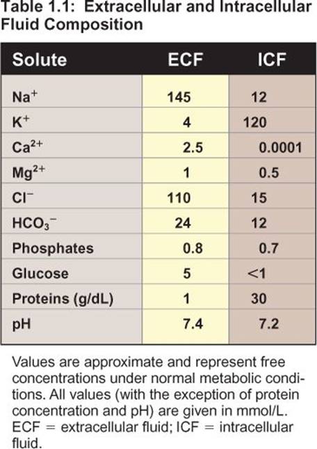
Cells are bathed in an extracellular fluid (ECF) that contains ionized sodium (Na+), potassium (K+), magnesium (Mg2+), chloride (Cl−), phosphate (PO43−), bicarbonate (HCO3−), glucose, and small amounts of protein (Table 1.1). It also contains around 2 mmol free calcium (Ca2+). Ca2+ is essential to life, but many of the biochemical reactions required of cells can only occur if free Ca2+ concentrations are lowered ten-thousandfold, to around 10−7 mol. Thus, cells erect a barrier that is impermeable to ions (the plasma membrane) to separate intracellular fluid ([ICF] or cytosol) from ECF and then selectively modify the composition of ICF to facilitate the biochemical reactions that sustain life. ICF is characterized by low Ca2+, Na+, and Cl− concentrations compared with ECF, whereas the K+ concentration is increased. Cells also contain more free protein than does the ECF, and the pH of ICF is slightly more acidic.

Figure 1.1
Differences in cell morphology.
III. MEMBRANE COMPOSITION
Membranes comprise lipid and protein (Figure 1.2). Lipids form the core of all membranes. Lipids are ideally suited to a barrier function because they are hydrophobic: They repel water and anything dissolved in it (hydrophilicmolecules). Proteins allow cells to interact with and communicate with each other, and they provide pathways that allow water and hydrophilic molecules to cross the lipid core.
A. Lipids
Membranes contain three predominant types of lipid: phospholipids, cholesterol, and glycolipids. All are amphipathic in nature, meaning that they have a polar (hydrophilic) region and a nonpolar (hydrophobic) region. The polar region is referred to as the head group. The hydrophobic region is usually composed of fatty acid “tails” of variable length. When the membrane is assembled, the lipids naturally gather into a continuous bilayer (Figure 1.3). The polar head groups gather at the internal and external surfaces where the two layers interface with ICF and ECF, respectively. The hydrophobic tail groups dangle down from the head groups to form the fatty membrane core. Although the two halves of the bilayer are closely apposed, there is no significant lipid exchange between the two membrane leaflets.

Figure 1.2
Membrane structure.
1. Phospholipids: Phospholipids are the most common membrane lipid type. Phospholipids comprise a fatty acid tail coupled via glycerol to a head group that contains phosphate and an attached alcohol. Dominant phospholipids include phosphatidylserine, phosphatidylethanolamine, phosphatidylcholine, phosphatidylinositol, and phosphatidylglycerol. Sphingomyelin is a related phospholipid in which glycerol has been replaced by sphingosine. The alcohol group in sphingomyelin is choline.
2. Cholesterol: Cholesterol is the second most common membrane lipid. It is hydrophobic but contains a polar hydroxyl group that draws it to the bilayer's outer surface, where it nestles between adjacent phospholipids (Figure 1.4). Between the hydroxyl group and the hydrocarbon tail is a steroid nucleus. The four steroid carbon rings make it relatively inflexible, so adding cholesterol to a membrane reduces its fluidity and makes it stronger and more rigid.
3. Glycolipids: The outer leaflet of the bilayer contains glycolipids, a minor but physiologically significant lipid type comprising a fatty acid tail coupled via sphingosine to a carbohydrate head group. The glycolipids create a carbohydrate cell coat that is involved in cell-to-cell interactions and that conveys antigenicity.

Figure 1.3
Membrane lipid bilayer.
B. Proteins
The membrane's lipid core seals the cell in an envelope across which only lipid-soluble materials, such as O2, CO2, and alcohol can cross. Cells exist in an aqueous world, however, and most of the molecules that they need to thrive are hydrophilic and cannot penetrate the lipid core. Thus, the surface (plasma) membrane also contains proteins whose function is to help ions and other charged molecules across the lipid barrier. Membrane proteins also allow for intercellular communication and provide cells with sensory information about the external environment. Proteins are grouped on the basis whether they localize to the membrane surface (peripheral) or are integral to the lipid bilayer (Figure 1.5).
1. Peripheral: Peripheral proteins are found on the membrane surface. Their link to the membrane is relatively weak and, thus, they can easily be washed free using simple salt solutions. Peripheral proteins associate with both the intracellular and extracellular plasma membrane surfaces.
a. Intracellular: Proteins that localize to the intracellular surface include many enzymes; regulatory subunits of ion channels, receptors, and transporters; and proteins involved in vesicle trafficking and membrane fusion as well as proteins that tether the membrane to a dense network of fibrils lying just beneath its inner surface. The network is composed of spectrin, actin, ankrin, and several other molecules that link together to form a subcortical cytoskeleton (see Figure 1.5).
b. Extracellular: Proteins located on the extracellular surface include enzymes, antigens, and adhesion molecules. Many peripheral proteins are attached to the membrane via glycophosphatidylinositol([GPI] a glycosylated phospholipid) and are known collectively as GPI-anchored proteins.
2. Integral: Integral membrane proteins penetrate the lipid core. They are anchored by covalent bonds to surrounding structures and can only be removed by experimentally treating the membrane with a detergent. Some integral proteins may remain localized to one or the other of the two membrane leaflets without actually traversing its width. Others may weave across the membrane many times (transmembrane proteins) as shown in Figure 1.6. Examples include various classes of ion channels, transporters, and receptors.

Figure 1.4
Cholesterol location with the membrane.
Clinical Application 1.1: Paroxysmal Nocturnal Hemoglobinuria
Paroxysmal nocturnal hemoglobinuria (PNH) is a rare, inherited disease caused by a defect in the gene that encodes phosphatidylinositol glycan A. This protein is required for synthesis of the glycophosphatidylinositol anchor used to tether peripheral proteins to the outside of the cell membrane. The gene defect prevents cells from expressing proteins that normally protect them from the immune system. The nighttime appearance of hemoglobin in urine (hemoglobinuria) reflects red blood cell lysis by immune complement. Patients typically manifest symptoms associated with anemia. PNH is associated with a significant risk of morbidity, in part, because patients are prone to thrombotic events. The reason for the increased incidence of thrombosis is not well delineated.
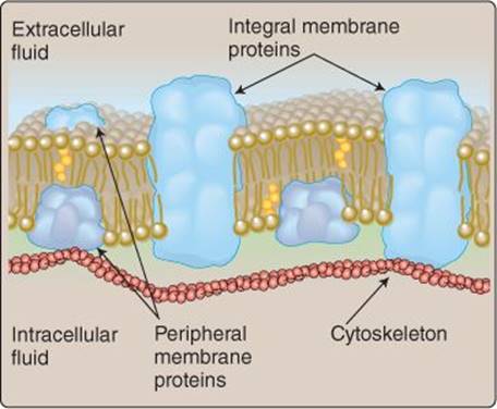
Figure 1.5
Membrane proteins.
IV. DIFFUSION
Movement across a membrane requires a motive force. Most substances cross the plasma membrane by diffusion, their movement driven by a transmembrane concentration gradient. When the concentration difference across a membrane is unfavorable, however, then the cell must expend energy to force movement “uphill” against the concentration gradient (active transport).

Figure 1.6
Membrane-spanning proteins.
A. Simple diffusion
Consider a container filled with water and divided into two compartments by a pure lipid membrane (Figure 1.7). Blue dye is now dropped into the container at left. Initially, the dye remains concentrated and restricted to its small entry area, but molecules of gas, water, or anything dissolved in water are in constant thermal motion. These movements cause the dye molecules to distribute randomly throughout the entire chamber, and the water eventually becomes a uniform color, albeit lighter than the original drop. The example shown in Figure 1.7 assumes that the dye is unable to cross the membrane, so the chamber on the right remains clear, even though the difference in dye concentrations across the barrier is very high. However, if the dye is lipid soluble or is provided with a pathway (a protein) that allows it to cross the barrier, diffusion will carry the molecules into the second chamber, and the entire tank will turn blue (Figure 1.8).
B. Fick law
The rate at which molecules such as blue dye cross membranes can be determined using a simplified version of the Fick law:
J = P × A (C1 − C2)
where J is diffusion rate (in mmol/s), P is a permeability coefficient, A is membrane surface area (cm2), and C1 and C2 are dye concentrations (mmol/L) in compartments 1 and 2, respectively. The permeability coefficient takes into account a molecule's diffusion coefficient, partition coefficient, and the thickness of the barrier that it must traverse.
1. Diffusion coefficient: Diffusion rates increase when a molecule's velocity increases, which is, in turn, determined by its diffusion coefficient. The coefficient is proportional to temperature and inversely proportional to molecular radius and the viscosity of the medium through which it diffuses. In practice, small molecules diffuse quickly through warm water, whereas large molecules diffuse very slowly through cold, viscous solutions.

Figure 1.7
Simple diffusion in water.
2. Partition coefficient: Lipid-soluble molecules, such as fats, alcohols, and some anesthetics can cross the membrane by dissolving in its lipid core, and they have a high partition coefficient. Conversely, ions such as Na+ and Ca2+ are repelled by lipids and have a very low partition coefficient. A molecule's partition coefficient is determined by measuring its solubility in oil compared with water.
3. Distance: Net diffusion rate slows when molecules have to traverse thick membranes compared with thin ones. The practical consequences of this relationship can been seen in the lungs (see 22·II·C) and fetal placenta (see 37·III·B), organs designed to maximize diffusional rates by minimizing diffusional distance between two compartments.
4. Surface area: Increasing the surface area available for diffusion also increases the rate of diffusion. This relationship is used to practical advantage in several organs. The lungs comprise 300,000,000 small sacs (alveoli) that have a combined surface area of ~80 m2 that allows for efficient O2 and CO2 exchange between blood and the atmosphere (see 22·II·C). The lining of the small intestine is folded into fingerlike villi (Figure 1.9), and the villi sprout microvilli that, together, create a combined surface area of ~200 m2 (see 31·II). Surface area amplification allows for efficient absorption of water and nutrients from the gut lumen. Efficient exchange of nutrients and metabolic waste products between blood and tissues is ensured by a vast network of small vessels (capillaries) whose combined surface area exceeds 500 m2 (see 19·II·C).
5. Concentration gradient: The rate at which molecules diffuse across a membrane is directly proportional to the concentration difference between the two sides of the membrane. In the example shown in Figure 1.8, the concentration gradient (and, thus, the dye diffusion rate) between the two compartments is high initially, but the rate slows and eventually ceases as the gradient dissipates and the two sides equilibrate. Note that thermal motion causes the dye molecules to continue moving back and forth between the two compartments at equilibrium, but net movement between the two is zero. If there were a way of removing dye continually from the chamber on the right, the concentration gradient and diffusion rate would remain high (we would also have to keep adding dye to the left chamber to compensate for movement across the barrier).
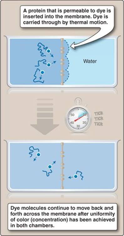
Figure 1.8
Diffusion through a lipid bilayer.
O2 movement between the atmosphere and the pulmonary circulation occurs by simple diffusion, driven by an air–blood O2 concentration gradient. Blood carries away O2 as fast as it is absorbed, and breathing movements constantly renew the O2 content of the lungs, thereby maintaining a favorable concentration gradient across the air–blood interface (see 23·V).

Figure 1.9
Intestinal villi.
C. Charged molecules
The prior discussion assumes that the dye is uncharged. The same basic principles apply to diffusion of a charged molecule (an electrolyte), but electrolytes are also influenced by electrical gradients. Positively charged ions such as Na+, K+, Ca2+, and Mg2+ (cations) and negatively charged ions such as Cl− and HCO3− (anions) are attracted to and will move toward their charge opposites. Thus, if the dye in Figure 1.8 carries a positive charge, and an electrical gradient is imposed across the container, dye molecules will move back through the membrane toward the negative electrode (Figure 1.10). The electrical gradient in Figure 1.10 was generated using a battery, but the same effect can be achieved by adding membrane-impermeant anions to Chamber 1. If Chamber 1 is filled with cations, the dye molecules will be repelled by the positive (like) charge and will accumulate in Chamber 2 (Figure 1.11). Note that the electrical gradient causes the dye concentration gradient between the two chambers to reform. Dye molecules will continue migrating from Chamber 2 back to Chamber 1 until the concentration gradient becomes so large that it equals and opposes the electrical gradient, at which point an electrochemical equilibriumhas been established. As discussed in Chapter 2, most cells actively expel Na+ ions to create an electrical gradient across their membranes. They then use the power of the combined electrical and chemical gradient (the electrochemical gradient) to move ions and other small molecules (e.g., glucose) across their membranes and for electrical signaling.

Figure 1.10
Charge movement induced by an electrical gradient.
V. PORES, CHANNELS, AND CARRIERS
Small, nonpolar molecules (e.g., O2 and CO2) diffuse across membranes rapidly, and they require no specialized pathway. Most molecules common to the ICF and ECF are charged, however, meaning that they require assistance from a pore, channel, or carrier protein to pass through the membrane's lipid core.
A. Pores
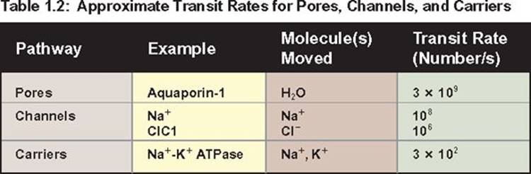
Pores are integral membrane proteins containing unregulated, water-filled passages that allow ions and other small molecules to cross the membrane. Pores are relatively uncommon in higher organisms because they are always open and can support very high transit rates (Table 1.2). Unregulated holes in the lipid barrier potentially can kill cells by allowing valuable cytoplasmic constituents to escape and Ca2+ to flood into the cell from the ECF. Aquaporin (AQP) is a ubiquitous water-selective pore. There are 13 known family members (AQP0–AQP12), three of which are expressed widely throughout the body (AQP1, AQP3, and AQP4). AQP is found wherever there is a need to move water across membranes. AQPs play a critical role in regulating water recovery from the renal tubule (see 27·V·C), for example, but they are also required for lens transparency in the eye (AQP0), keeping skin moist (AQP3), and mediating brain edema following insult (AQP4). Because AQP is always open, cells must regulate their water permeability by adding or removing AQP from the membrane.

Figure 1.11
Repellent effects of like charges.
Clinical Application 1.2: Staphylococcus aureus
Staphylococcus aureus is a skin-borne bacterium that is a leading cause of community- and hospital-acquired, bloodborne “staph” infections (bacteremia). The incidence of such infections has been rising steadily in recent decades and is of growing concern because of the high associated mortality rates and the increasing prevalence of antibiotic-resistant strains such as MRSA (methicillin-resistant Staphylococcus aureus).1 The bacterium's lethality is due to its producing a toxin (α-hemolysin) that kills blood cells. The toxin targets the plasma membrane, where toxin monomers assemble into a multimeric protein with an unregulated pore at its center. The cell loses control of its internal environment as a result. The ion gradients dissipate, and the cell swells and then blisters and ruptures, releasing nutrients that are believed to nourish the bacterium as it proliferates.
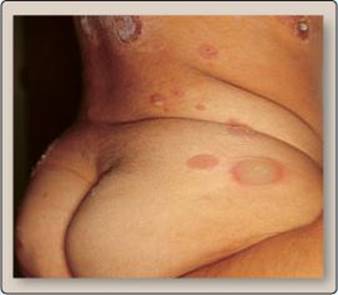
Skin lesions caused by
Staphylococcus aureus.
B. Channels
Ion channels are transmembrane proteins that assemble so as to create one or more water-filled passages across the membrane. Channels differ from pores in that the permeability pathways are revealed transiently (channel opening) in response to a membrane-potential change, neurotransmitter binding, or other stimulus, thereby allowing small ions (e.g., Na+, K+, Ca2+, and Cl−) to enter and traverse the lipid core (Figure 1.12). Ion movement is driven by simple diffusion and powered by the transmembrane electro-chemical gradient. Ions are forced to interact with the channel pore briefly so that their chemical nature and suitability for passage can be established (a selectivity filter), but the rate at which ions traverse the membrane via channels can be as high as 108 per second (see Table 1.2). All cells express ion channels, and there are numerous types, including the voltage-gated Na+channel that mediates nerve action potentials and voltage-gated Ca2+ channels that mediate muscle contraction. Ion channels are discussed in detail in Chapter 2.
 1For more information on Staphylococcus aureus and MRSA, see LIR Microbiology, 3e, Chapter 8.

Figure 1.12
Ion channel opening.
C. Carriers
Larger solutes, such as sugars and amino acids, are typically assisted across the membrane by carriers. Carriers can be considered enzymes that catalyze movement rather than a biochemical reaction. Translocation involves a binding step, which slows transport rate considerably compared with pores and channels (see Table 1.2). There are three principal carrier modes: facilitated diffusion, primary active transport, and secondary active transport.
1. Transport kinetics: Carriers, like enzymes, show substrate specificity, saturation kinetics (Michaelis-Menten kinetics), and susceptibility to competition. A general scheme for carrier-mediated transport envisions a solute-binding step, a change in carrier conformation that reveals a conduit through which the solute may pass, and then release on the opposite side of the membrane (Figure 1.13). When solute concentrations are low, carrier-mediated transport is more efficient than simple diffusion, but a finite number of solute binding sites means that a carrier can saturate when substrate concentrations are high (Figure 1.14). The transport rate at which saturation occurs is known as the transport maximum (Tm) and is the functional equivalent of Vmax that defines maximal reaction velocity catalyzed by an enzyme.1
2. Facilitated diffusion: The simplest carriers use electrochemical gradients as a motive force (facilitated diffusion) as shown in Figure 1.15A. They simply provide a selective pathway by which organic solutes, such as glucose, organic acids, and urea, can move across the membrane down their electrochemical gradients. The binding step ensures selectivity of passage. Common examples of such carriers includes the GLUT family of glucose transporters and the renal tubule urea transporter (see 27·V·D). The GLUT1 transporter is ubiquitous and provides a principal pathway by which all cells take up glucose. GLUT4 is an insulin-regulated glucose transporter expressed primarily in adipose tissue and muscle.
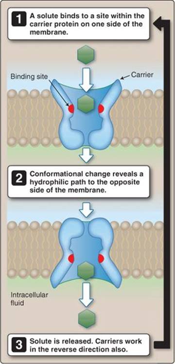
Figure 1.13
Model for transport by a carrier protein.
3. Primary active transport: Moving a solute uphill against its electrochemical gradient requires energy. Primary active transporters are ATPases that move or “pump” solutes across membranes by hydrolyzing adenosine triphosphate (ATP) as shown in Figure 1.15B. There are three main types of pump, all related P-type ATPase family members: a Na+-K+ ATPase, a group of Ca2+ ATPases, and a H+-K+ ATPase.
a. Na+-K+ ATPase: The Na+-K+ ATPase (Na+-K+ exchanger or Na+-K+ pump) is common to all cells and uses the energy of a single ATP molecule to transport three Na+ out of the cell, while simultaneously bringing two K+ back from the ECF. Movement of both ions occurs uphill against their respective electrochemical gradients. The physiologic importance of the Na+-K+ ATPase cannot be overstated. The Na+ and K+ gradients it establishes permit electrical signaling in neurons and myocytes, for example, and is used to drive passage of other solutes into and out of virtually all cells by secondary active transport (see below).

Figure 1.14
Carrier saturation kinetics.
1For further discussion of enzymatic maximal velocity, see LIR Biochemistry, 5e, p. 56.
b. Ca2+ ATPases: All cells express a plasma membrane Ca2+ ATPase (PMCA) that pumps Ca2+ out of the cytoplasm and is primarily responsible for maintaining intracellular Ca2+ concentrations at submicromolar levels. A related sarco (endo) plasmic reticulum Ca2+ ATPase (SERCA) is expressed in the sarcoplasmic reticulum of myocytes and the ER of other cells. SERCA sequesters Ca2+ in intracellular stores.
c. H+-K+ ATPase: The H+-K+ ATPase pumps acid and is responsible for lowering stomach pH, for example (see 30·IV·C). It is also found in the kidney, where it is involved in pH balance (see 27·IV·D).
4. Secondary active transport: A second class of active transporters use the energy inherent in the electrochemical gradient of one solute to drive uphill movement of a second solute (secondary active transport). Such carriers do not hydrolyze ATP directly, although ATP may have been used to create the gradient being harnessed by the secondary transporter. Two transport modes are possible: countertransport and cotransport.
a. Countertransport: Exchangers (antiporters) use the electrochemical gradient of one solute (e.g., Na+) to drive flow of a second (e.g., Ca2+) in the opposite direction to the first (see Figure 1.15C). The Na+-Ca2+exchanger helps maintain low intracellular Ca2+ concentrations by using the inwardly directed Na+ gradient to pump Ca2+ out of the cell. Other important exchangers include a Na+-H+ exchanger and a Cl−-HCO3−exchanger.
b. Cotransport: Cotransporters (symports) use the electro-chemical gradient of one solute to drive flow of a second or even a third solute in the same direction as the first (see Figure 1.15D). For example, cotransporters use an inwardly directed Na+ gradient to recover glucose and amino acids from the intestinal lumen and renal tubule (Na+-glucose and Na+–amino acid cotransporters, respectively), but other examples include a Na+-Cl−cotransporter, a K+-Cl− cotransporter, and a Na+-K+-2Cl− cotransporter.

Figure 1.15
Principal modes of membrane transport. ATP = adenosine triphosphate.

Figure 1.16
Chemical signaling pathways.
VI. INTERCELLULAR COMMUNICATION
The body's various organs each have unique properties and functions, but they must work closely together to ensure the well-being of the individual as a whole. Cooperation requires communication between organs and cells within organs. Some cells contact and communicate with each other directly via gap junctions (Figure 1.16A). Gap junctions are regulated pores that allow for exchanging chemical and electrical information (see 4·II·F) and that play a vital role in coordination of cardiac excitation and contraction, for example. Most intercellular communication occurs using chemical signals, which have traditionally been classified according to the distance and route they have travel to exert a physiologic effect. Hormones are chemicals produced by endocrine glands and some nonendocrine tissues that are carried to distant targets by the vasculature (see Figure 1.16B). Insulin, for example, is released into the circulation by pancreatic islet cells for carriage to muscle, adipose tissue, and the liver. Paracrines are released from cells in very close proximity to their target (see Figure 1.16C). For example, the endothelial cells that line blood vessels release nitric oxide as a way of communicating with the smooth muscle cells that make up the vessel walls (see 20·II·E·1). Paracrines typically have a very limited signaling range because either they are degraded or are taken up rapidly by neighboring cells. Autocrine messengers bind to receptors on the same cell that released them, creating a negative feedback pathway that modulates autocrine release (see Figure 1.16D). Autocrines, like paracrines, have a very limited signaling range.
VII. INTRACELLULAR SIGNALING
Once a chemical message arrives at its destination, it must be recognized as such by the target cell and then transduced into a form that can modify cell function. Most chemical messengers are charged and cannot permeate the membrane, so recognition has to occur at the cell surface. Recognition is accomplished using receptors, which serve as cellular switches. Hormone or neurotransmitter binding trips the switch and elicits a preprogrammed instruction set that culminates in a cellular response. Receptors are typically integral membrane proteins such as ligand-gated channels, G protein–coupled receptors(GPCRs), or enzyme- associated receptors. Lipophilic messengers can cross the plasma membrane and are recognized by intracellular receptors.
A. Channels
Ligand-gated ion channels facilitate communication between neurons and their target cells, including other neurons (see 2·VI·B). For example, the nicotinic acetylcholine (ACh) receptor is a ligand-gated ion channel that allows skeletal muscle cells to respond to excitatory commands from α-motor neurons. Neurotransmitter binding to its receptor causes a conformational change that opens the channel and allows ions such as Na+, K+, Ca2+, and Cl− to flow across the membrane through the pore (Figure 1.17A). Charge movement across the membrane constitutes an electrical signal that influences target cell activity directly, but channel-mediated Ca2+ influx can have additional and potent effects on cell function by activating various Ca2+-dependent signal transduction pathways (see below).

Figure 1.17
Neurotransmitter and hormone receptors. ACh = acetylcholine.
B. G protein–coupled receptors
GPCRs sense and transduce a majority of chemical signals, and the GPCR family is large and diverse (the human genome contains >900 GPCR genes). They are found in both neural and nonneural tissues. Common examples include the muscarinic ACh receptor, α-and β-adrenergic receptors, and odorant receptors. GPCRs all share a common structure that includes seven membrane-spanning regions that weave back and forth across the membrane (Figure 1.18). Receptor binding is transduced by a G protein (guanosine triphosphate [GTP]-binding protein), which then activates one or more second messengerpathways (see Figure 1.17B). Second messengers include cyclic 3′5′-adenosine monophosphate (cAMP), cyclic 3′5′-guanosine monophosphate (cGMP), and inositol trisphosphate (IP3). Multistep signal relay pathways allow for profound amplification of receptor-binding events. Thus, one occupied receptor can activate several G proteins, each of which can yield multiple second messenger molecules that, in turn, can activate multiple effector pathways (Figure 1.19).

Figure 1.18
G protein–coupled receptor structure.

Figure 1.19
Signal amplification by second messengers. GDP = guanosine diphosphate; GTP = guanosine triphosphate.
1. G proteins: G proteins are small membrane-associated proteins with GTPase activity. Two types of G protein have been described. The class that associates with hormone and neurotransmitter receptors are assemblies of three subunits: α, β, and γ. The GTPase activity resides in the α-subunit (Gα), which is normally bound to GDP. Receptor binding causes a conformational change that allows it to interact with its G-protein partner. The α-subunit then releases GDP, binds GTP, and dissociates from the protein complex (Figure 1.20). An occupied receptor can activate many G proteins before the hormone or transmitter dissociates. Active Gα subunits can interact with a variety of second messenger cascades, the principal ones being the cAMP and IP3 signaling pathways. The duration of Gα’s effects are limited by the protein's intrinsic GTPase activity. The rate of hydrolysis is slow, but, once GTP has been converted to GDP (and inorganic phosphate), the subunit loses its ability to signal. It then redocks with the Gβγassembly in the surface membrane and awaits a further opportunity to bind to an occupied receptor.
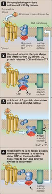
Figure 1.20
The cyclic adenosine monophosphate (cAMP) signaling pathway.
ATP = adenosine triphosphate;
Gs = stimulatory G protein; GDP = guanosine diphosphate; GTP = guanosine triphosphate; Pi = inorganic phosphate; PPi = pyrophosphate.
At least 16 different Gα subunits have been described. They can be classed according to their effects on a target pathway. Gαs subunits are stimulatory. Gαi subunits are inhibitory, meaning that they suppress second messenger formation when active.

Figure 1.21
The inositol trisphosphate (IP3) signaling pathway. Gq = stimulatory G protein; GDP = guanosine diphosphate; GTP = guanosine triphosphate.
2. cAMP signaling pathway: cAMP is a second messenger that is synthesized from ATP by adenylyl cyclase. Adenylyl cyclase is regulated by G proteins. Gαs stimulates cAMP formation, whereas Gαi inhibits it. cAMP activates protein kinase A (PKA), which phosphorylates and modifies the function of a variety of intracellular proteins, including enzymes, ion channels, and pumps. The cAMP signaling pathway is capable of tremendous signal amplification, so two checks are in place to limit its effects. Protein phosphatases counter the effects of the kinase by dephosphorylating the target proteins. The effects of the adenylyl cyclase are countered by a phosphodiesterase that converts cAMP to 5′-AMP.
3. IP3 signaling pathway: Gα q is a G-protein subunit that liberates three different second messengers via activation of phospholipase C (PLC) as shown in Figure 1.21. The messengers are IP3, diacylglycerol(DAG), and Ca2+. PLC catalyzes the formation of IP3 and DAG from phosphatidylinositol 4,5-bisphosphate (PIP2), a plasma membrane lipid. DAG remains localized to the membrane, but IP3 is released into the cytoplasm and binds to a Ca2+ release channel located in the ER. Ca2+ then floods out of the stores and into the cytosol, where it binds to calmodulin (CaM) as shown in Figure 1.22. CaM mediates Ca2+-activation of many enzymes and other intracellular effectors. Ca2+ also coordinates with DAG to activate protein kinase C (see Figure 1.21), which phosphorylates proteins involved in muscle contraction and salivary secretion, for example.

Figure 1.22
Ca2+-calmodulin (CaM)-dependent enzyme activation.

Figure 1.23
Tyrosine receptor kinase activation.
C. Catalytic receptors
Some ligands bind to membrane receptors that either associate with an enzyme or that have intrinsic catalytic activity (see Figure 1.17C). For example, natriuretic peptides influence renal function via a receptor guanylyl cyclaseand cGMP formation. Most catalytic receptors are tyrosine kinases (TRKs), the most common example being the insulin receptor. The insulin receptor is tetrameric, but most TRKs are single-peptide chains that associate only after ligand binding.
1. Receptor activation: Hormones and other messengers bind extracellularly to one of the peptide chains, causing a conformational change that favors dimerization (Figure 1.23). The intracellular portion of each monomer contains a kinase domain. Dimerization brings the two catalytic domains into contact, and they phosphorylate each other, thereby activating the receptor complex, which begins signaling.
2. Intracellular signaling: Active TRKs influence cell function via a number of transduction pathways, including the MAP (mitogen-activated protein) kinase cascade. Communication with these pathways first requires an adapter protein that mediates between the receptor and its intracellular effector. There are many different adapter proteins, but they all contain Src homology domains named SH2 and SH3. The SH2 domain recognizes the phosphorylated tyrosine domains on the activated TRK and allows the adapter protein to bind to the signaling complex.
D. Intracellular receptors
A fourth receptor class is located intracellularly and includes receptors for thyroid hormone and a majority of steroid hormones (see Figure 1.17D). All are transcription factors that influence cell function by binding to DNA and altering gene expression levels. Some of the receptors are cytoplasmic, whereas others are nuclear and may be associated with DNA. The cytoplasmic receptors are normally bound to a “heat shock” protein, which is displaced by the conformational change caused by steroid binding. The occupied receptor then translocates to the nucleus and binds to a hormone response element within the promoter region of the target gene. Nuclear receptors act in a similar way. Once bound, the receptor induces gene transcription, and the product alters cell function.
Chapter Summary
• All cells erect a lipid barrier (the plasma membrane) to separate the inside of the cell from the outside, and then selectively modify the ionic composition in the intracellular environment to facilitate the biochemical reactions that sustain life. Intracellular fluid contains very low concentrations of Ca2+ compared with extracellular fluid. Na+ concentrations are also lower inside, but K+ levels are higher.
• The plasma membrane contains three principal lipid types: phospholipids, cholesterol, and glycolipids. Phospholipids dominate the structure, cholesterol adds strength, and glycolipids mediate interactions with other cells.
• Movement across membranes occurs primarily by diffusion. Diffusion rate is dependent on the transmembrane concentration difference, molecular size, membrane thickness and surface area, temperature, viscosity of the solution through which the molecule must diffuse, and the molecule's solubility in lipid (partition coefficient).
• Integral membrane proteins such as pores, channels, and carriers provide pathways by which hydrophilic molecules may cross the lipid barrier.
• Pores are always open and are rare, the principal example being aquaporin, a ubiquitous water channel. Channels are regulated pores that open transiently to allow passage of small ions, such as Na+, Ca2+, K+, and Cl−. Movement through pores and channels occurs by simple diffusion down electrical and chemical concentration gradients (electrochemical gradient).
• Carriers selectively bind ions and small organic solutes, carry them across the membrane, and then release them on the opposite side. Carriers operate by two modes of transport: facilitated diffusion and active transport. Facilitated diffusion moves solutes “downhill” in the direction of the electrochemical gradients (e.g., glucose transport by the GLUT transporter family). Active transport uses energy to move solutes “uphill” from an area containing low solute concentration to an area of higher concentration.
• Primary active transporters, or pumps, use adenosine triphosphate to drive solutes uphill against their electrochemical gradient. Pumps include the Na+-K+ ATPase that is present in all cells, Ca2+ATPases, and the H+-K+ATPase.
• Secondary active transporters move solutes uphill by harnessing the energy inherent in electrochemical gradients for other ions. Exchangers move two solutes across the membrane in opposite directions (e.g., the Na+-Ca2+exchanger). Cotransporters (e.g., Na+-K+-2Cl− cotransporter and the Na+-glucose cotransporter) move two or more solutes in the same direction.
• The plasma membrane also contains receptor proteins that allow cells to communicate with each other using chemical messages. Signaling can occur over long distances via the release of hormones (e.g., insulin) into the bloodstream. Cells that are in close proximity to each other communicate using paracrines (e.g., nitric oxide). Autocrines are chemical signals that target the same cell that released them.
• Receptor binding is transduced in a variety of ways. Ligand-gated ion channels transduce binding using changes in membrane potential. Other receptor classes release G proteins to activate or inhibit second messengerpathways. Many receptors possess intrinsic kinase activity and signal occupancy through protein phosphorylation. A fourth class of receptor is located inside the cell. Intracellular receptors affect levels of gene expression when a message binds.
• G proteins modulate two major second-messenger cascades. The first involves cyclic adenosine monophosphate (cAMP) formation by adenylyl cyclase. cAMP acts primarily through regulation of protein kinase A and protein phosphorylation.
• Other G proteins activate phospholipase C and cause the release of inositol trisphosphate (IP3) and diacylglycerol (DAG). IP3, in turn, initiates Ca2+ release from intracellular stores. Ca2+ then binds to calmodulin and activates Ca2+-dependent transduction pathways. Ca2+ and DAG together activate protein kinase C and cause phosphorylation of target proteins.
• Receptors with intrinsic tyrosine kinase activity autophosphorylate when the message binds. This allows them to complex with adapter proteins that initiate signal cascades affecting cell growth and differentiation.
• Intracellular receptors translocate to the nucleus and bind to hormone response elements within the promoter region of target genes. Cell function is altered through increased levels of target gene expression.