2
I. OVERVIEW
All cells selectively modify the ionic composition of their internal environment to support the biochemistry of life (see 1·II) as shown in Figure 2.1. Moving ions into or out of a cell creates a charge imbalance between the intracellular fluid (ICF) and the extracellular fluid (ECF) and thereby allows a voltage difference to form across the surface membrane (a membrane potential, or Vm). This process creates an electrochemical driving force for diffusion that can be used to move charged solutes across the membrane or that can be modified transiently to create an electrical signal for intercellular communication. For example, nerve cells use changes in Vm (action potentials) to signal to a muscle that it needs to contract. The muscle cell, in turn, uses a change in Vm to activate Ca2+ release from its internal stores. Ca2+release then facilitates actin and myosin interactions and initiates muscle contraction. Neuronal and muscle action potentials both involve carefully coordinated sequences of ion channel events that allow selective transmembrane passage of ions (e.g., Na+, Ca2+, and K+) between ICF and ECF.

Figure 2.1
Intracellular fluid (ICF) modification by ion transporters. ATP = adenosine triphosphate; ECF = extracellular fluid.
II. MEMBRANE POTENTIALS
The term “membrane potential” refers to the voltage difference that exists across the plasma membrane. By convention, the ECF is considered to be at zero volts, or electrical “ground. ” Inserting a fine electrode across the surface membrane reveals that the cell interior is negative with respect to the ECF by several tens of millivolts. A typical nerve cell has a resting potential of − 70 mV, for example (Figure 2.2). Vm is established by membrane-permeant ions traveling down their respective concentration gradients and generating diffusion potentials.
A. Diffusion potentials
Imagine a model cell in which the plasma membrane is composed of pure lipid, the ICF is rich in potassium chloride (KCl, which dissociates into K+ and Cl−), and ECF is pure water (Figure 2.3). Although there is a strong KCl concentration gradient for diffusion across the membrane, the lipid barrier prevents both K+ and Cl− from leaving the cell and thereby constrains both ions to the ICF. The charges carried by K+ and Cl− cancel each other out and, thus, there is no voltage difference between ECF and ICF. If a protein that permits passage of K+ alone is inserted into the lipid barrier, K+ is now free to diffuse down its concentration gradient from ICF to ECF, and the membrane is said to be semipermeable (Figure 2.4). Because potassium ions carry charge, their movement causes a diffusion potential to form across the membrane in direct proportion to the magnitude of the concentration gradient. The potential may be significant (tens of millivolts) but involves relatively few ions.
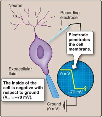
Figure 2.2
Membrane potential (Vm).

Figure 2.3
Charge distribution in a model cell with an impermeable membrane.
ECF = extracellular fluid;
ICF = intracellular fluid.

Figure 2.4
Origin of a diffusion potential. ECF = extracellular fluid; ICF = intracellular fluid.
The principle of bulk electroneutrality notes that the number of positive charges in a given solution is always balanced by an equal number of negative charges. The ICF and ECF are also subject to this rule, even though all cells create a negative Vm by altering charge distribution between the two compartments. In practice, Vm is established by just a few charges moving in the immediate vicinity of the cell membrane and their net effect on overall charge distribution within the bulk of the ICF and ECF is negligible.
B. Equilibrium potentials
When K+ crosses the membrane down its concentration gradient, it leaves a negative charge in the form of Cl− behind. Net charge magnitude builds in direct proportion to the number of ions leaving the ICF (see Figure 2.4), but, because opposite charges attract, K+ movement slows and eventually stops when the attraction of the negative charges inside the cell precisely counters the outward driving force created by the concentration gradient (electrochemical equilibrium). The potential at which equilibrium is established is known as the equilibrium potential for K+.
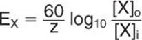
1. Nernst equation: Equilibrium potentials can be calculated for any membrane-permeant ion assuming that the ion's charge and concentrations on either side of the membrane are known:
|
Equation 2.1 |
|

Figure 2.5
Equilibrium potentials for Na+ (ENa), Ca2+ (ECa), and K+ (EK). ECF extracellular fluid; ICF = intracellular fluid.
where EX is the equilibrium potential for ion X (in mV), T is absolute temperature, z is the valence of the ion, R and F are physical constants (the ideal gas constant and the Faraday constant), and [X]o and [X]iare ECF and ICF concentrations of X (in mmol/L), respectively. Equation 2.1 is known as the Nernst equation. If T is assumed to be normal human body temperature (37°C), Equation 2.1 can be simplified:
|
Equation 2.2 |
|
Most of the common inorganic ions (Na+, K+, Cl−, HCO3−) have an electrical valence of 1 (monovalents). Ca2+ and Mg2+ have a valence of 2 (divalents).
Example 2.1
A cell has an intracellular free Mg2 concentration of 0.5 mmol/L and is bathed in a saline solution with a Mg2 composition that approximates plasma (1.0 mmol/L). The saline is held 37°C. If the cell has a membrane potential (Vm) of − 70 mV, and the membrane contains a gated channel that is Mg2+ permeable, will Mg2+ flow into or out of the cell when the channel opens?
We can use Equation 2.2 to calculate the Mg2+ equilibrium potential for (EMg+):
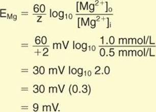
EMg tells us that Mg2+ will flow into the cell, its positive charges tending to drive Vm toward 9 mV.
2. Equilibrium potentials: The ICF and ECF are both strictly regulated, and their ionic composition is well known (see Figure 2.1, also see Table 1.1). Using known values for concentrations of the common ions, we can use the Nernst equation to predict that, for most cells in the body, EK = −90 mV, ENa = +61 mV, and ECa = +120 mV. Intracellular Cl concentrations can vary considerably, but ECl usually lies very close to Vm. If any of these ions are provided with a pathway that allows them to diffuse across the plasma membrane, they will drag Vm toward the equilibrium potential for that ion (Figure 2.5).
C. Resting potential
The plasma membranes of living cells are rich in ion channels that are permeable to one or more of the ions mentioned above, and some of these channels are open at rest. Resting Vm (resting potential) thus reflects the sum of the diffusion potentials generated by each of these ions flowing through open channels. Vm can be calculated mathematically as follows:

where gT is total membrane conductance (membrane conductance is the reciprocal of membrane resistance, in Ohms−1); gNa, gK, gCa, and gCl are individual conductances for each of the common ions (Na+, K+, Ca2+, and Cl−, respectively); and ENa, EK, ECa, and ECl are equilibrium potentials for these ions (in mV). Vm can also be calculated using the Goldman-Hodgkin-Katz (GHK) equation, which is similar in form to the Nernst equation above (Equation 2.1). The GHK equation derives Vm using relative membrane permeabilities for each of the ions that contribute to membrane potential.
In practice, most cells at rest have a negligible permeability to either Na+ or Ca2+. Cells do have a significant resting K+ conductance, however. Thus, Vm typically rests close to the equilibrium potential for K+(Figure 2.6). The approximate value for resting potential in neurons is −70 mV, −90 mV in cardiac myocytes, −55 mV in smooth muscle cells, and −40 mV in hepatocytes, for example.

Figure 2.6
Resting potential origins. ECF = extracellular fluid; ICF = intracellular fluid.
D. Extracellular ion effects
The ionic composition of the ECF is regulated within a fairly narrow range, but significant disturbances can occur through inadequate or excessive ingestion of salts or water. Because resting membrane permeability to Na+ and Ca2+ is low, Vm is relatively insensitive to changes in ECF concentration of either ion. Vm is sensitive to changes in extracellular K+ concentration, however, because resting potential is closely tied to the equilibrium potential for K+ (see Figure 2.6). Increasing extracellular K+ concentration (hyperkalemia) reduces the electrochemical gradient that drives K+ efflux, causing the membrane to depolarize (Figure 2.7). Conversely, lowering the extracellular K+ concentration (hypokalemia) steepens the gradient, and Vm becomes more negative.
E. Transporter contribution
The Na+-K+ ATPase that resides in the plasma membrane of all cells drives three Na out of the cell while simultaneously transferring two K+ from ECF to ICF. The three-for-two exchange results in an excess of positive charges being removed from the cell. Because the transporter creates a charge imbalance across the membrane, it is said to be electrogenic. The direct contribution of this exchange to Vm is insignificant, however. The main role of the Na+-K+ ATPase is to maintain a K+ concentration gradient across the membrane, because it is the K+ gradient that ultimately determines Vm via the K+ diffusion potential.

Figure 2.7
Membrane-potential (Vm) dependence on K+ concentration in the extracellular fluid (ECF). ICF = intracellular fluid.
Clinical Application 2.1: Hypokalemia and Hyperkalemia
Excitable cell function critically depends on maintaining membrane potential within a narrow range, so plasma levels normally range between 3.5 and 5.0 mmol/L. Hypokalemia and hyperkalemia are both commonly encountered clinically, however. Hypokalemia is generally of less concern than hyperkalemia, although some individuals with a rare inherited disorder (hypokalemic periodic paralysis) can experience muscle weakness when plasma K+concentrations dip such as following a meal. Hyperkalemia is, potentially, a more serious condition. The slow depolarization caused by rising plasma K+ levels inactivates Na+ channels that are required for muscle excitation, resulting in skeletal muscle weakness or paralysis and cardiac arrhythmias and conduction abnormalities. Hyperkalemia usually results from kidney failure and impaired ability to excrete K+. Treatment typically requires either diuresis or dialysis to remove excess K+ from the body.
III. EXCITATION
Many cell types use changes in Vm and transmembrane ion fluxes as a means of signaling or initiating intracellular events. Sensory cells (e.g., mechanosensors, olfactory receptors, and photoreceptors) transduce sensory stimuli by generating a Vm change called a receptor potential. Neurons signal to each other and to effector tissues using action potentials. Myocytes and secretory cells also use changes in Vm to increase intracellular Ca2+ concentration, thereby facilitating contraction and secretion, respectively. All such cells are said to have excitable membranes.
A. Terminology
The electrical changes caused by increased membrane permeability to ions do not consider the permeant ion's species (e.g., Na+ versus K+ or Cl−), only the charge that it carries.
1. Membrane potential changes: The inside of a cell at rest is always negative with respect to the ECF. When a positively charged ion (cation) flows into a cell, negative charges (anions) are neutralized, and the membrane loses polarization. The influx is said to have depolarized the cell, or caused membrane depolarization. By convention, depolarization is shown as an upward pen deflection on a voltage record (Figure 2.8). Conversely, if a cation leaves the cell, Vm becomes more negative: The efflux hyperpolarizes the cell (membrane hyperpolarization) and yields a downward pen deflection on a recording device.
2. Currents: When positive charges flow into a cell, they generate an inward current. By convention, recording devices, such as oscilloscopes and chart recorders, are configured so that inward currents cause a downward deflection (see Figure 2.8). Positive charges leaving the cell cause an outward current and an upward deflection on a recording device.

Figure 2.8
Membrane potential changes and ion currents.
Anions and cations are equally effective in changing Vm, but, because anions carry negative charges, their effects are opposite to those of cations. When an anion enters the cell from the ECF, it hyperpolarizes the membrane and yields an outward current. Conversely, anions leaving a cell create an inward current, and the cell depolarizes.
B. Action potentials
Action potential size, shape, and timing may vary widely between the different cell types, but there are several common characteristics, including the existence of a threshold for action potential formation, all-or-nothingbehavior, overshoots, and afterpotentials (Figure 2.9). The discussion below focuses on a nerve action potential whose upstroke is mediated by voltage-dependent Na channels, but voltage-dependent Ca2+ channels can support action potentials also (e.g., see 17·IV·B·3).
1. Threshold potential: Because action potentials are explosive membrane events that have consequences (e.g., initiating muscle contraction), they must be triggered with care. Vm normally fluctuates over a range of a few millivolts with changes in extracellular K+ concentration and other variables, even at rest, but such changes do not trigger spikes. Neurons only fire action potentials when Vm depolarizes sufficiently to cross the voltage threshold for action potential formation (Vth), which, in a neuron, usually resides at around 60 mV. Vth corresponds to the voltage needed to open the number of voltage-dependent Na+ channels required to trigger an action potential.
2. All or nothing: Voltage-dependent Na+ channels that mediate action potentials are typically present in the membrane in high numbers. When Vm crosses threshold, they open to allow a massive inward current, and the membrane depolarizes in a self-perpetuating (regenerative) fashion toward ENa (61 mV). This “all-or-nothing” behavior can be likened to breaching a dam wall. Once depolarization begins, it does not stop until the ionic flood is complete.
3. Overshoot: The action-potential peak typically does not reach ENa, but it often “overshoots” the zero-potential line, and the inside of the cell becomes positively charged with respect to the ECF.
4. Afterpotentials: Action potentials are transient events. The down-stroke is caused in part by voltage-dependent K+ channels that open to allow K+ efflux, causing Vm to repolarize. In some cells, the action potential may be followed by an afterpotential of varying size and polarity. A hyperpolarizing afterpotential takes the membrane negative to Vm for a period before eventually settling at the normal resting potential.

Figure 2.9
An action potential. Vm membrane potential; Vth threshold potential.
C. Action potential propagation
When neurons fire an action potential, the electrical event does not instantaneously involve the entire cell, but rather the spike begins at one end of the cell and then propagates at speeds of up to 120 m/s to the far end (Figure 2.10). Muscle cells behave similarly, although conduction velocities are typically lower in muscle than in nerve (~1 m/s). The advantage to propagation is that it allows a message to be carried unlimited distances. By way of analogy, the travel distance of a written message within a hollow baton thrown to a recipient is limited by the strength of the throw. Pass the message to a team of relay runners, however, and travel distance is limited only by the number of runners available. In practice, signal propagation allows spinal neurons to communicate with the feet, which are typically a meter away!

Figure 2.10
Action potential propagation.
Neuronal signaling involves a number of sequential steps, including membrane excitation, action potential initiation, signal propagation, and recovery.

Figure 2.11
Passive current spread and degradation in a neuron. Vm = membrane potential.
1. Excitation: Action potentials are typically initiated by sensory receptor potentials or dendritic postsynaptic potentials, for example. These are minor membrane events whose amplitude is graded with input intensity. Their reach is limited, much as throwing a baton is limited by arm muscle strength. The potential spreads passively and instantaneously (electrotonically), using the same physical principles by which electricity travels in a wire. Its reach is limited because the local currents created by the receptor potential are short-circuited by leak channels, which are found in all excitable membranes (Figure 2.11). Leak channels (typically K+ channels) are open at rest, allowing voltage changes to fizzle before they can travel far by “leaking” current across the membrane.
Electrical impulses travel through conductive materials like shock waves. A Newton cradle (i.e., classic desk toy comprising five silver balls suspended side by side within a frame) provides a good visual analogy. When a ball at one end is lifted and released, it impacts its neighbor and imparts its kinetic energy via a shock wave to the ball at the opposing end without disturbing the three intervening balls. The ball rises on its nylon line with little apparent energy loss. Electricity similarly creates shock waves between adjacent metal atoms within a copper wire that are transmitted at close to the speed of light. Electrical currents cause electrons to move also, but they travel at speeds closer to that of cold molasses.
2. Initiation: If the receptor potential is sufficiently large to cause Vm in a region of the membrane that contains voltage-dependent Na+ channels to cross threshold, it will trigger a spike.
3. Propagation: Na+-channel opening allows Na+ to flow into the cell, driven by the electrochemical gradient for Na+ and generating an active current (Figure 2.12). The current then spreads electrotonically and causes a Vmdepolarization that extends some distance down the axon. If the distant region contains Na+ channels and the change in Vm is sufficiently large to cross threshold, the channels in this region open and regenerate the signal, much as a relay runner picks up a baton. The cycle of Na+ influx, electronic spread, and regenerative Na+-channel opening is repeated (propagates) down the length of the cell.
4. Recovery: Na+ channels are inactivated by depolarization within a few milliseconds, which temporarily inhibits further excitation and prevents action potentials endlessly boomeranging back and forth along an axon. Excitation is followed by a period of recovery, during which time ion gradients are renormalized by ion pumps and channels recover from excitation.

Figure 2.12
Regenerative signal propagation in a neuron.
All cells have a membrane potential but not all are excitable. By definition, nonexcitable cells do not generate action potentials, but many do show functional changes in Vm. For example, glucose causes the membrane potential of pancreatic β cells to oscillate in a sustained, rhythmic fashion. The electrical events correlate with insulin release.
D. Currents
Action potentials are gross membrane events reflecting net charge movements through many thousands of individual ion channels. Each channel-opening event generates a unitary current, the size of which is directly proportional to the number of charges moving through its pore (Figure 2.13). The sum of individual Na+ channel-opening events yields a whole-cell Na+ current. Similarly, the sum of individual K+channel events yields a whole-cell K+ current. Because there are many different classes of ion channel with selective permeabilities for all of the common inorganic ions, a whole-cell K+ current (for example) may represent K+ efflux through two or more discrete K+-channel types. Such currents can be dissected into their individual components based on their physical properties using voltage-clamp (whole-cell recordings) and patch-clamp (recordings made from small membrane patches) techniques.

Figure 2.13
Single-channel and whole-cell ion currents. C = closed; Im = membrane current; O = open.
IV. ION CHANNELS
Ion channels are integral membrane proteins containing one or more hydrophilic pores that open transiently to allow ions to cross to the membrane. Channels have several distinguishing features that identify them as such, including an activation mechanism, a selectivity filter, and a finite conductance. Many channels also inactivate with time during prolonged stimulation.
A. Activation
Ion channels create holes in the lipid barrier separating the ICF from ECF. If they were unregulated, ions would continue to flow across the membrane and collapse their respective concentration gradients, along with Vm. Thus, most channels have activation gates that regulate passage through the pore (Figure 2.14). When a channel is in its closed state, the gate seals the pore, and ions cannot pass. Channel activation (e.g., in response to a change in Vm; see below) initiates a change in protein conformation that opens the gate and allows ions to pass (i.e., the open state). Some channels transition between the open and closed states thousands of times per second, with the net open time (or, open probability) increasing in direct proportion to the strength of the activating stimulus.
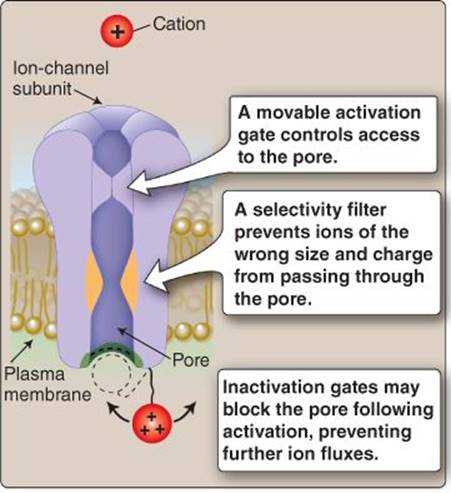
Figure 2.14
Ion-channel structure.
B. Selectivity
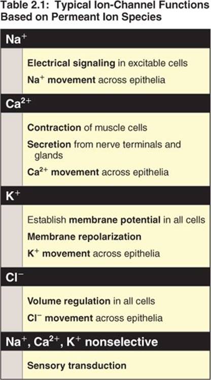
Before an ion can traverse the membrane, it must pass through a selectivity filter that determines its suitability for passage (see Figure 2.14). Selectivity filters reside within the pore and comprise regions where the permeant ion is forced to interact with one or more charged groups that limit passage based on molecular size and charge density. Thus, Na+ channels only pass Na+, K+ channels are selective for K, Ca2+ channels are Ca2+ selective, and Cl−channels pass only Cl− (Table 2.1). Other nonselective channels may allow passage of two or more different ions. Note that there are many different classes of each type of channel, differentiated by their mode of activation, kinetics, conductance, regulatory mechanisms, tissue specificity, and pharmacology.
The human genome encodes genes for over 400 ion channels. Almost half of these are K+ channels. Although they all have selectivity for K+ over other ions, the individual members of this large family all have unique properties (mode of activation, activation kinetics, conductance, regulatory mechanism, for example) and roles to play in membrane physiology (membrane repolarization, absorption, and secretion, for example).
C. Conductance
When a channel opens, membrane resistance falls because the channel allows current to flow across the lipid barrier. The extent to which resistance falls is dependent on the number of ions flowing through its pore per unit time, or its conductance. A channel's maximum conductance is one of its distinguishing hallmarks and is usually expressed in picoSiemens (pS).
D. Inactivation
Some channels possess an “inactivation gate” that is tripped upon activation, causing it to seal the channel pore and prevent further passage of ions (see Figure 2.14). The timescale of inactivation can vary from milliseconds to many tens of seconds, depending on channel class. Regardless, once a channel has been inactivated, it remains unresponsive to new stimuli, no matter how large the activating stimulus might be. Reactivation can only occur once the inactivation gate has been reset, a process that also has a variable timescale depending on channel type.
V. CHANNEL STRUCTURE
There are many ways in which a protein can be configured so as to create a transmembrane channel. Most mammalian channels follow a similar design principle, however, in which up to four to six subunits assemble around a central, water-filled pore. Tetrameric channels are the most common form (see below), as shown in Figure 2.15, but many ligand-gated channels are pentameric, and connexin channels are hexameric (see 4·II·F). Tetrameric channel subunits typically comprise six membrane-spanning domains (S1–S6). The S5 and S6 domains include charged residues that fashion a pore and selectivity filter when the subunits are assembled. Voltage-dependent Na+ and Ca2+channels are products of a single gene incorporating four subunit-like domains, but most channels are assembled from independent proteins. The advantage to a modular approach to channel design is that it allows for infinite channel diversity. Changes in a single subunit can cause a voltage-gated channel to become a second messenger–gated channel, for example, or change its selectivity or its regulatory mechanism.
VI. CHANNEL TYPES
Channels are usually identified on the basis of their ion selectivity and their gating (activation) mechanism. Thus, a “voltage-dependent Na+ channel” is activated by membrane depolarization and is Na+selective. Several different gating mechanisms are known.
A. Voltage gated
Voltage-gated Na+ channels, Ca2+ channels, and K+ channels all belong to the same channel superfamily related by a common tetrameric structure. The voltage-gated Na+ channel responsible for the action-potential upstroke in nerve and muscle cells comprises a single pore-forming subunit that associates with several smaller regulatory subunits. The subunit contains four related subunit-like domains that assemble around a central pore, as discussed in section V above. Each domain includes a highly charged peptide sequence (the S4 region) that functions as a voltage sensor. Membrane depolarization alters the charge distribution between the inner and outer membrane surfaces, and the voltage sensor shifts within the membrane, initiating a conformational change that opens the gate and reveals the channel pore.
B. Ligand gated
Ligand-gated channels transduce chemical signals and are the principal means by which neurons communicate with their targets. The diversity of the ligand-gated channel family is discussed in more detail in Chapter 6, but there are six principal classes that can be placed in three groups: cys-loop receptors, ionotropic glutamate receptors, and adenosine triphosphate (ATP) receptors.
1. Cys-loop superfamily: The cys-loop family includes the nicotinic acetylcholine receptor (nAChR), the 5-hydroxytryptamine (5-HT) receptor, the γ-aminobutyric acid (GABA) receptor, and the glycine receptor. All family members share a short, highly conserved amino acid sequence that gives the family its name, and all comprise five subunits arranged around a central pore (Figure 2.16). The nAChR and serotonin receptors are relatively nonspecific cation channels that support a mixed Na, K+, and Ca2+ influx upon ligand binding. The resultant membrane depolarization is excitatory. GABA and glycine receptors are anion channels that mediate Cl fluxes. These fluxes tend to stabilize Vm around resting potential and thereby inhibit membrane excitation. The nAChR and other family members have two ligand-binding sites that must be occupied simultaneously before the channel opens.

Figure 2.15
Voltage-gated ion-channel structure.
1. Cys-loop superfamily: The cys-loop family includes the nicotinic acetylcholine receptor (nAChR), the 5-hydroxytryptamine (5-HT) receptor, the γ-aminobutyric acid (GABA) receptor, and the glycine receptor. All family members share a short, highly conserved amino acid sequence that gives the family its name, and all comprise five subunits arranged around a central pore (Figure 2.16). The nAChR and serotonin receptors are relatively nonspecific cation channels that support a mixed Na, K+, and Ca2+ influx upon ligand binding. The resultant membrane depolarization is excitatory. GABA and glycine receptors are anion channels that mediate Cl fluxes. These fluxes tend to stabilize Vm around resting potential and thereby inhibit membrane excitation. The nAChR and other family members have two ligand-binding sites that must be occupied simultaneously before the channel opens.
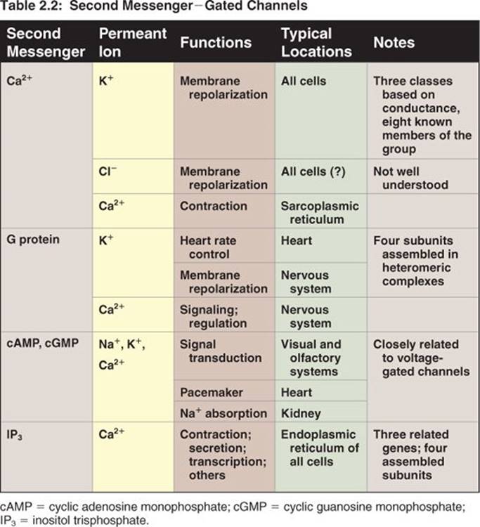

Figure 2.16
Cys-loop ligand-gated channel structure.
2. Ionotropic glutamate receptors: Ionotropic glutamate receptors are common in the central nervous system, where they play a critical role in learning and memory. All are tetrameric structures that support relatively nonselective Na+ and K+ fluxes when active. There are three principal groups that are differentiated pharmacologically (see Table 5.2): AMPA (-amino-3-hydroxy-5-methyl-4- isoxazolepropionic acid receptor) receptors, kainate receptors, and NMDA (N-methyl-D-aspartate) receptors.
3. Adenosine triphosphate receptors: ATP-gated channels are P2X-family purinoreceptors that are activated by ATP and support a nonspecific Na+, K+, and Ca2+ flux when open. They are believed to form trimeric channels in vivo. ATP receptors are involved in taste transduction (see 10·II).
C. Second messenger gated
A third class of channel opens or closes in response to changes in intracellular messenger concentration (Table 2.2). Ca2+-gated channels are ubiquitous, opening any time intracellular Ca2+ levels rise, regardless of whether the source of Ca2+ is an intracellular store or the ECF via a voltage-gated Ca2+ channel. Other channels are activated by G proteins, cyclic nucleotides, IP3, and a number of additional messengers.
D. Sensory channels
Transient receptor-potential channels (TRPs) form a large and diverse group of channels that function as cellular sensors transducing temperature, taste, pain, and mechanical stress (cell swelling and shear stress), for example. TRPs are also required for Ca2+ and Mg2+ reabsorption from the renal tubule (see 27·III). TRPs are currently the subject of intense study, and many aspects of their behavior in vivo have yet to be delineated, but they are known to be tetrameric assemblies similar to the voltage-gated channels described above. Most family members are weakly cation selective, passing Na+, K+, and Ca2+, the net result being membrane depolarization. The TRP family comprises six structurally distinct groups whose functions are summarized in Table 2.3.

Chapter Summary
• All cells modify their internal ionic environment using ion pumps (ATPases), which causes chemical concentration gradients to form across their surface membrane. Ions diffusing back down these concentration gradients create diffusion potentials. Membrane potential represents the sum of the diffusion potentials for all permeant ions (Na+, Ca2+, K+, Mg2, and Cl−).
• Ions are also influenced by electrical gradients, so their tendency to cross a membrane is governed by the net electrochemical gradient. The potential at which the chemical and electrical gradients balance precisely (the equilibrium potential) can be calculated using the Nernst equation.
• Most cells are impermeable to Na+ and Ca2+ at rest, but the presence of a significant resting K+ conductance causes resting potential to settle at close to the K+ equilibrium potential. The resting K+conductance makes resting potential highly susceptible to changes in extracellular K+ concentration (hypokalemia and hyperkalemia).
• Excitable cells use changes in membrane potential (action potentials, or spikes) to communicate with each other and to trigger cellular events, such as muscle contraction and secretion. Action potentials are effected by the sequential opening and closing of ion channels. Voltage-dependent Na+-channel opening facilitates an inward Na+ current to cause membrane depolarization. Membrane repolarization is effected (in part) by an outward K+ current through voltage-dependent K+ channels.
• Action potentials are initiated locally at the site of stimulation and then propagate in a self-sustaining, regenerative fashion along the length of a cell.
• Most cells express many different ion-channel classes in their surface membrane, which can be distinguished on the basis of their mode of activation (gating), ion selectivity, activation and inactivation kinetics, conductance, and pharmacology.
• Voltage-dependent Na+ channels, K+ channels, Ca2+ channels, and Cl channels are activated by changes in membrane potential. Ligand-gated channels are activated by neurotransmitters, including acetylcholine, -aminobutyric acid, and glutamate. Second messenger–gated channels are sensitive to intracellular Ca2+, G proteins, cyclic nucleotides, and inositol trisphosphate. Transient receptor-potential channels are cellular sensors, mediating responses to chemicals, hot and cold temperatures, and mechanical stress.