
13
I. OVERVIEW
Cardiac muscle shares much in common with skeletal muscle. Skeletal and cardiac muscle sarcomeres are organized similarly, so the two muscle types give similar banding patterns when viewed under polarized light (Figure 13.1). The essential principles and molecular components of contraction are the same also. There are some key differences, however, because the tasks required of the two muscles are unique. Skeletal muscle fibers contract independently of each other, each responding to individualized commands from the motor cortex. By contrast, cardiac muscle functions independently of somatic motor control, and the fibers are extensively interconnected so as to form a single functional unit. The impetus for cardiac muscle contraction comes from within the musculature itself and then spreads from myocyte to myocyte, which necessitates well-developed pathways for communication between all muscle fibers within a heart. Cardiac muscle design also incorporates a different mechanism for regulating contractile force compared with skeletal muscle because myocardial excitation always involves all fibers, and there is no option of recruiting motor units to increase overall contractile force as there is in skeletal muscle. Cardiac muscle contractility is, instead, regulated through changes in membrane Ca2+ permeability, which is controlled primarily by the autonomic nervous system (ANS).

Figure 13.1
Cardiac muscle structure.
II. STRUCTURE
Cardiac myocytes contain contractile arrays that are structurally similar to those of skeletal muscle (see 12·II). The significant differences between skeletal and cardiac muscle begin with the regulatory protein troponin.
A. Troponin
Troponin is a Ca2+-sensitive heterotrimer associated with the actin thin filament. Increases in sarcoplasmic Ca2+ concentration cause a conformational change in troponin that rolls tropomyosin away from myosin-binding sites on the thin filament, thereby facilitating actin–myosin interaction. In skeletal muscle, troponin must bind two Ca2+ ions before contraction can begin. In cardiac muscle, only one Ca2+ ion is required for contraction.
Clinical Application 13.1: Cardiac Markers
Cardiac troponin I (cTnI) and troponin T (cTnT) are troponin isoforms unique to heart muscle. cTnI and cTnT both appear in the circulation within 3 hours of acute myocardial infarction (MI), reaching a peak after 10–24 hours. Their appearance in blood indicates ischemia- induced necrosis and loss of cardiac myocyte integrity and, thus, can be used clinically to detect an acute MI. Blood tests also measure creatine kinase (CK) levels, but CK is not specific to cardiac muscle. Elevated plasma CK levels can be caused by cocaine use or exercise-induced skeletal muscle trauma in the absence of any cardiac damage.
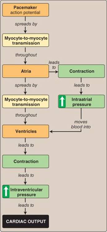
Figure 13.2
Concept map for cardiac function.
B. Communication pathways
Skeletal muscle fibers are under voluntary control and contract only when commanded by an α-motor neuron. Cardiac muscle is regulated by the nervous system, but a command to contract normally originates within an area of the myocardium (the sinoatrial node) that is specialized to function as a pacemaker. Pacemaker cells periodically generate spikes that spread from myocyte to myocyte until every fiber within the organ is involved (Figure 13.2), made possible by gap junctions and extensive cellular branching.
1. Pacemaker: The cardiac pacemaker is located high in the wall of the right atrium. Myocytes in this region are noncontractile, and their membranes contain ion channels that conduct a “funny current” ([If] see 17·IV·A) that causes them to depolarize spontaneously and generate action potentials ~100 times per minute. Once initiated, an action potential spreads regeneratively across both atria and ultimately involves the ventricles, with contraction following in its wake. The rate at which nodal cells depolarize and initiate spikes is modulated by the ANS as a way of controlling heart rate (see 17·IV·C).
2. Gap junctions: Gap junctions link adjacent myocytes and provide pathways for direct electrical and chemical communication (see 4·II·F). They are located at specialized sarcolemmal contact regions known as intercalated discs (Figure 13.3), which also contain structural elements (desmosomes and fascia adherens) that fuse the cells together mechanically and allow them to withstand the tension that is generated during muscle contraction. This contrasts with the complete lack of pathways for communication between adjacent skeletal-muscle fibers.
3. Cellular branching: When a command to contract is issued by the pacemaker, it must spread rapidly throughout the entire heart. Whereas skeletal muscle fibers are long, thin, and unbranching, cardiac myocytes branch extensively. The combination of branching cells and gap junctions creates a vast interconnected cellular network that functions as a single unit (a syncytium).
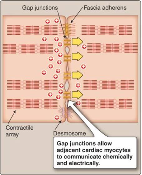
Figure 13.3
Structure of the intercalated disc.
Clinical Application 13.2: Hypertrophic Cardiomyopathy
Because the heart is a muscle tissue, inherited disorders of sarcomeric function that cause skeletal muscle wasting may lead to sudden cardiac death (SCD) when cardiac muscle is affected. Hypertrophic cardiomyopathy (HCM) is an inappropriate myocardial enlargement that is a leading cause of SCD in young athletes. More than half of these deaths result from mutations in genes that encode sarcomeric proteins and, most often, the gene encoding the cardiac isoform of the myosin heavy chain. HCM alleles disrupt the normal organization of myocytes within the myocardium, causing the myocytes to be disarrayed, the fibers hypertrophied, and the cellular structures distorted (compare the micrograph at right with Figure 13.1). Extensive deposition of connective tissue within the interstitium contributes to gross ventricular wall thickening associated with HCM. Patients with HCM are prone to atrial and ventricular arrhythmias, which accounts for the high risk of SCD in affected individuals.
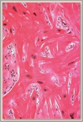
Myocyte disorganization.
C. Transverse tubules and sarcoplasmic reticulum
The action potential generated by the cardiac pacemaker is a signal that is conveyed into the core of each muscle fiber by transverse (T) tubules and then transmitted to the sarcoplasmic reticulum (SR) for electromechanical transduction. Although the SR provides the bulk of the Ca2+ needed for contraction, the membrane system is less extensive in cardiac muscle than in skeletal muscle. There are two other notable differences in cardiac T-tubule and SR design compared with skeletal muscle.
1. Location: In skeletal muscle, the T tubules align with the ends of the thick filaments, two per sarcomere. In cardiac muscle, there are fewer tubules, and they run along the Z line. T tubules tend to be wider in cardiac muscle and branch less extensively.
2. Diads: T tubules carry action potentials to the SR. In skeletal muscle, the tubules interface with two SR cisternae at junctional complexes known as triads. In cardiac muscle, tubules associate with a single extension of the SR at an analogous structure called a diad (see below).
III. CONTRACTILITY REGULATION
The molecular mechanisms underlying contraction are essentially the same in skeletal and cardiac muscle (see 12·III). The key difference between skeletal and cardiac muscle relates to the degree to which sarcoplasmic Ca2+ rises during excitation because Ca2+ availability is used to regulate contractility in cardiac myocytes.
A. Calcium-induced calcium release
When an action potential arrives at a diad, it encounters and activates L-type Ca2+ channels (also known as dihydropyridine receptors) in the T-tubule membrane (Figure 13.4). In skeletal muscle, this opening event forces Ca2+-release channels (also known as ryanodine receptors) in the SR to open also. Ca2+ then floods into the sarcoplasm, and contraction ensues. In cardiac muscle, L-type Ca2+–channel opening creates a Ca2+ trigger flux that is alone responsible for activating Ca2+-induced Ca2+ release (CICR) from the SR. There are far fewer CICR channels in cardiac muscle than in skeletal muscle, reflecting the differences between the principal way that force is regulated in the two muscle types. In skeletal muscle, excitation always triggers maximal Ca2+ release from the SR, and, therefore, the amount of force developed is always maximal also. In cardiac muscle, the size of the trigger flux and the amount of force generated is regulated by the ANS. An approximate 1:1 relationship between the number of L-type Ca2+ channels and dependent release channels permits fine control over sarcoplasmic Ca2+ concentration and contractility.
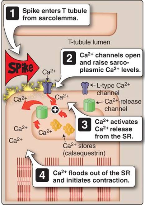
Figure 13.4
Ca2+-induced Ca2+ release. SR = sarcoplasmic reticulum; T tubule = transverse tubule.
B. Contractility regulation
The ANS controls both the rate and force with which cardiac muscle contracts. Rate control involves the coordinated actions of both the sympathetic and parasympathetic branches of the ANS (see 17·IV·C), but contractility is regulated primarily by the sympathetic nervous system (SNS). The two main targets of this regulation are the L-type Ca2+ channels and the Ca2+ ATPase that sequesters Ca2+ within the SR (SERCA; Figure 13.5).
1. Sympathetic activation: The SNS typically activates when changes in tissue activity increase the need for arterial blood supply. In practice, such needs are met by SNS-stimulated increases in cardiac output (CO). CO is increased by increasing heart rate (HR) and by making the heart a more effective pump (i.e., increased contractility). SNS terminals release norepinephrine at the cardiac neuromuscular junction and stimulate epinephrine release from the adrenal medulla. Epinephrine travels via the circulation to the heart. Neurotransmitter and hormone both bind to β1-adrenergic receptors on cardiac sarcolemma and increase protein phosphorylation by protein kinase A through activation of adenylyl cyclase and cyclic adenosine monophosphate formation.
2. Calcium channels: L-type Ca2+ channel phosphorylation increases channel open probability and thereby increases the size of the Ca2+ trigger flux. Increasing the trigger flux increases the magnitude of CICR from the SR and increases the number of Ca2+ ions available to bind troponin and initiate crossbridge cycling.

Figure 13.5
Regulation of Ca2+ release and contractility. ATP = adenosine triphosphate; cAMP = cyclic adenosine monophosphate; SNS = sympathetic nervous system.
3. Calcium pump: Cardiac muscle relaxation relies on two transporters to clear Ca2+ from the sarcoplasm. SERCA returns Ca2+ to the SR, whereas a Na+-Ca2+ exchanger in the sarcolemma transports Ca2+out of the cell. SERCA is associated with a small integral SR membrane protein called phospholamban, which functions as a pump limiter. When phospholamban is phosphorylated, its ability to inhibit the pump is curtailed, which allows the pump to cycle faster. There are two important consequences: faster relaxation times and increased amounts of Ca2+ stored for release on the next heartbeat.
a. Relaxation: When the pump runs faster, intracellular Ca2+ concentration drops to resting levels (0.1 μmol/L) more quickly. A rapid decrease in sarcoplasmic Ca2+ concentration allows the contractile machinery to relax faster, which prolongs the time available for ventricular filling during HR increases (see 39·V·B).
b. Calcium release: When the SR pump runs faster, more Ca2+ is stored in the SR, and less is returned to the extracellular space compared with previously. Stocking the stores with additional Ca2+ makes more available for release on the next contraction, and contractile force increases as a result.
Clinical Application 13.3: Beta Blockers and Calcium-Channel Blockers
Beta blockers and Ca2+-channel blockers are two important classes of drug used to control cardiac function and blood pressure.1 Beta blockers (e.g., propranolol) are β-adrenergic receptor antagonists that prevent norepinephrine- and epinephrine- mediated increases in myocardial Ca2+ concentration, thereby reducing heart rate and contractility and decreasing blood pressure. Ca2+-channel blockers prevent Ca2+influx through L-type Ca2+channels. Verapamil and diltiazem are both relatively myocardium-specific. Nifedipine is a dihydropyridine Ca2+-channel blocker that reduces blood pressure by relaxing vascular smooth muscle.
IV. PRELOAD DEPENDENCE
Skeletal and cardiac muscle show a similar inverted U-shaped relationship between sarcomere length and force generation. Skeletal muscle is designed such that thick- and thin-filament overlap is optimized when the muscle is at rest, and further stretching decreases contractility. Cardiac muscle is designed to take advantage of the length–force relationship to match its performance to the volume of blood entering its chambers.
A. Cardiac output
Cardiac muscle makes up the walls of the heart's four chambers (Figure 13.6). When the heart fills with blood, the chambers and cardiac myocytes contained within their walls stretch (preload), as shown in Figure 13.7. Cardiac muscle is designed such that in the absence of preload (i.e., an empty heart), sarcomeric length is minimal, and the possibility for further shortening and force development is small. Preloading pushes the sarcomere progressively higher up the left arm of the inverted U. In practice, this means that if the amount of blood returning from the vasculature between beats is high, the chamber walls and sarcomeres are stretched to a greater extent. Stretching increases the amount of force that the muscle is able to generate on the next beat, but this force is actually needed to expel the additional blood volume (preload) on the next beat. The length–tension relationship thereby provides the heart with a near-perfect way of matching contractile force to the volume of blood contained within its chambers. This phenomenon is known as the Frank-Starling law of the heart.
B. Length-dependent activation
The relationship between preload and force has traditionally been explained in terms of optimizing overlap between thick and thin filaments. However, preloading also causes length-dependent activation of the contractile apparatus. Activation sensitizes the crossbridge cycle to Ca2+, allowing for increased force generation even though the Ca2+ trigger flux and amount of Ca2+ released from the SR remains constant. The velocity of contraction increases during activation also. The molecular mechanisms of length-dependent activation are uncertain at present.
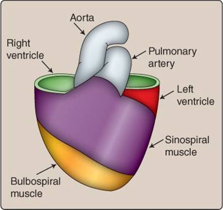
Figure 13.6
Bands of cardiac muscle.
 1For a discussion of beta blockers and Ca2+-channel blockers see LIR Pharmacology, 5e, p. 222.
C. Preload limits
Excessive preloads could potentially stretch the sarcomere to the point where force generation is impaired and CO is compromised. Thus, cardiac muscle is designed with structural components that strongly curtail stretching beyond an optimal range (i.e., the peak of the inverted “U” in Figure 13.7). Structural enhancements include intracellular elastic elements, increased amounts of connective tissue in the extracellular matrix, and a fibrous sac around the heart (pericardium) that limits its filling volume.
V. ENERGY SOURCE
Cardiac muscle contracts about once a second for 80 or more years, but the contractions are always brief (<1 s). This contrasts with skeletal muscle in which isometric contractions can last for minutes and can be sustained to the point of fatigue. A heart capable of even one prolonged (tetanic) contraction would quickly kill its owner! Thus, cardiac muscle has lost the ability to sustain adenosine triphosphate (ATP) production and contraction for more than a few seconds. It maintains modest ATP stores that support short contractions and then regenerates these stores using aerobic pathways when relaxed. The limited anaerobic capability creates a high dependence on O2. If O2 supply is limited due to reduced arterial blood supply, a creatine phosphate pool can sustain ATP levels for several tens of seconds, and then lactate acid begins to be produced. Prolonged O2 deprivation (minutes) causes irreversible hypoxic muscle damage and myocardial infarction.

Figure 13.7
Preloading effects on the ventricular myocardium.
Chapter Summary
• Cardiac muscle shares many features in common with skeletal muscle. Both have highly organized and aligned sarcomeres that give them a striated appearance under polarized light.
• Whereas skeletal muscle is under voluntary control, cardiac muscle functions autonomously. A specialized pacemaker periodically generates action potentials that spread from myocyte to myocyte to involve the entire heart. The autonomic nervous system regulates the timing and force of contraction but does not initiate it.
• Gap junctions that connect adjacent myocytes facilitate action-potential propagation. The myocytes also branch extensively to maximize interactions with adjacent cells.
• Force generation is regulated by the autonomic nervous system via β-adrenergic receptors and cyclic adenosine monophosphate–dependent phosphorylation by protein kinase A. Kinase targets include the L-type Ca2+ channelin the sarcolemma and sarcoplasmic reticular Ca2+ pump. Contractile force rises as a consequence.
• Force generation by cardiac muscle is highly preload dependent. Increasing sarcomeric length increases the force of contraction, thereby providing a way for the heart to increase contractility when required to expel increased filling volumes.
• Cardiac muscle relies on aerobic pathways to supply the adenosine triphosphate needed for contraction. The limited anaerobic capability makes heart muscle vulnerable to decreasing O2 availability during interruptions in blood supply.