
14
I. OVERVIEW
Skeletal and cardiac muscle are both designed to contract rapidly. Rapid kinetics facilitate locomotion (skeletal muscle) and sustain a cardiac output (cardiac muscle) that supports flow to dependent organs, even at rest. However, the human body performs many other functions that require muscular involvement on a less urgent time scale. The tasks are varied but are ably fulfilled by the highly adaptable smooth muscle. Smooth muscle is found in all regions of the body. It is layered within the walls of blood vessels and airways (Figure 14.1), functioning to adjust lumen diameter to regulate blood and air flow, respectively. Smooth muscle mixes and propels food and secretions through the gastrointestinal (GI) tract. Smooth muscle also regulates pupil diameter as a way of regulating the amount of light that enters the eye, and it regulates lens shape to adjust visual focus. Some of smooth muscle's diverse functions are summarized in Table 14.1. The varied demands made of smooth muscle have necessitated many organ- specific adaptations to its structure and function. For example, whereas striated muscle is activated by a handful of neurotransmitters and hormones, smooth muscle is modulated by hundreds of chemical signals. Contractile filaments within resting striated muscle typically have a standard length that does not vary over time. Contractile filaments in smooth muscle do not appear to have a standard length and may be disassembled and reassembled even as a contraction ensues. Plasticity of smooth muscle sarcomeres and cytoskeletal framework is necessary to maintain contractility during changes in hollow organ luminal volume (e.g., urinary bladder, GI tract, gallbladder, and uterus). Many details of smooth muscle structure and function are currently unresolved, but, like striated muscle, its core function is to generate force, accomplished using the same basic principles as in skeletal and cardiac muscle. Contraction begins when intracellular Ca2+ concentrations rise, and force is generated when head groups extending from a myosin thick filament bind to and pull on actin thin filaments.
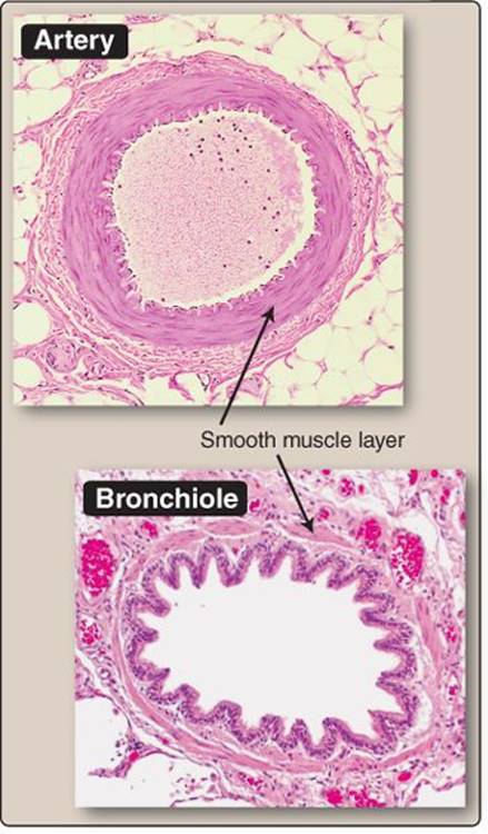
Figure 14.1
Arterial and airway smooth muscle.
II. STRUCTURE
Smooth muscle develops force through actin–myosin interactions, but the contractile filaments are organized less formally than they are in striated muscle.
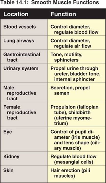
A. Contractile units
Smooth muscle cells are densely packed with actin and myosin filaments, which are organized into contractile units that have been referred to as “minisarcomeres.” However, because the contractile units are not aligned by Z disks as they are in skeletal and cardiac muscle, there are no visible striations when viewed under polarized light. The lack of striations gives “smooth muscle” its name.
1. Thick filaments: Smooth muscle myosin has a similar tertiary structure to that of striated muscle, but its amino acid sequence is quite distinct. Smooth muscle myosin is hexameric like that of other muscle types, containing two heavy chains that include head and neck regions and two pairs of light chains (a 17–kDa essential light chain and a 20–kDa regulatory light chain, also known as MLC20). The myosin heads contain ATPase activity and an actin-binding site. Although the ultrastructural basis for smooth muscle contraction has not been elucidated, one suggestion is that the myosin head groups within a thick filament have a sided or “sidepolar” arrangement that allows two actin filaments to be pulled simultaneously in different directions (Figure 14.2). Such an arrangement could account for observations that smooth muscle myocytes can shorten to a far greater degree than can striated muscle fibers.
2. Thin filaments: Thin filaments in striated muscle are associated with troponin, which imparts Ca2+ dependence to contraction. Smooth muscle does not contain troponin, but it does associate with two muscle-specific regulatory proteins called caldesmon and calponin. Their properties have been studied extensively in vitro, but their roles in vivo are still debated.
a. Caldesmon: Caldesmon is an actin-associated myosin ATPase inhibitor. Inhibition is relieved by high Ca2+- calmodulin (CaM) concentrations or by phosphorylation by Ca2+-CaM–dependent protein kinase(or several other endogenous kinases).
b. Calponin: Calponin is an abundant actin-associated myosin ATPase inhibitor that is regulated by Ca2+-CaM–dependent protein kinase–dependent phosphorylation.
3. Assembly: For a muscle cell to exert a useful force, its contractile units have to align roughly with the long axis of the cell. In smooth muscle, this seems to involve parallel assemblies of 3–5 thick filaments, each surrounded by numerous (10–12) thin filaments. Both thick and thin filaments appear to have variable lengths. This contrasts with striated muscle, where filament length is standardized and strictly controlled. The filaments are anchored by α-actinin–rich dense bodies (desmosomes) found scattered throughout the sarcoplasm (Figure 14.3) and believed to be the functional equivalent of Z disks in striated muscle (see 12·II·C·1). Dense bodies do not align as they do in striated muscle, but the general arrangement of thin filaments, thick filaments, and dense bodies does resemble a sarcomere.

Figure 14.2
A possible model for smooth muscle contraction.
B. Organization
Smooth muscle cells do not contain myofibrils as such, but long assemblies of contractile filaments and associated dense bodies appear to stretch the length of the cell. Contractile arrays are suspended within the cytoskeletal framework by a network of intermediate filaments comprising vimentin and desmin, the relative amounts depending on smooth muscle type. The arrays are tethered to the sarcolemma by dense plaques, which are related to dense bodies. The plaques are distributed over the entire cell surface and link adjacent cells mechanically (adherens junctions; see 4·II·G and Figure 14.3) so that smooth muscle may exert directed force upon contraction.
C. Membrane systems
Crossbridge cycling begins when intracellular Ca2+ concentrations rise above rest (0.1 μmol/L). In striated muscle, this involves L-type Ca2+ channels (dihydropyridine receptors) in transverse (T)-tubule membranes and Ca2+release from the sarcoplasmic reticulum (SR) via ryanodine receptors (see 12·III). Although the structural relationships between contractile arrays and SR is less well organized in smooth muscle compared with striated muscle, smooth muscle employs similar mechanisms to initiate contraction.
1. Caveolae: Smooth muscle cells do not contain T tubules that carry action potentials deep into the body of a muscle fiber, but they do show linear arrays of caveolae that may have a related function. Caveolae are 50–100 nm flask-shaped sarcolemmal pockets that form narrow junctions (15–30 nm) with the underlying SR. Caveolae are enriched in L-type Ca2+ channels, suggesting that the junctions serve a role similar to that of diads and triads in striated muscle.
2. Sarcoplasmic reticulum: Smooth muscle cells contain extensive tubular networks of SR that store Ca2+ until contraction begins. Unlike striated muscle, smooth muscle SR contains two types of Ca2+-release channels, one activated by Ca2+, the other by inositol trisphosphate (IP3) (Figure 14.4).
a. Calcium-induced calcium-release channels: Ca2+-induced Ca2+ release (CICR) channels are opened by Ca2+ entering the myocyte via voltage-dependent Ca2+ channels in the surface membrane. Although CICR is the primary means by which contraction is initiated in striated muscle, its role in smooth muscle is less clear.
b. Inositol trisphosphate–gated calcium channels: Smooth muscle SR also contains an IP3-gated Ca2+ channel. IP3 is a second messenger that communicates binding of one or more chemical signals, including many hormones and neurotransmitters (e.g., norepinephrine and acetylcholine), at the cell surface.

Figure 14.3
Suggested contractile filament alignment within a myocyte.
D. Neuromuscular junction
Most smooth muscle types are controlled by the autonomic nervous system (ANS). Depending on function and location within the body, smooth muscle may receive inputs from the sympathetic nervous system, parasympathetic nervous system, and enteric nervous system. The ANS neuromuscular junction is less developed than in skeletal muscle, but pre- and postsynaptic structures are arranged similarly. ANS efferents may contact multiple smooth muscle cells via a series of varicosities (swellings) spaced along the length of an axon, each of which is the site of a neuromuscular junction (see Figure 7.6).
Clinical Application 14.1: Irritable Bowel Syndrome
Irritable bowel syndrome is a gastrointestinal (GI) disorder associated with intestinal cramping, increased flatulence, and altered bowel habits. It has no known cause or cure. Treatment options are limited, but include oral antispasmodics such as L-hyoscamine, an atropine analog.1 Atropine is an alkaloid cholinergic receptor antagonist that blocks parasympathetic nervous system–induced increases in smooth muscle contractility in the GI tract and urinary bladder. The muscle relaxes as a result, which decreases cramping-associated pain. Natural remedies include peppermint oil, which has a similarly relaxing effect on smooth muscle. It is believed to act by blocking Ca2+channels in the smooth muscle plasmalemma, thereby reducing contractility and relaxing the muscle. Because peppermint oil relaxes sphincters also, it is contraindicated in patients with gastroesophageal reflux disease (GERD). GERD is associated with lower esophageal sphincter (LES) dysfunction. Because the LES is composed entirely of smooth muscle, peppermint oil further impairs sphincter contractility and exacerbates GERD symptoms.

Figure 14.4
Excitation–contraction coupling in smooth muscle. ATP = adenosine triphosphate; CICR = Ca2+-induced Ca2+ release; IP3 = inositol trisphosphate; PIP2 = phosphatidylinositol 4,5-bisphosphate; PLC = phospholipase C.
III. EXCITATION–CONTRACTION COUPLING
The number of competing signals that vie for control of smooth muscle function is immense (hundreds). Some of these signals may promote contraction and others relaxation, but, ultimately, most converge on Ca2+, just as in other muscle types. The main difference between smooth muscle and striated muscle is that the Ca2+-sensitive step has been transferred from the thin filament (via troponin and tropomyosin) to the thick filament (via myosin phosphorylation).
A. Calcium source
Contraction begins when sarcoplasmic Ca2+ concentrations rise. There are three potential mechanisms by which this can occur in smooth muscle: Ca2+ influx across the sarcolemma, CICR from the SR, and IP3-mediated Ca2+release from the SR. The relative contributions of these pathways to the net rise in intracellular Ca2+ concentration varies according to smooth muscle type.
1. Calcium influx: Smooth muscle cells express at least two types of Ca2+ channels. L-type Ca2+ channels that are found concentrated in caveolae are voltage-gated. The channels open in response to membrane depolarization to mediate Ca2+ influx and the upstroke of an action potential commonly seen in detrusor muscle and visceral muscle, for example. Most smooth muscle cells also express one or more receptor-operated Ca2+ channels(ROCCs). For example, visceral smooth muscle expresses muscarinic ROCCs, whereas vascular smooth muscle expresses adrenergic ROCCs. Although ROCC-mediated Ca2+ fluxes are relatively minor and generally insufficient to support contraction on their own, they do depolarize cells to potentiate (or initiate) Ca2+ influx and contraction via L-type Ca2+ channels.
 1For a discussion of antispasmodics, see LIR Pharmacology, 5e, p. 61.
2. Calcium-induced calcium release: CICR can be visualized as “Ca2+ sparks” in fluorescent imaging studies of smooth muscle, but their role is still being investigated. In some smooth muscle types (e.g., detrusor muscle), CICR may potentiate contraction, whereas in others (e.g., vascular smooth muscle), Ca2+ release relaxes the muscle by opening Ca2+-dependent K+ channels that hyperpolarize the membrane and decrease L-type Ca2+ channel open probability.
3. Inositol trisphosphate: Most smooth muscle types express a rich variety of G protein–coupled receptors (GPCRs) that modulate contraction through phospholipase C and IP3 formation. When intracellular IP3concentrations rise, the IP3-gated Ca2+ channel in the SR opens, and the Ca2+ stores are released (see Figure 14.4). IP3-mediated Ca2+ release and smooth muscle contraction can (and frequently does) occur independently of an action potential or other membrane potential change and is known as pharmacomechanical coupling. The IP3 pathway is a primary means of initiating contraction in smooth muscle.
B. Contraction
In striated muscle, rising intracellular Ca2+ concentrations prompt troponin to move tropomyosin away from myosin-binding sites on the actin filament (see 12·III·B). Striated muscle myosin has a high intrinsic ATPase activity, and, with the binding sites exposed, contraction proceeds rapidly. In smooth muscle, the ATPase remains inactive until the MLC20 regulatory light chain is phosphorylated. A rise in sarcoplasmic Ca2+ is sensed by CaM, which then activates myosin light-chain kinase (MLCK) as shown in Figure 14.5. MLCK phosphorylates MLC20 and, in the presence of ATP, the myosin head group is now able to reach forward and attach to a binding site on the actin thin filament. Crossbridge cycling then proceeds by essentially the same mechanism as described for striated muscle (see 12·III·B). Because myosin phosphorylation is required for smooth muscle contraction, it is said to be thick-filament regulated as opposed to thin-filament regulated in striated muscle. The myosin isoform's kinetics and the multistep nature of the activation process means that smooth muscle contracts 10–20 times slower than does skeletal muscle.
C. Relaxation
Muscle relaxation occurs when sarcoplasmic Ca2+ concentrations renormalize. Because smooth muscle contraction involves a kinase and a phosphorylation step, relaxation requires a phosphatase.
1. Calcium renormalization: When excitatory signaling ends, Ca2+ is expelled from the cell by Ca2+ ATPases and Na+-Ca2+ exchangers, and returned to the SR by a sarco (endo) plasmic reticulum Ca2+ATPase ([SERCA] Figure 14.6). MLCK then deactivates.
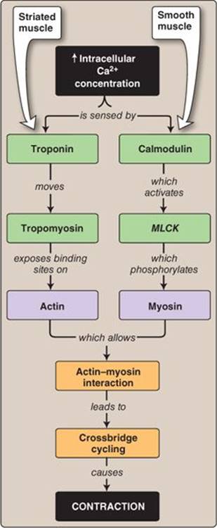
Figure 14.5
Differences between events facilitating crossbridge cycling in striated and smooth muscle. MLCK = myosin light-chain kinase.
Clinical Application 14.2: Erectile Dysfunction
Penile erection during male sexual arousal results from increased blood flow and engorgement of three sinusoidal tissue cavities within the penis (the corpora cavernosae and corpus spongiosum). Blood flow to these cavities is regulated in part by parasympathetic nerves that signal via nitric oxide (NO) release. NO relaxes intracavernosal trabeculae, which comprise smooth muscle cells lined by endothelium. Relaxation allows the erectile tissue to fill with blood in preparation for sexual activity. NO is a short-lived signaling molecule that exerts its effects through activation of guanylyl cyclase, cyclic guanosine monophosphate (cGMP) formation, and phosphorylation of proteins involved in regulation of intracellular Ca2+ concentration by cGMP-dependent protein kinase. Men who have difficulty achieving or sustaining an erection have benefited from the availability of drugs such as sildenafil and tadalafil, which are phosphodiesterase (PDE) inhibitors.1 PDEs degrade NO and terminate signaling. Inhibiting PDEs increases local NO levels, allowing an erection to be sustained.
Compounds that modulate smooth muscle contractility through protein kinase A and protein kinase G–dependent pathways (e.g., nitric oxide) do so by influencing SERCA and L-type Ca2+-channel activity.
2. Store refill: The plasmalemmal Ca2+ transporters compete with SERCA for available Ca2+ during relaxation, so the stores have to be topped off with Ca2+ from the outside of the cell via a store-operated Ca2+ channel(SOC). This is a critical step that must be completed for continued smooth muscle contractility. SOCs are common to many cell types, including lymphocytes, where a need for topping off is sensed by Stim1, a Ca2+ sensor, and requires Orai, a Ca2+ channel or Ca2+-channel subunit.
3. Dephosphorylation: Once MLC20 is phosphorylated, cross-bridge cycling continues for as long as ATP is available to power contraction. Relaxation of smooth muscle is, therefore, dependent on myosin phosphatase, which is constitutively active and always undoing the work of MLCK. Myosin phosphatase is a protein trimer comprising a protein phosphatase catalytic domain (PP1c), a myosin binding subunit (MYPT1), and a small subunit of unknown function. When sarcoplasmic Ca2+ concentrations fall and MLCK deactivates, myosin phosphatase quickly strips MLC20 of phosphate groups and the muscle relaxes.

Figure 14.6
Ca2+ handling during smooth muscle relaxation. ATP = adenosine triphosphate; IP3 = inositol trisphosphate.
1For a discussion of phosphodiesterase inhibitors, see LIR Pharmacology, 5e, p. 363.
D. Latchbridge formation
If myosin is dephosphorylated when still attached to actin, it locks or “latches” in place. Latchbridges have an intrinsically slow cycling rate that allows blood vessels, sphincters, and hollow organs such as the bladder to sustain contractions for prolonged periods with minimal ATP use (~1% of the amount required by skeletal muscle to achieve the same effect). Because latchbridges are contractile events, they can only occur if intracellular Ca2+ levels remain minimally elevated above background. Although many details of latchbridge formation, termination, and regulation have yet to be elucidated, the ability to form latchbridges is a unique and fundamental property of smooth muscle.
E. Regulation
In striated muscle, force regulation usually occurs through control of intracellular Ca2+ concentration. In smooth muscle, force is regulated via changes in MLC20 phosphorylation state. Force development is dependent on crossbridge formation, which can only occur when MLC20 is phosphorylated. Because MLC20 phosphorylation state is dependent on both MLCK and myosin phosphatase, there are multiple potential control points. Two principal regulatory pathways involve Rho-kinase (ROCK) and protein kinase C (PKC) as shown in Figure 14.7.
1. Rho-kinase: ROCK is a serine–threonine protein kinase regulated by RhoA, a GTP-binding protein. RhoA is activated indirectly following GPCR binding by, for example, norepinephrine, angiotensin II, endothelin, or any of a number of other ligands. ROCK has a several targets, including the MYPT1 myosin phosphatase myosin binding subunit. Phosphorylation inhibits myosin phosphatase activity and thereby promotes contraction. ROCK also has MLCK-like activity and stimulates contraction through direct effects on MLC20. Note that this pathway acts independently of any changes in intracellular Ca2+concentration.
2. Protein kinase C: Agonists that activate phospholipase C and promote contraction via IP3 simultaneously activate PKC via diacylglycerol release. PKC also phosphorylates many proteins that regulate contraction, including CPI-17. CPI-17 is a endogenous 17-kDa protein that becomes a potent myosin phosphatase inhibitor when phosphorylated, thereby promoting contraction. ROCK also phosphorylates CPI-17 in vitro, but the possible significance for smooth muscle contraction function in vivo is unresolved.
The ROCK- and PKC- mediated contractility increases account for a phenomenon known as “Ca2+ sensitization.” Ca2+ sensitization refers to an observed shift in the Ca2+-dependence of contractility toward lower intracellular free Ca2+ values following hormone or transmitter binding.
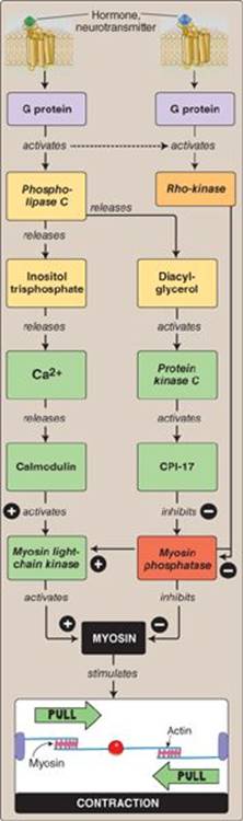
Figure 14.7
Pathways regulating smooth muscle contraction.
Clinical Application 14.3: Rho-kinase and Hypertension
Essential hypertension is a vascular smooth muscle disease. The underlying cause and cellular pathways involved are still largely unknown, despite hypertension's prevalence and cost, both financially and in terms of its effects on the health of the individual. High blood pressure reflects an inappropriate systemic vasoconstriction, forcing the left ventricle to generate higher arterial pressures to force blood through the narrowed vessels. Because rho-kinase(ROCK) is a primary regulator of smooth muscle contractility, the ROCK signaling pathway has come under scrutiny as a possible cause of and means of treating hypertension. Fasudil is a ROCK- specific kinase inhibitor currently used in Japan to treat the cerebral vasospasm that commonly follows subarachnoid hemorrhage. The inhibitor relaxes most smooth muscle types and is currently being investigated as a possible treatment option for essential and pulmonary hypertension.
IV. MECHANICS
Striated muscle and smooth muscle use the same basic principles for force generation so their mechanics are similar. Optimizing the degree of thick and thin filament overlap (preloading) maximizes force development, and contraction velocity slows as the afterload increases. There are notable differences, however, as discussed below.
A. Contraction rate
Smooth muscle is capable of contracting rapidly when the need arises; when eye pupil size must be decreased to reduce the amount of light falling on the retina, for example. The myosin isoform found in smooth muscle has inherently slow ATPase activity, however, which limits maximum contraction velocity to only a fraction of that seen in skeletal muscle.
B. Length adaptation
Smooth muscle regulates wall tone in many hollow organs whose lumen size is variable. For example, airways and blood vessels rhythmically dilate and constrict in time with the respiratory and cardiac cycles, respectively, yet the smooth muscle contained within their walls maintains a constant tone throughout the cycle. Similarly, urinary bladder luminal volume increases from ~6 to ~500 mL when full, yet the detrusor muscle within its walls can contract and expel urine any point during filling cycle. During bladder filling, the muscle “minisarcomeres” are stretched to their physiologic limits. Stretching reduces the degree of actin–myosin overlap and limits contractility, as in other muscle types, but smooth muscle is unique in that it adapts to this stress over a period of minutes and regains full contractility even at an increased length. This phenomenon is known as length adaptation and is a fundamental property of smooth muscle. The pathways involved have not been well delineated, but the phenomenon represents one of the key differences between striated muscle and smooth muscle. Whereas the length and number of contractile filaments in striated muscle is largely fixed and the contractile arrays ordered so as to optimize force production along a constant vector, smooth muscle is a fundamentally plastic cell type. When stretched, myocytes are believed to replicate contractile units and insert them in series with existing assemblies (Figure 14.8). Preexisting filaments are probably lengthened also. When the muscle returns to normal length, it again adapts, probably by removing the additional contractile units or shortening the contractile arrays. Note that the entire cytoskeletal framework must necessarily be remodeled to accommodate such changes. The pathways and proteins involved may be the same as those involved in initiating and regulating contraction.

Figure 14.8
Length adaptation during urinary bladder filling.
V. TYPES
Smooth muscle is a diverse tissue type that can be classified in many ways, but one of the more useful is based on function. Phasic smooth muscle contracts transiently when stimulated. Examples include muscles that make up the walls of the GI tract (stomach, small intestine, large intestine), and urogenital tract (ureters, urinary bladder, vas deferens, fallopian tube, uterus). Tonic smooth muscle is capable of sustained contractions, a feature that is often used to maintain a constant muscular tone. Examples include vascular and airway muscle, sphincters (e.g., lower esophageal sphincter, pyloric sphincter), and eye ciliary and iris muscles.
Most smooth muscle types are a blend of phasic and tonic, which allows them to respond to a range of stimuli.
A. Phasic
Phasic smooth muscle often functions like cardiac muscle. Specialized pacemaker cells generate action potentials that spread via gap junctions from myocyte to myocyte until the whole tissue is involved.
1. Pacemakers: Some smooth muscle cells are capable of generating spontaneous membrane potential (Vm) changes. In the GI tract, these may manifest as “slow waves” with a periodicity of ~3–5 per minute. If the waves achieve an amplitude sufficient to cross threshold, action potentials may be initiated that spread via gap junctions throughout the entire muscle body. A wave of contraction follows in its wake (Figure 14.9A). Because all myocytes within phasic muscle are excited as a single unit, phasic muscle may also be referred to as “unitary” smooth muscle.
2. Action potentials: Smooth muscle action potentials are characteristically slow and their form and time course highly variable. The upstroke is mediated by L-type Ca2+ channels, as is the upstroke of cardiac nodal cell “slow” action potential (see 17·IV·B). Ca2+ influx simultaneously initiates contraction and opens Ca2+-activated K+ channels (“BK” channels). Ca2+-channel inactivation and BK-mediated K+ efflux together help repolarize the membrane and yield the action potential downstroke.

Figure 14.9
Contractility control in phasic and tonic smooth muscle.
B. Tonic
Tonic smooth muscle (also known as multiunit smooth muscle) resembles skeletal muscle in that individual myocytes or groups of myocytes function independently of their neighbors (see Figure 14.9B). This feature allows for fine control over movements, which is advantageous for precise control of pupillary diameter and eye lens shape, for example. Multiunit control also allows force to be ramped up through recruitment, much as skeletal muscle force is controlled using combinations of discrete motor units (see 12·IV·D). Tonic smooth muscle typically does not generate action potentials and, indeed, contraction may occur in the absence of any Vm change through pathways described in Section III above.
Chapter Summary
• Smooth muscle serves many diverse functions in essentially all areas of the body. It is found in the walls of many hollow organs, including blood vessels, airways, intestines, and urogenital tract. Smooth muscle contracts slowly compared with striated muscle but is able to maintain a steady tone with minimal energy expenditure.
• Smooth muscle contraction involves interaction between actin and myosin, but the thick and thin filaments are loosely organized compared with striated muscle.
• Contraction is usually initiated when intracellular Ca2+ concentrations rise. Ca2+ may originate extracellularly and enter the cell via Ca2+ channels, or from Ca2+ stores in the sarcoplasmic reticulum (SR). The SR releases its Ca2+ stores either in response to Ca2+ influx from the outside of the cell (Ca2+-induced Ca2+ release) or a rise in intracellular inositol trisphosphate (IP3) concentration. The latter occurs as a result of surface receptor binding and acts via an IP3-gated Ca2+ channel in the SR.
• Control of crossbridge cycling in smooth muscle is through myosin phosphorylation. Rising intracellular Ca2+ concentrations cause Ca2+–calmodulin-dependent activation of myosin light-chain kinase (MLCK). MLCKphosphorylates the myosin head group, and crossbridge cycling begins.
• Relaxation of smooth muscle occurs when Ca2+ levels fall, and myosin light-chain kinase deactivates. Myosin phosphatase then dephosphorylates myosin and allows relaxation to occur. When Ca2+ levels are barely above baseline, smooth muscle may enter a latch state in which muscle tone is maintained for prolonged periods with minimal energy use.
• Smooth muscle contractile state usually represents a balance between myosin light-chain kinase and myosin phosphatase activity, and external ligands are able to modulate contractility by manipulating this balance. Myosin phosphatase is regulated by pathways that include protein kinase C and Rho-kinase, both of which potentiate contraction.
• Smooth muscle is required to maintain a steady tone in hollow organs whose internal volume changes appreciably over time. This is made possible by length adaptation, a process that allows the length–tension relationship to shift in parallel with organ expansion or contraction. Length adaptation may involve sarcomeric and cytoskeletal remodeling.
• There are two broad groups of smooth muscle. Phasic (unitary) smooth muscle functions as a single unit, much like cardiac muscle. Adjacent myocytes are connected via gap junctions, which allows for waves of excitation and contraction to propagate from one cell to the next. Tonic (multiunit) smooth muscle is composed of myocytes that function and are controlled i ndependently of each other, an organization reminiscent of skeletal muscle.