
16
I. OVERVIEW
Individual cells erect a membrane around their periphery to create a barrier between the extracellular and intracellular environments, which allows them to regulate their cytoplasmic composition. The body similarly encloses its tissues within skin, a multilayered covering comprising epidermis, dermis, and a functionally linked hypodermis (Figure 16.1). Skin forms a physical barrier that excludes microorganisms and other foreign substances, while simultaneously helping the body retain vital fluids. Skin has several important functions. The outermost layers protect underlying tissues from abrasion and other mechanical insults, chemicals, pathogens, and ultraviolet (UV) light. It contains active microbial defense mechanisms that bolster its barrier function when breached. Skin also serves a vital thermoregulatory role. It secretes aqueous solutions that enhance heat loss by evaporation. Also, the amount of blood traveling through the skin is modulated as a way of conserving or offloading body heat to the environment. Finally, skin is a sensory organ that contains a variety of nerves and specialized receptors that collect sensory information about the external environment and interactions with foreign bodies. Skin and its associated appendages form the integument, or body covering.
II. ANATOMY
Skin comprises two anatomical layers: a superficial epidermis and the dermis. Combined epidermal and dermal thickness varies from 0.5–5 mm, depending on location. This measurement does not include subcutaneous tissue (the hypodermis), which is often considered a part of the skin (see Section V below). Skin is the body's largest organ, accounting for 15%–20% of total body mass. Functionally, it is helpful to distinguish between hairy skin (nonglabrous) and areas without hair follicles (glabrous skin [derived from glaber, meaning “bald” or “hairless” in Latin]).
A. Hairy skin
Most areas of the body are covered with hairy skin. Although these areas do contain sensory receptors related to touch, pressure, temperature, and pain, their density is lower than that of glabrous skin. Nonglabrous skin is important for thermoregulation.

Figure 16.1
Skin structure and associated appendages.
B. Glabrous skin
Glabrous skin is smooth and hairless. Examples include the lips, soles of the feet, palms of the hand, and fingertips. The palms and soles of the feet are contact points for grasping and locomotion. Hair here would interfere with motor functions and would decrease the ability to discern the texture and temperature of surfaces. Glabrous skin contains a high density of sensory fibers to aid taction and thick structural adaptations to help protect against abrasions. Glabrous skin does not play a large role in thermoregulation, but skin blood flow does both increase and decrease as well as become erythemic due to elevated estrogen levels in pregnancy and chronic alcoholic liver disease (Figure 16.2).

Figure 16.2
Palmar erythema.
III. EPIDERMIS
The external environment is inherently hostile. It contains a number of elements capable of causing tissue damage, so the body must erect a barrier to protect itself. This barrier is called the epidermis, a reinforced skin layer located at the interface with the external world. Its function is primarily protective, but it also helps minimize water loss from underlying tissues.
A. Structure
The epidermis is a stratified squamous epithelium that is shed and renewed constantly. It lacks blood vessels, obtaining nutrients by diffusion from the deeper layers. It is composed mainly of keratinocytes, but it also contains melanocytes, Langerhans cells, and Merkel cells (Figure 16.3).
1. Keratinocytes: Keratinocytes are the most abundant cells in the epidermis. They are created by the division of basal cells in the stratum basale. They differentiate as they progress toward the surface and ultimately become inert (the stratum corneum). The primary products of keratinocytes are keratins and lipids.
a. Keratin: Keratin is a resilient, fibrous protein that eventually fills (keratinizes) the cells during a process in which the nucleus and organelles are removed. A cornified cell layer develops (the stratum corneum, composed of corneocytes) that has a turnover rate of ~14 days.
b. Lipids: Keratinocytes synthesize and secrete a lipid mixture that contains cholesterol, fatty acids, and ceramides. Lipid precursors are deposited and stored in lamellar bodies prior to secretion.
2. Melanocytes: Melanocytes produce melanin, a photoprotective pigment that is synthesized in membrane-bound organelles called melanosomes. Melanin is then passed to keratinocytes and other adjacent cells through pigment donation. Melanin biosynthesis is regulated through melanocortin receptors via α-melanocyte-stimulating hormone (α-MSH) and adrenocorticotropic hormone (ACTH). The amount and type of melanin determines skin hue.
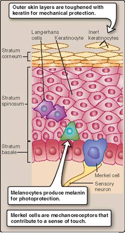
Figure 16.3
Epidermal structure.

Figure 16.4
Skin barrier functions. UV = ultraviolet.
3. Langerhans cells: Langerhans cells are antigen-presenting cells that are integral to adaptive immune responses. They ingest foreign matter, digest it, and then present fragments of the material on the cell surface. Presentation allows other immune cells to recognize the fragments. Langerhans cells are also important in delayed (type IV) hypersensitivity reactions.
4. Merkel cells: Merkel cells are slow-adapting mechanoreceptors located in skin areas that have a high tactile sensitivity. Merkel cells are also located at the base of hair follicles to further aid in touch sensation. Merkel cells associate with a nerve terminal to form a Merkel disk receptor (see Section VII below).
B. Barrier functions
The epidermis forms a barrier that both protects tissues from damage and minimizes evaporative water loss. The epidermis shields the body in four principal areas: it is resistant to mechanical and other physical assaults, photoprotective, antimicrobial, and also water repellent (Figure 16.4).
1. Physical shield: The superficial layers of the epithelium are keratinized, which creates a tough, multilayered, physical barrier around the body. The barrier resists mechanical abrasion and mild penetrating insults that inevitably occur during physical contact with solid objects. The barrier also resists chemical attack and prevents underlying tissue exposure to toxins and allergens.
2. Photoprotective shield: UV radiation naturally originates from the sun. UV light can be highly deleterious to biologic tissues because it breaks chemical bonds and disrupts the structure of DNA and protein. Melanocytes synthesize and donate melanin to adjacent cells to create a photoprotective barrier that absorbs UV radiation and causes it to dissipate safely as heat. Repeated exposure to the sun or other sources of UV radiation can stimulate melanocyte proliferation and melanin production, causing the skin to darken, which increases the level of photoprotection.
3. Antimicrobial shield: The superficial keratinized layers provide a physical barrier to microbes. If the physical barrier is breached, Langerhans cells and other immune components provide a rapid response to microbial invasion, warding off infection until the barrier can be repaired.
4. Water-resistive shield: The cornified cell envelope of keratinocytes creates a water-repellent shield that serves a dual function.
a. Structure: The shield is composed of lipids that are synthesized by keratinocytes and then secreted onto the surface. The lipid mixture forms a coating approximately 5-nm thick that is connected to the cell membrane via ester bonds. The cellular component is thicker (>15 nm) and is composed of cross-linked, insoluble proteins, including loricrin and keratin.
b. Function: The shield functions much like car wax. It causes water to bead on the epithelial surface and thereby prevents solutes from being washed out of the layers below. The waxy shield also minimizes evaporative water loss from underlying tissues.
Clinical Application 16.1: Abrasions and Burns
Abrasions and burns caused by ultraviolet radiation, heat, and fire can impair one or more skin barrier functions. For example, burn patients have increased transepithelial water loss from the skin that can challenge fluid homeostasis if a sufficiently large area of the body surface is involved. Burn patients are also at an increased risk of developing infections, which is why burned areas are often wrapped with bandages to supplement the physical barrier, and why topical antibiotics are applied as an antimicrobial shield.

Scalding Burn.
IV. DERMIS
The epidermal barrier is supported and maintained by the dermis. The dermis does not contribute to barrier function directly, but it gives skin strength and elasticity. It also contains immune cells that react to pathogens that may have breached the barrier.
A. Structure
The dermis comprises a meshwork of connective tissue. Primarily, type 1 collagen fibers provide structural support to the skin, whereas elastin fibers provide elasticity (see 4·VI·B·2). Within this matrix are nerve roots and sensory receptors, the cutaneous vasculature, and most skin specializations.
B. Cellular components
The principal cellular components of the dermis include mast cells, macrophages and dermal dendritic cells, and fibroblasts.
1. Mast cells: Mast cells are involved in both immune and inflammatory responses. Activated mast cells release histamine, prostaglandins, leukotrienes, cytokines, and chemokines (Figure 16.5). These agents increase cutaneous blood flow and capillary permeability.
2. Macrophages and dermal dendritic cells: Macrophages are phagocytic and aid in a number of immune-related responses. Dermal dendritic cells are antigen-presenting cells similar to the Langerhans cells of the epidermis. Dermal dendritic cells are integral to cutaneous adaptive immunity responses.
3. Fibroblasts: Fibroblasts are responsible for both the synthesis and degradation of fibrous and nonfibrous connective tissue proteins. These cells are also important in wound healing and scarring.

Figure 16.5
Mast cell activation effects.
V. HYPODERMIS
The hypodermis lies beneath the dermis (see Figure 16.1). It consists primarily of subcutaneous fat, blood and lymph vessels, and nerves. Around 50% of total body fat is located within the hypodermis of an average person. Thus, it is possible to take caliper measures of skin-fold thickness to estimate peripheral fat stores. The hypodermis cushions the skin, allows it to slide over underlying structures, and anchors skin to the tissues below.
Clinical Application 16.2: Triple Response
Wheal, erythema, and flare (known as a “triple response”) denote a classic reaction to abrasion or histamine-releasing stimuli. First, an erythemic (red) spot develops that spreads outward for a few millimeters, reaching maximal size in about 1 minute. Second, a brighter flush spreads slowly in an irregular flare around the original spot. Third, an edemic wheal forms over the original spot. Mast-cell histamine release can account for the triple response, mediating vasodilation and fluid extrusion into the interstitial space and stimulating nerve endings to give the sensation of itch. The triple response to histamine is often used as a positive control for a skin prick allergy test. Histamine is pricked into the skin followed by a row of other potential allergens such as pet dander, dust mites, and pollens.

VI. SPECIALIZED SKIN STRUCTURES
Skin contains several specialized glands and appendages that are protective or that participate in thermoregulation. An “appendage” is traditionally defined as a structure that protrudes from the body. Dermatologists use the term to indicate any specialized skin structure, including glands that originate in the dermis or hypodermis and then protrude through the epidermis to the skin surface. Appendages include hairs, sebaceous glands, sweat glands, and nails.
A. Hair
The base of each hair is attached to a piloerector muscle that is controlled by the sympathetic nervous system ([SNS] Figure 16.6). When stimulated to contract, piloerector muscles cause hair erection and produce “goose bumps.”

1. Structure: Hairs are constructed from three layers of fused keratinized cells (see Figure 16.6). The outermost protective layer (cuticle) is colorless. A middle layer (cortex) imparts strength and contains two types of melanin, the relative proportions of which give hair its natural color. Larger hairs also contain an inner medulla. The portion that protrudes beyond the epidermis is known as the hair shaft. The shaft emerges from a hair follicle, a specialized skin structure containing the hair bulb, keratinocytes, and associated glands (apocrine and sebaceous).
Some forms of cancer can be treated using chemotherapy, which targets rapidly dividing cells. The treatment is nondiscriminatory, however, also affecting rapidly proliferating keratinocytes, which causes hair thinning and loss.
Clinical Application 16.3: Acne
Acne is a common adolescent disorder that may also occur in adults. Closed comedo acne pimples (whiteheads) occur when skin cells block a hair follicle's external opening. Sebum becomes trapped but continues to be produced. Bacteria may colonize the accumulating sebum, causing local inflammation. Drugs that reduce sebum secretion (e.g., retinoids) help control acne occurrence and spreading.
2. Hair cycle: The hair follicle cycle consists of growth, rest, regression, and shedding. Growth-phase duration determines hair length and can vary from one area of the body to the next, which explains why head hairs are typically much longer than in other regions.
3. Associated structures: Hair follicles may also contain sebaceous and sweat glands (described below). Follicles also contain a sensory nerve fiber network (root plexus) that provides information about touch, pressure, and pain (described in Section VII below).
B. Sebaceous glands
Sebaceous glands produce sebum, a lipid-based secretion. The gland comprises keratinocytes and sebocytes, the latter of which are responsible for sebum synthesis. The gland duct empties sebum directly into the hair follicle, coating the shaft and then flowing onto the epidermal surface. Sebum functions are likely related to its antioxidant, antimicrobial, and hydration properties. Sebum secretion is continuous, but gland output is modulated by sex hormones. Androgens and growth hormone increase secretion rate, whereas estrogens inhibit it.
C. Sweat glands
Sweat glands secrete fluids of variable composition at the epidermal surface.
1. Types: There are three classes of sweat glands: apocrine, eccrine, and apoeccrine (Figure 16.7).
a. Apocrine: Apocrine sweat glands are restricted to the axillae and perineum. They activate in response to emotional stimuli and secrete a viscous, milky fluid into the follicle. Bacterial action on these secretions produces odors that may be involved in pheromonal signaling and are the reason that underarm antiperspirants and deodorants were developed. The pathologically foul-smelling sweat in these areas is known as bromhidrosis.
b. Eccrine: Eccrine sweat glands are widespread, the greatest concentrations being found on the palms of the hands and soles of the feet. The glands secrete a hypotonic fluid directly onto the skin surface. Fluid evaporation cools the skin and is important for thermoregulation. The skin is capable of producing copious amounts of sweat (1.0–1.5 L/hr on the low end and more than 3 L in heat-acclimated individuals). Such high levels of fluid loss from the body can compromise fluid balance and cardiovascular function.

Figure 16.7
Eccrine sweat gland activation.
c. Apoeccrine: Apoeccrine sweat glands are located primarily in the axillae, where they make up 50% of the total sweat gland number, and perineum. These glands are of a mixed archetype and function: Their pores typically open into the hair follicle, but their sweat composition is comparable to that of an eccrine gland. Apoeccrine glands tend to produce copious amount of sweat when stimulated appropriately in a continuous rather than pulsatile fashion.
2. Structure: Sweat glands can be functionally subdivided into a pore opening (acrosyringium), a secretory coil, a duct, and a layer of myoepithelial cells that allows sweat to be discharged onto the skin surface.
a. Secretory coil: When active, sweat glands secrete a precursor fluid into the secretory coil lumen that comprises a protein-free plasma filtrate. Osmotic forces drive the fluid through the duct toward the skin surface. Sweating is stimulated by SNS postganglionic cholinergic nerves (see Figure 16.7). Acetylcholine (ACh) binds to muscarinic type-3 receptors (G protein–coupled receptor superfamily) on the secretory cells (clear cells), which increases cytosolic Ca2+ from both extracellular and endoplasmic reticular sources to stimulate Na+-K+-2Cl− cotransporter activity. The resultant cation influx is balanced by K+ leakage and Na+ extrusion by a basolateral Na+-K+ ATPase. Apical membrane Cl− permeability increases also by a less understood mechanism, thereby increasing luminal Cl− concentrations and causing paracellular Na+ transport (Figure 16.8). The combined increase in Na+ and Cl− in the coil lumen osmotically draws water into the lumen via aquaporin-5 channels.

Figure 16.8
Sweat formation by clear cells. ATP = adenosine triphosphate; M3 = muscarinic type-3 receptor; PLC = phospholipase C.
Clear cells also express adrenergic receptors, which causes them to be activated by catecholamines during SNS activation.
b. Reabsorbing duct: Ions, principally Na+ and Cl−, are reabsorbed as the precursor fluid passes through the duct by channels such as the epithelial Na+ channel (ENaC) and the cystic fibrosis transmembrane conductance regulator (CFTR). The resultant fluid is sweat, which is hypotonic. Disorders such as cystic fibrosis reduce Cl− reabsorption through the CFTR, leading to increased ion loss in sweat.
c. Myoepithelial cells: The secretory coil is surrounded by myoepithelial cells, which contract upon cholinergic stimulation. Contraction does not force sweat out of the coil but rather provides the structural support that allows high osmotic forces (up to 500 mm Hg) to develop within the coil. These forces eventually propel fluid to the skin's surface in pressure pulses.
D. Nails
Nails are hard, scaly epidermal extensions that shield the posterior fingertips. The nail (known as the nail plate) is a hardened keratinized structure that mechanically protects the underlying skin (nail bed). Nail keratin contains a high number of disulphide bonds, which give it its strength and rigidity. Skin keratin is softer, reflecting fewer disulphide bonds.
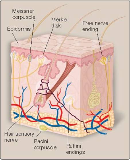
Figure 16.9
Cutaneous tactile receptors.
VII. CUTANEOUS NERVES
The cutaneous senses are a part of the somatosensory system. Every square millimeter of skin represents an opportunity to interact with and analyze the external environment, and, thus, it is dense with sensory nerve fibers. Not only do these nerves provide us with a sense of touch (taction), they also sense pain (nociception), itch (pruritoception), and temperature (thermoreception; see 38·II·A·2).
A. Touch
Physical contact can take many forms. Sometimes it might be a light touch, such as dragging a feather across the skin. Other times it might be the intense pressure of holding a plastic grocery bag full of cans. The ability to sense such disparate stimuli requires mechanoreceptors that are attuned to varying aspects of stimulus intensity, frequency, and duration. Their depth below the skin surface partly determines the size of their receptive field. A receptive field defines the area that a sensory receptor monitors. Receptors that collect stimuli over a wide receptive field have an increased chance of recording events but are unable to locate the source of the stimulus precisely. Receptors with small receptive fields are able to pinpoint the source of the stimulus with a high degree of accuracy and are usually clustered in large numbers to ensure adequate coverage over a wide surface area.
1. Tactile receptors: Skin contains several different types of mechanoreceptors that transduce tactile stimuli (Figure 16.9). Transduction occurs when a sensory nerve ending is deformed. The endings may be bare or encased in accessory structures that modify their sensitivity and responsiveness to different types of stimuli. Glabrous skin contains rapidly adapting Pacini and Meissner corpuscles. Hairy skin contains slowly adapting Merkel disksand Ruffini endings as well as rapidly adapting hair plexus neurons and sensory fibers around hairs.
a. Pacini corpuscles: Pacini corpuscles are rapidly adapting mechanoreceptors ~1 mm long that sense high-frequency vibrations in glabrous skin (Figure 16.10A). They reside deep in the skin, and their receptive field is wide. The corpuscles consist of a sensory nerve ending wrapped in numerous layers of fibrous tissue with gelatinous fluid between, so that they resemble an onion in cross section. The entire structure is then wrapped in a connective tissue capsule. The gelatinous layers cushion the nerve so that only transient stimuli are able to deform and excite the nerve membrane. The afferent nerves are myelinated for most of their length, which allows for rapid relay of sensory signals.
b. Meissner corpuscles: Meissner corpuscles also adapt rapidly (see Figure 16.10B). They are exquisitely sensitive to touch and low-frequency vibrations, which produce a fluttering sensation. They are smaller than Pacini corpuscles, but their construction and skin distribution is similar, in that a sensory nerve ending meanders between stacked layers of flattened support cells, all enclosed within a capsule.

Figure 16.10
Rapidly adapting cutaneous tactile receptors.
c. Ruffini endings: These afferents are slowly adapting receptors (Figure 16.11A) located in the deeper layers of the skin. The nerve endings branch and weave between bundles of collagen fibers to form a long, thin, spindle-shaped structure. The fibers are enclosed within a connective tissue capsule that is firmly tethered to surrounding tissues. When skin is stretched, the capsule and structures within are distorted also.
d. Merkel disks: Merkel disks are also slowly adapting receptors (see Figure 16.11B) that respond best to low-frequency stimulation and light touch. They lie just below the skin surface, which gives them a very narrow receptive field. Fingertips are endowed with very large numbers of Merkel disks, which allows for fine discrimination of form and texture.
e. Hair cells: Every hair on the body surface functions as a mechanosensor, made possible by the presence of a sensory nerve that wraps around its follicle (see Figure 16.9). When the hair bends, the nerve ending distorts and signals.
f. Free nerve endings: Free sensory nerve endings can be found throughout the skin. They can also contribute to taction, in addition to other sensations including pain, itch, and temperature.
2. Mechanosensory transduction: Deformation of a tactile nerve ending opens a Na+ channel, causing a depolarizing receptor potential (also known as a generator potential). The Na+ channel may be an ENaC family member.
3. Sensory nerve fibers: Sensory nerve afferents are classified according to how fast they relay signals to the central nervous system ([CNS] Table 16.1). All of the tactile sensory afferents are myelinated (type Aβ) and conduct at relatively high velocity. These signals then travel via the spinal cord to the CNS for processing (see 6·II). The area perceived by a particular mechanoreceptor is dependent on receptor type and body area. The hands are more discriminatory than the upper arms, for example (Figure 16.12).

Figure 16.11
Slowly adapting cutaneous tactile receptors.
B. Pain
Mechanical and thermal stimuli that are innocuous or even pleasurable at low intensities can cause significant cellular damage at higher levels. The function of pain is to alert the CNS of local damage and to initiate a motor reflex that causes the body to either avoid or pull back from the source of stimulation (see 11·III·D).
1. Nociception: A number of nociceptor types transduce painful stimuli into a membrane potential change.
a. Mechanical: Mechanical nociceptors respond to intense pressure or mechanical deformation of the skin. They also respond to sharp objects that stab or cut the skin. These receptors are likely very high threshold mechanoreceptors that only respond to mechanical stimuli when they reach noxious levels.

b. Thermal: Temperature extremes (freezing cold and burning heat) cause tissue damage. Cold stimuli become noxious at ~20°C, with intensity of perceived pain increasing linearly to ~0°C. Cold responses are also sensitive to the rate of cooling, with rapid cooling producing more intense responses. The threshold for noxious heat sensation is ~43°C.
c. Chemical: Because nociceptive fibers are free nerve endings, they are accessible to chemicals that cross the epidermal barrier or that are released by damaged tissues (Table 16.2). Capsaicin, the active ingredient in hot chili peppers, produces a burning sensation via activation of nociceptors when applied topically.
d. Polymodal: A subpopulation of nociceptors is sensitive to two or more stimuli and is known as polymodal.
2. Nociceptive stimulus transduction: The precise mechanisms by which nociceptive stimuli are sensed and signaled are not understood fully, but transduction of many noxious stimuli involves transient receptor potential (TRP) channel family members (see 2·VI·D).
a. Receptors: Heat activates TRPV1, a member of the vanilloid class of TRP receptors. It is also activated by capsaicin. Skin cooling activates TRPM8. Activating either TRP channel class results in Na+ and Ca2+ influx and excitation. Hydrogen ions excite nociceptive neurons by permeating an acid-sensing channel of the ENaC family. Other channels may be involved in pain sensation also.
b. Nociceptive fibers: Nociceptor activation is relayed to the CNS by fast (myelinated) Aδ fibers and slower C fibers. The Aδ fibers mediate sensations of sharp, intense, pricking pain (first pain), followed by a more prolonged dull, throbbing, burning pain associated with C-fiber activation (second pain).
C-fibers are particularly sensitive to lidocaine, a local anesthetic that is applied topically to relieve skin itching and pain.1 It blocks the Na+ channel that mediates the nerve action potential. Lidocaine is also commonly injected to anesthetize teeth prior to dental surgery or is combined with prilocaine (a related Na+-channel blocker) in an ointment. Lidocaine is sometimes combined with a vasoconstrictor to reduce local blood flow and thereby reduce drug washout effects. Local anesthesia is prolonged as a result.

Figure 16.12
Receptive fields of two receptor types in the hand and sensory discrimination along the arm.

 1For additional discussion of amide local anesthetics such as lidocaine, see LIR Pharmacology, 5e, pp. 147–149.
3. Sensitization: Tissue damage initiates a chain of events that sensitizes surrounding afferent nerve endings to innocuous stimuli, thereby causing them to be perceived as painful (hyperalgesia). Sensitization initially remains localized to the site of damage (primary hyperalgesia) but spreads within minutes to involve surrounding areas (secondary hyperalgesia). Sensitization follows the progress of swelling and inflammation and involves many of the common inflammatory mediators. Its effects may persist for months after recovery from the initial injury.
C. Itch
Pruritis (derived from prurire, the Latin word for “itch”) is the most recently recognized member of the cutaneous senses. Itches appear designed to trigger reflex scratching or rubbing to remove an insect or other irritant. The sensation is mediated by two populations of C-type nerve fibers. One fiber type responds optimally to histamine, whereas the other (nonhistamine) type is activated by a wide range of pruritogens, such as prostaglandins, interleukins, proteases, and ACh. Itch sensations can be suppressed by painful stimuli (such as scratching) and by antihistamines and potentiated by analgesics. The mechanisms by which painful and pruritic sensations interact are not understood fully.
D. Dermatomes
Sensory information from skin receptors is relayed by the CNS via afferent nerves. The nerves have a limited area of coverage, which can be mapped onto the body surface as a series of discrete bands called dermatomes (Figure 16.13). Each band corresponds to a single spinal segment. There is overlap in coverage between the bands, so that cutting a single pair of posterior nerve roots does not result in complete sensory loss in the corresponding dermatome. Pain that localizes to a particular dermatome may be helpful in identifying the site of spinal cord injury, for example.

Figure 16.13
Dermatomes.
Chapter Summary
• The epidermis provides the majority of skin barrier functions. The water barrier is provided by a combination of a thin lipid layer and a thicker protein layer. The ultraviolet (UV) barrier is provided by melanin, which absorbs a portion of UV radiation.
• The dermis is the location of the majority of the functional aspects of the skin and is home to most skin appendages. The dermal layer contains mast cells, which are involved in local inflammatory responses.
• Hair follicles provide a pore for glandular secretions of both sebaceous and apocrine sweat glands. Hair growth is a complex process involving active growth, regression, and then shedding of the hair.
• Eccrine sweat glands participate in thermoregulation and involve the formation of isotonic precursor sweat in the secretory coil and then ion reabsorption in the duct. This produces a hypotonic solution that can evaporate depending on environmental conditions.
• The cutaneous senses are a part of the somatosensory system, which monitors events occurring within or at the surface of the body.
• Tactile receptors provide information resulting from physical contact with objects. They activate in response to touch, the sensations being transduced by mechanical deformation of a sensory neuron. Some receptors (Pacini and Ruffini) respond best to high-frequency vibration or stretch. Others (Meissner and Merkel receptors) are more sensitive to pressure and low-frequency events.
• Sensory afferents from tactile receptors are myelinated and conduct impulses at high speed.
• Pain receptors activate in response to intense and noxious mechanical, thermal, or chemical stimuli.
Study Questions
Choose the ONE best answer.
III.1 Which of the following cytoskeletal proteins functions like a spring, limiting the extent to which the sarcomere can be stretched?
A. α-Actinin
B. Dystrophin
C. Nebulin
D. Titin
E. Z disk
Best answer = D. Titin is a massive, thick filament–associated structural protein that limits sarcomere length when a muscle is stretched (12·II·C). Thin filament–associated proteins do not act as springs but, rather, provide structural integrity. For example, α-actinin binds the ends of thin filaments to Z disks (structural plates that serve as attachment points for thin filaments); dystrophin anchors the contractile array within the cytoskeletal framework; and nebulin, which extends the length of the actin filament, is believed to establish thin-filament length.
III.2 When two acetylcholine molecules bind to a nicotinic receptor on skeletal muscle, the channel opens and allows transmembrane passage of ions. The resulting ion flux is dominated by which of the following choices under normal physiologic conditions?
A. Ca2+
B. Mg2+
C. H+D. Cl−E. Na+
Best answer = E. The nicotinic acetylcholine receptor (nAChR) is a nonspecific cation channel that allows passage of Na+, K+, and Ca2+ (2·VI·B). The Ca2+ flux is small and not physiologically significant. The electrochemical gradients for Na+ and K+ are normally configured such that the nAChR supports simultaneous Na+ influx and K+ efflux. Na+ influx dominates this exchange, however, and the myocyte depolarizes (12·II·G). Membrane depolarization then opens voltage-dependent Na+ channels, which allows an action potential to propagate across the sarcolemma and down the T-tubule system to initiate muscle contraction. The nAChR does not support significant H+, Cl−, or Mg2+ fluxes under normal, physiologic conditions.
III.3 A 22-year-old woman receives botulinum toxin type A (a cholinergic presynaptic release inhibitor) injections to treat palmar hyperhidrosis (excess sweating). Her grasp is weakened by the treatments, through a decrease in the synaptic levels of what substance?
A. Acetylcholinesterase B. Acetylcholine
C. Calsequestrin
D. Myoglobin
E. Nicotinic receptors
Best answer = B. Botulinum toxin is a protease that prevents exocytosis and release of neurotransmitters from nerve terminals (5·IV·C). Botulinum toxin type A is commonly used clinically to inhibit acetylcholine (ACh) release at the neuromuscular junction, which reduces synaptic ACh levels. It reduces eccrine sweat gland activity through a similar mechanism. Acetylcholinesterase, which normally degrades ACh and terminates neuromuscular signaling (12·II·G), would not be affected by the toxin. Myoglobin is a sarcoplasmic O2 storage and transport molecule, whereas calsequestrin is a Ca2+-binding protein found in the sarcoplasmic reticulum. Neither is directly involved neuromuscular transmission.
III.4 Phospholamban is a regulatory protein associated with the cardiac sarcoplasmic reticulum Ca2+ ATPase. Phospholamban phosphorylation would most likely increase the rate of which of the following events?
A. Relaxation
B. Ca2+ influx
C. Crossbridge cycling
D. Electrical conduction
E. Nodal cell depolarization
Best answer = A. Phospholamban normally acts as a rate limiter on sarcoplasmic reticulum (SR) Ca2+ ATPase (SERCA) function (13·III·B). Phospholamban phosphorylation reduces its inhibitory effects, allowing the pump to speed up. SERCA normally helps remove Ca2+ from the sarcoplasm following excitation. Increasing pump speed causes sarcoplasmic free Ca2+ levels to fall faster than normal, promoting decreased relaxation times. Ca2+ influx occurs during excitation and probably would not be appreciably affected by changes in SERCA. Crossbridge cycling rate is dependent on actin–myosin interactions. Electrical conduction between myocytes is dependent on gap junction function. Although faster relaxation times do facilitate heart rate increases, the rate of nodal cell depolarization is controlled through ion channel modulation.
III.5 Cardiac muscle contraction is dependent on a rise in sarcoplasmic Ca2+ concentration. The bulk of the Ca2+ required for full force generation flows through which of the following Ca2+ channel types?
A. Dihydropyridine receptors
B. Ryanodine receptors
C. Inositol trisphosphate–gated channels
D. Transient receptor-potential channels
E. Stretch-activated channels
Best answer = B. Full force development by a cardiac myocyte relies on Ca2+ release from stores in the sarcoplasmic reticulum ([SR] 13·III·A). Release is mediated by Ca2+-induced Ca2+ release (CICR) channels, also known as ryanodine receptors. Dihydropyridine receptors are L-type Ca2+ channels that mediate voltage-gated Ca2+ fluxes across the T-tubule membrane. Ca2+ influx via this pathway acts as a trigger for CICR. Inositol trisphosphate mediates Ca2+ release from the SR in smooth muscle. Transient receptor-potential channels are found in many tissues, often mediating cellular sensory stimulus transduction (2·VI·D). Stretch-activated channels are also widespread but ryanodine receptors are the principal pathway for Ca2+ fluxes during contraction.
III.6 What type of smooth muscle Ca2+ channels localize to plasma membrane caveolae and are gated primarily by membrane potential change?
A. Ca2+-induced Ca2+ release channels
B. Receptor-operated Ca2+ channels
C. Store-operated Ca2+ channels
D. Inositol trisphosphate–gated Ca2+ channels.
E. L-type Ca2+ channels
Best answer = E. L-type Ca2+ channels are voltage gated, opening in response to membrane depolarization. They are found in many cell types, including smooth muscle, where they are concentrated within plasma membrane pockets called caveolae (14·II·C). Receptor-operated Ca2+ channels open when a ligand binds to the associated receptor, rather than a voltage change. Ca2+-induced Ca2+ release channels and inositol trisphosphate–gated Ca2+channels are located on the sarcoplasmic reticulum membrane and mediate Ca2+ store release. Store-operated Ca2+ channels are used to top off intracellular Ca2+ stores with extracellular Ca2+ during muscle relaxation (14·III·C). Channel opening is controlled by a store Ca2+ sensor (Stim1).
III.7 A pharmaceutical company is intent on developing a drug that decreases smooth muscle–induced vasospasm. Which of the following enzymes normally antagonizes smooth muscle contraction and might, thus, make a suitable target for modulation (stimulation or upregulation) by a pharmaceutical product?
A. Rho-kinase
B. Myosin phosphatase
C. Myosin light-chain kinase
D. Protein kinase C
E. Phospholipase C
Best answer = B. Myosin phosphatase normally dephosphorylates a myosin regulatory light chain (MLC20) to inhibit myosin ATP-ase activity, thereby blocking smooth muscle contraction (14·III·C). Upregulating this enzyme would decrease contractility and potentially reduce vasospasm. Myosin phosphatase activity is regulated by at least two different pathways. Rho-kinase and protein kinase C are components of separate pathways that normally inhibit myosin phosphatase activity and promote contraction. Myosin light-chain kinase (MLCK) phosphorylates and activates smooth muscle myosin, thereby facilitating contraction. Phospholipase C also promotes contraction through a pathway that simultaneously stimulates MLCK and inhibits myosin phosphatase.
III.8 What are the mineral crystals that resist compression and give bones their characteristic strength and resilience?
A. Urate
B. Hydroxyapatite
C. Glycosaminoglycan
D. Creatinine
E. Calcium oxalate
Best answer = B. Hydroxyapatite is a crystalline mineral containing calcium and phosphate (15·II·A). Hydroxyapatite crystals are cemented within collagen fibers and then bundles of mineralized fibers embedded in ground substance to create a material that has a high resistance to compression and tensile stress. Urate, creatinine, and calcium oxalate are found at high concentrations in urine. When sufficiently concentrated, they form crystals that may be observed in urine sediments. Glycosaminoglycan is a mucopolysaccharide found in ground substance, which fills the spaces between all cells, including bone.
III.9 What type of precursor cells found in bone express RANKL (receptor activator of nuclear factor κB ligand) on their surface to facilitate bone resorption?
A. Osteoblast
B. Osteoclast
C. Osteocyte
D. Bone lining cell
E. Hematopoietic
Best answer = A. Bone resorption and remodeling involves several cell types that work together within a bone-remodeling compartment (15·IV·C). Osteoblast precursors express receptor activator of nuclear factor κB (RANK) ligand (RANKL) on their surface. RANKL binds to RANK, a receptor expressed on the surface of osteoclast precursors (these are hematopoietic precursors), causing several such cells to fuse and form large multinucleated osteoclasts. Osteoclasts digest bone and release its mineral content for return to blood. Osteocytes are cells embedded in the bone matrix that monitor stress and integrity. Bone lining cells are found on the bone surface and signal a need for remodeling as needed.
III.10 A 36-year-old woman has a parathyroid hormone (PTH)-secreting tumor. Which of the following might be expected to increase as a result of chronic PTH elevation?
A. Bone resorption
B. Bone deposition
C. Ca2+ excretion from the intestines
D. PO43− absorption from the intestines
E. PO43− reabsorption from the kidneys
Best answer = A. Parathyroid hormone (PTH) release from parathyroid glands is normally regulated by plasma Ca2+ levels (15·V·A; 35·V·B). When plasma Ca2+ (or Mg2+) levels decrease, PTH is secreted to stimulate Ca2+resorption from bone. PTH's effects are mediated by PTH receptors on osteoblasts, which then recruit osteoclasts to a bone-remodeling site. PTH does not stimulate bone deposition. PTH causes Ca2+ reabsorption (not excretion) by the renal distal tubule (27·III·C). PTH also inhibits PO43− reabsorption by the renal proximal tubule, thereby increasing excretion rates (26·VI·A).
III.11 A 4-year-old boy with a family history of cystic fibrosis has been presenting with mild respiratory and gastrointestinal symptoms. If cystic fibrosis is suspected, his sweat composition might best be described as which of the following, compared with that of a healthy boy his age?
A. Hypotonic
B. Isotonic
C. Hypertonic
D. Copious
E. Scant
Best answer = C. The cystic fibrosis (CF) transmembrane conductance regulator (CFTR) is an adenosine triphosphate–binding cassette transporter that functions as a Cl− channel in many epithelia. CFTR defects prevent Cl−secretion by respiratory and gastrointestinal epithelia. Secretion creates an osmotic gradient that is used to draw water onto the apical surface, so CF patients typically form mucus that is thick, which makes it difficult to expel from the lungs, for example. In sweat glands, CFTR is used to reabsorb Cl− from the ductal lumen during sweat's passage to the skin surface, causing sweat to become hypotonic (16·VI·C·2·b). In CF patients, a CFTR defect prevents Cl− (and Na+) reabsorption, so their sweat is hypertonic. CFTR defects do not cause major changes in sweat volume.
III.12 A 42-year-old jackhammer operator presents with decreased high-frequency vibration sensitivity in the glabrous skin of the hands. Which receptor is most likely being affected?
A. Ruffini endings
B. Merkel disks
C. Free nerve endings
D. Pacini corpuscles
E. Hair sensory fibers
Best answer = D. Pacini corpuscles are rapidly adapting tactile receptors responsible for sensing vibrations in the 40–500 Hz range (16·VII·A). Glabrous skin does not have hair and, therefore, no hair sensory fibers. Ruffini endings are slowly adapting tactile receptors that sense skin stretch rather than vibrations. Merkel disks sense light skin pressure. Glabrous skin also contains free nerve endings, but these are less sensitive to vibration than are Pacini corpuscles.