
15
I. OVERVIEW
The brain and many other soft tissues of the body are enclosed within a protective framework made of bone. Bone also fashions limbs that, together with skeletal muscles, facilitate locomotion and allow objects to be manipulated. The 206 bones that make up the human skeleton work in conjunction with cartilage, ligaments, tendons, and skeletal muscle. These tissues help maintain skeletal structure and control bone movement. Bone is a connective tissue (see 4·IV·A) that has been mineralized to give it high resilience to stress and trauma. The mineral component of bone persists long after the body has died and the soft tissues have decomposed, but bones are not lifeless structures. Bone is honeycombed with tunnels and cavities that are teeming with cells and that provide channels through which blood vessels and nerve fibers permeate the matrix. Many bones also contain a central chamber filled with marrow that manufactures blood cells and stores fat (Figure 15.1). Finally, bone is a highly dynamic tissue that is constantly being remodeled and turned over. Remodeling is partly an adaptive response to mechanical stress, but it also reflects bone's immense repositories of calcium and phosphate (>99% and 80% of body total, respectively) that can be mobilized when plasma concentrations fall below optimal.
II. FORMATION
Bone structure reflects two competing needs. Bones must be strong to protect the soft tissues from mechanical trauma and to support body weight during locomotion. Bone's resilience is afforded in part by minerals, which makes bones heavy. On the other hand, bones must be sufficiently light to allow for rapid responses to external threats (e.g., from predators). Light bones are prone to fracture, however. Bone breakage is a potentially lethal event (through predation, hemorrhage, or circulatory shock) and, therefore, must be avoided at all cost. Thus, bone design represents a compromise between strength and weight. In practice, bones contain just enough minerals to withstand normal mechanical stress limits plus an added safety margin.

Figure 15.1
Bone structure.

Limb bones typically fracture when subjected to stresses that cause them to deform by three to four times that experienced during normal physiologic activity. If stress levels increase chronically (e.g., during weight training), bone remodels to compensate for the added stress (the Wolff law) and to reestablish normal safety margins.
Most of a bone's mineral content and strength is concentrated in a thin, highly compacted outer layer. This construction is similar to the hollow steel tubing used to fashion the legs of chairs and tables. Bone's interior is not air filled but is composed of a light, highly porous matrix. Resistance to compression and mechanical shear is accomplished using a resilient mix of minerals and proteins (Table 15.1).
A. Mineral component
Resistance to compression is achieved using thin, tabular hydroxyapatite crystals measuring ~50 nm long. Hydroxyapatite is a mineral comprising calcium and phosphate (Ca10 [PO4]6 [OH]2) that occurs naturally in stalagmites and mineral crusts.
B. Protein scaffolding
Hydroxyapatite crystals do not readily compress, but they do shear. Taking advantage of their natural properties requires that they be cemented and tethered within collagen fibers (Figure 15.2). The cement is made from mucopolysaccharides and is rich in Mg2+ and Na+ (~25% of total body Na+ is contained within bone). Collagen is the principal component of tendons, tissues notable for their flexibility and high resistance to tensile (stretching) and shear stress. When the collagen fibers with their embedded hydroxyapatite crystals are cemented together by ground substance (see 4·IV·B·3), they create a material that is able to support heavy loads and resist mechanical impact yet is flexible enough to torque and bend without fracturing. Similar construction techniques are used to make adobe (a mix of straw and mud) and reinforced concrete (a mix of steel rods or “rebar” and cement).
C. Assembly
Bone is formed by osteoblasts, which are related to fibroblasts (see 4·IV·B). Bone formation can be divided into four steps: collagen deposition; secretion of ground substance; crystal seeding; and, finally, maturation.
1. Collagen deposition: Bone formation begins with the creation of collagen scaffolding. Collagen molecules comprise chains of tropocollagen subunits, each composed of three polypeptide chains braided into a helix. Monomers join end to end and then spontaneously assemble into collagen fibrils, which are the cellular equivalent of steel rebar in concrete. Monomers within the fibrils are crosslinked extensively and staggered like bricks in a wall for additional stability and strength.

Figure 15.2
Mineralized collagen formation.
Clinical Application 15.1: Osteogenesis Imperfecta
Osteogenesis imperfecta, or “brittle bone disease,” results from a group of inherited defects in two type I collagen genes. The most common is a point mutation that replaces glycine with a bulkier amino acid at a site where the three strands of the helix normally come close together. The result is a molecular “bleb” that interferes with normal fibril formation and packing of hydroxyapatite crystals. Bones formed with such collagen are weak and prone to fracture.1

Fractures in an infant with osteogenesis imperfecta.
2. Ground substance: Ground substance is an amorphous, gel-like matrix of glycosaminoglycans, proteoglycans, glycoproteins, salts, and water that fills the space between cells in all tissues. Bone ground substance differs from that found in other tissue in that it is saturated with Ca2+ and PO43−. The combination of collagen fibrils and ground substance is called osteoid (Figure 15.3).
3. Crystal seeding: Osteoblasts continue secreting Ca2+ and PO43− into ground substance until it becomes supersaturated, at which point the minerals begin precipitating as calcium phosphate crystals. Osteoblasts also secrete seed crystals that are cemented to collagen fibrils to provide nucleation sites for continued growth. The fibrils slowly become encrusted with amorphous mineral deposits.
4. Maturation: In succeeding months, the calcium phosphate crystals are remodeled by osteoblasts to form mature hydroxyapatite. The tabular hydroxyapatite crystals are tethered to collagen fibrils by proteoglycans, and the fibrils themselves become extensively interlinked to give bone the tensile strength that approaches that of structural steel.
D. Immature
Bone deposition is, by nature, a very slow process, which presents a problem when bone is damaged and must be repaired. Even though bone fracture is an uncommon event in healthy people, most individuals break a bone at some point in their lives. Breaking a proximal phalanx in a finger is painful and inconvenient, but breaking a major limb bone (e.g., a femur) is more serious because it impairs motion. In the wild, breaking a leg can leave an animal extremely vulnerable to predation, so deposition of new bone usually occurs with a view to speed rather than strength. The result is woven bone, which, although weaker than the mature form, allows breaks to heal in weeks rather than months (Figure 15.4A). In time, woven bone is replaced through remodeling with the mature lamellar form (see Figure 15.4B). Woven bone has the appearance of fabric when viewed in section, reflecting the fact that osteoblasts deposit collagen fibers at random within osteoid. The random orientation provides resistance to stress in all directions and, therefore, is an excellent all-purpose patch for a broken bone. Woven bone is also found at bone growth plates.
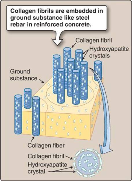
Figure 15.3
Osteoid.
 1For additional discussion of osteogenesis imperfecta, see LIR Biochemistry, 5e, p. 49.
Clinical Application 15.2: Paget Disease
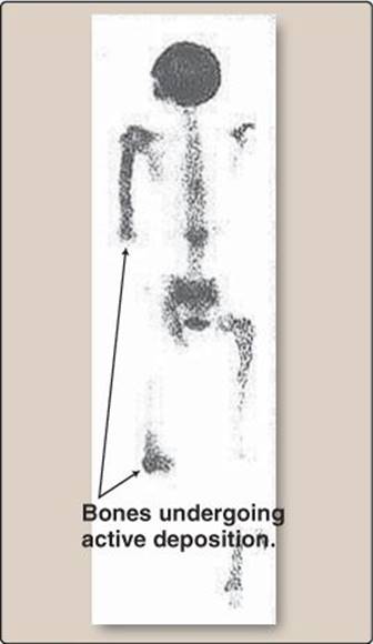
Paget disease is the second most common bone disorder after osteoporosis. The underlying causes are unknown. Paget disease manifests as inappropriately high levels of osteoclast activity, often affecting solitary bones. Osteoclasts normally digest bone during remodeling. Osteoblasts compensate for the resulting bone loss by laying down new, woven bone, but the rate of turnover in affected bones is so high that it never has time to mature and strengthen. Most Paget disease patients remain asymptomatic. Others present clinically with bone deformities, arthritis, bone pain, symptoms caused by peripheral nerve compression, and increased incidence of fractures.
Bones undergoing active deposition.
E. Mature
Bone takes up to 3 years to mature fully. During this time, collagen fibers are aligned with predominant stress lines to provide maximal strength. Lamellar bone is laid down in 10–30 concentric rings that form cylinders ~200 μm wide and a few millimeters long (~1–3 mm) known as osteons, or Haversian systems (Figure 15.5). At the center of each cylinder is a Haversian canal that provides a thoroughfare for blood vessels and nerve fibers. Two types of lamellar bone can be distinguished based on density and porosity, compact bone and trabecular bone.
1. Compact: Compact bone is extremely dense and is configured for strength. Also known as cortical bone, it is found at the periphery of all bones.
2. Trabecular: Trabecular bone (cancellous, or spongy bone) lines the marrow cavity at the center of a bone. It has a lacy, porous structure that gives it a very high surface area. When plasma Ca2+ levels fall, trabecular bone is the first to be sacrificed in order to release its mineral content to the circulation. When Ca2+ and PO43− levels renormalize, the bone is rebuilt. Trabecular bone also provides critical mechanical support, particularly in the vertebrae.

Figure 15.4
Woven and lamellar bone.

Figure 15.5
Haversian systems in compact bone.
F. Vasculature
Bone is a living tissue that must be supplied with blood. Blood vessels reach bone by way of the periosteum, a fibrous membrane that covers the nonarticulating surfaces of bones and serves as an attachment point for blood vessels and nerves. Supply arteries penetrate the bone cortex and terminate in the medulla. Smaller arterial vessels course through the marrow cavity and then reenter bone to supply the cells within. Vessels travel longitudinally in Haversian canals and outward toward the cortex via Volkmann canals (see Figure 15.5).
G. Stress-monitoring system
When bone formation is complete, osteoblasts either undergo apoptosis, or they persist as either osteocytes or bone lining cells. Together, these two cell types form a vast sensory network that monitors bone stress and integrity.
1. Osteocytes: Some osteoblasts become entombed in the osteoid of their own making during bone formation and persist, potentially for years, as osteocytes. Osteocytes reside in small (~10–20 μm) cavities called lacunae, where they remain to monitor bone stress levels and signal the need for remodeling if microfractures appear (see Figure 15.5). The cells extend long, thin processes (dendrites) in all directions through microscopic channels (canaliculi) that permeate the entire bone matrix (Figure 15.6). Canaliculi allow osteocytes and bone lining cells to communicate with each other via gap junctions at the site of contact.
2. Bone lining cells: Bone lining cells form a monolayer covering every bone surface. They communicate with and provide an interface between osteocytes and the bone exterior.
3. Nutrient supply: Canaliculi have a diameter of <0.5 μm, which is too small to carry blood vessels, but they do provide pathways for extracellular fluid (ECF) flow that carries O2 and nutrients to the osteocytes. Flow is driven by hydrostatic pressure originating in the vasculature. Osteocytes may detect bone stress levels by monitoring changes in ECF flow rates caused by bone deformation.

Figure 15.6
Osteocytes visualized with a fluorescent stain.
III. ANATOMY AND GROWTH
The bones that comprise a human skeleton come in a number of different shapes and sizes. They are usually classified based on their shape.
A. Classification
Bones can be divided into five groups on the basis of anatomy: long, short, flat, irregular, and sesamoid bones. Long bones are found in arms (humerus, radius, ulna) and legs (femur, tibia, fibula). The bone shaft (diaphysis) is a long, thin tube of cortical bone with trabecular bone at the center (see Figure 15.1). They typically widen (the metaphysis) toward the end (epiphysis) to form a site of articulation with another bone. The ends are broadened to spread the load, and they are filled with trabecular bone that acts as a shock absorber during locomotion. In children, the region between the metaphysis and epiphysis contains a growth plate that is the active site of new bone formation, also called the physis, or epiphyseal plate.
B. Growth
Bone growth (widening and lengthening) during fetal development and throughout childhood is effected by chondrocytes. Chondrocytes are derived from the same mesenchymal stem cell line that gives rise to osteoblasts and are arranged in 10–20 columns within a growth plate. Chondrocytes nearest the ends of the bone divide rapidly (Figure 15.7). Further down the column, the chondrocytes enlarge and push the ends apart. Chondrocytes also secrete cartilage, which becomes mineralized by osteoblasts and forms a template for further ossification. Older chondrocytes eventually undergo apoptosis, leaving spaces within the matrix that are invaded by nerves, blood vessels, and additional osteoblasts, which complete the task of bone maturation. When skeletal growth is complete, the growth plate dwindles, and the two epiphyses are united with the shaft (epiphyseal closure).
C. Bone marrow
Bone marrow is a soft tissue located in the center of some bones. There are two types: red and yellow.
1. Red: Red marrow is the source of virtually all blood cells, including red cells, most white cells, and platelets. Blood cells are formed from hematopoietic multipotent stem cells. Marrow is also the location of mesenchymal stem cells, which give rise to osteoblasts and chondrocytes, among others.

Figure 15.7
Bone growth.
2. Yellow: Yellow marrow stores fat. Yellow marrow appears and accumulates in long bones during adulthood. It is derived from and can be converted back to red marrow if there is a need to increase blood cell production.
IV. REMODELING
Bone is a dynamic tissue that is constantly being turned over at a rate of about 20% per annum in young adults, slowing to 1%–4% per annum in older adults. Remodeling is partly a response to mechanical stress but also reflects bone's vital role as a Ca2+ and PO43− repository. Remodeling involves four bone cell types that, together, comprise a basic multicellular unit (BMU). Osteocytes signal the need for remodeling, bone lining cells facilitate and coordinate remodeling, osteoclasts digest old bone, and osteoblasts lay down new bone (Figure 15.8). BMUs are roving demolition and construction crews replacing bone constantly for the life of an individual.
A. Causes
There are three main forces driving bone remodeling: mechanical stress, microdamage, and Ca2+ and PO43− homeostatic needs.
1. Mechanical stress: Many bones are subject to repeated mechanical stress associated with lifting and carrying weight. For example, the arm bones form a lever system powered by skeletal muscles. When the muscles contract to lift a weight, the levers are stressed. Bones are designed to withstand such stresses within a normal physiologic range, but a muscle that is exercised repeatedly grows stronger and increases the stress on the levers. Thus, bones are also designed to sense mechanical stress and lay down additional bone mass to compensate if necessary. Conversely, when the stress on bones is reduced, they lose mass.
The stroke (playing) arm of professional tennis players is subjected to years of repeated mechanical stress. The bones of the forearm respond by increasing bone density, diameter, and length. Individuals who have been freed from gravity and its associated mechanical stresses lose bone mass. Astronauts who remain in space for prolonged periods exercise daily to help offset the effects of bone unloading, but they still lose pelvic bone mass at a rate of 1%–2% per month.

Figure 15.8
Concept map for the basic multicellular unit.
2. Microdamage: Bones constantly develop microscopic damage as a result of mechanical stress, either acutely or as the result of normal actions that are performed repeatedly over time. The organic component of bone also deteriorates with time, which increases the likelihood of microfissures and microscopic cracks forming. Because fissures and cracks can ultimately result in fracture, damaged areas are replaced with new bone through remodeling.
Clinical Application 15.3: Remodeling Disorders
At any one time, around 1 to 2 million basic multicellular units are at work remodeling bone in the adult skeleton. In the absence of external influences, such as mechanical stress or disease, total bone mass remains constant. This exact matching of bone deposition to bone resorption requires tight functional coupling between osteoblasts and osteoclasts. A disturbance in the balance between the activities of the two cell types causes loss of bone mass or abnormal bone deposition.
Osteoporosis is a common term for a group of disorders in which the balance between bone resorption and formation is tipped in favor of osteoclasts. The osteoblasts fail to keep up with osteoclast activity, and the bone becomes increasingly porous and fragile as a result. Fracture is common in patients with osteoporosis. Bone loss associated with aging (type II osteoporosis) is common in both women and men, but postmenopausal women are at particular risk. Estrogen limits osteoclast activity, so when circulating levels of this hormone fall after menopause, osteoclasts become increasingly active, and bone is resorbed faster than it is replaced. Osteoporosis affects all bones, but the effects are most dramatic on trabecular bone, which is the primary site of remodeling in healthy individuals. Excessive resorption thins all trabeculae and truncates many of them, which destroys the template that is required for renewed bone deposition. It also seriously compromises their mechanical functions and greatly increases the likelihood of fracture. Treatment options for both men and women include oral bisphosphonates (e.g., alendronate, trade name Fosamax), which inhibit bone mineral breakdown.1
Osteopetrosis, or “stone bone,” results from a heterogeneous group of rare inherited disorders that impair osteoclast function. The most common form (~60%) results from a mutation in a subunit of the V-type ATPase that secretes acid from the ruffled border of an osteoclast onto the bone surface, but other mutations affect genes encoding Cl− channels, intracellular H+ pumps, and RANK+ (receptor activator of nuclear factor κB); the role of these proteins in normal osteoclast function is discussed in section C below. The mutations tip the bone resorption–deposition balance in favor of the osteoblasts, resulting in bones that are dense yet brittle. Affected individuals may show skeletal deformities, an increased likelihood of fractures, and secondary effects related to incursion of bone into the marrow space and the vascular and nerve supply to the bone matrix.

Comparison of normal and osteoporetic trabecular bone.

Abnormal bone density in an infant with osteopetrosis.
1For a discussion of agents used to treat osteoporosis, see LIR Pharmacology, 5e, p. 365.
3. Hormones: Bone contains immense reservoirs of Ca2+ and PO43− that can be mobilized and circulated if plasma levels fall. The balance between bone resorption and deposition is controlled by two hormones (parathyroid hormone [PTH] from the parathyroid gland and calcitonin from the thyroid) and by vitamin D. The pathways involved are summarized briefly in Section V below and are considered in more detail in Chapter 35.
B. Signaling
Bone remodeling involves extensive signaling between the various cellular participants. Few of the pathways or signals involved have been characterized fully, although there are many candidates. When microdamage occurs, osteocytes at the fissure site undergo apoptosis, and bone lining cells then initiate the remodeling sequence (Figure 15.9, step 1).
C. Remodeling sequence
The bone remodeling sequence lasts ~200 days in total. The sequence can be divided into four phases: activation, resorption, reversal, and formation.
1. Activation: During the activation phase, bone lining cells recruit osteoclast precursors to the remodeling site, expose underlying mineral, and then form a canopy over the BMU worksite.
a. Osteoclast precursors: Bone is absorbed by osteoclasts, a blood cell line related to macrophages (“-clast” is derived from the Greek word klastos, meaning “broken”). Osteoclasts are large, multinucleate, phagocytic cells formed by fusion of many hematopoietic precursors. The precursors are called to action from the vasculature by chemoattractant chemokines, including macrophage colony–stimulating factor (see Figure 15.9, step 2).
b. Mineral exposure: Osteoclasts digest osteoid very slowly, so bone lining cells lend assistance by releasing collagenase and other enzymes onto the bone surface to expose the mineral.
c. Canopy: Bone lining cells then lift off the bone surface as a single sheet and form a canopy over the worksite. This canopy creates a bone-remodeling compartment (BRC) whose environment can be optimized for remodeling. The canopy becomes highly vascularized, which allows for recruitment of osteoclast and osteoblast precursors from the vasculature and from marrow (see Figure 15.9, step 3).
2. Resorption: The resorption phase takes about 2 weeks to complete. Osteoclast precursors fuse to form mature osteocytes and then digestion and resorption begins.
a. Fusion: Osteoclast precursors express a receptor on their surface that is related to the tumor necrosis factor receptor (RANK [receptor activator of nuclear factor κB]). When precursors arrive at the BRC, they encounter osteoblast precursors (bone marrow stromal cells), which express RANKL (RANK+ ligand) on their surface. Contact between the two sets of precursors allows RANK–RANKL binding, and an intracellular cascade is then initiated within the osteoclast precursors that culminates in the synthesis of a variety of fusion proteins. Four or five osteoclast precursors then fuse to form large, multinucleate osteoclasts (see Figure 15.9, step 4).

Figure 15.9
Bone remodeling cycle. RANK+ = receptor activator of nuclear factor κB; RANKL = RANK+ ligand.
b. Digestion: The osteoclasts seal to the exposed bone matrix at their periphery and polarize. The surface area of the apical membrane increases to form a ruffled border, which pumps acid onto the bone surface using a vacuolar-type H+ ATPase. Lysosomes fuse with the apical membrane and empty their contents onto the bone surface also. Constituents include acid and cathepsin K, a protease that specifically digests collagen and other connective tissue components in osteoid. Acid degrades hydroxyapatite and releases Ca2+ and HPO43− for transfer to the circulation. Osteoclasts usually create a simple pit on the surface of trabecular bone, but in cortical bone, they work deep below the surface, creating tunnels that run for several millimeters through the matrix. These tunnels are eventually replaced with a new Haversian system.
3. Reversal: Once the resorption phase is complete, bone digestion stops, and the worksite must be refilled. The reversal phase involves several concurrent steps, including osteoclast apoptosis, surface cleaning, and recruitment of osteoblast precursors.
a. Apoptosis: Osteoblasts determine when sufficient bone has been removed, at which point they release osteoprotegerin (OPG). OPG is a decoy receptor that binds to and masks RANKL on the osteoblast surface, thereby preventing activation of additional osteoclast precursors. The osteoclasts detach and undergo apoptosis.
b. Cleaning: Mononuclear cells arrive at the worksite and clean it with proteases in preparation for new bone deposition.
c. Osteoblast precursor gathering: Bone resorption liberates numerous growth factors from the matrix, including insulin-like growth factor (IGF). The growth factors recruit osteoblast precursors from blood and bone marrow. The growth factors are incorporated into the bone matrix by osteoblasts during bone formation. Precursors arriving at the worksite migrate into the BRC and differentiate into osteoblasts (see Figure 15.9, step 5).
4. Formation: Bone regrowth is slow compared with resorption, and it takes several months for the cavity to be refilled. The steps involved in osteoid deposition and mineralization are outlined in Section II·C above. Once the cavity has been filled with osteoid, the osteoblasts cease work and either die or differentiate into bone lining cells or remain in place as osteocytes (see Figure 15.9, step 6). The osteocytes inhibit further bone deposition by releasing sclerostin into the canalicular system. Sclerostin diffuses to the surface and prevents osteoblast formation by blocking receptors that mediate osteoblast differentiation.

V. REGULATION
The balance between bone resorption and formation is regulated locally and by hormones. The pathways involved are complex and remain undefined. Local regulatory factors include nitric oxide, prostaglandins, and IGF. The resorption–formation cycle is also influenced by hormones involved in Ca2+ and PO43− homeostasis and by estrogens and androgens.
A. Calcium and phosphate homeostasis
Plasma Ca2+ and PO43− levels are regulated through the concerted actions of vitamin D, calcitonin, and PTH (see Chapter 35). Vitamin D and calcitonin have minimal direct effects on bone remodeling. Chronic elevation in plasma PTH levels causes bone resorption, thereby increasing Ca2+ and PO43− availability. PTH binds to osteoblasts and stimulates release of factors that recruit and activate osteoclast precursors.
B. Sex hormones
Estrogens and androgens are both required for an individual to maintain a stable skeletal mass. Circulating levels of these hormones decline with age in both men and women, and, hence, both sexes may develop osteoporosis in later years (see Clinical Application 15.3). However, because males generally achieve a greater bone mass during development than do females, age-related effects have less of an impact on skeletal integrity in men. These hormones stimulate OPG synthesis and reduce RANKL expression by osteoblasts, which limits osteoclast precursor activation and bone resorption. Estrogen also directly increases osteoclast apoptosis, which further favors bone mass retention.
Chapter Summary
• Bone is a connective tissue comprising mineralized collagen fibers cemented within an amorphous ground substance. Calcium phosphate crystals (predominantly hydroxyapatite) give bone strength, whereas collagen imparts flexibility and resistance to tensile stress that allows bones to torque and bend without fracturing.
• Bone is formed by osteoblasts. Bone is created by embedding collagen fibers within a matrix that is supersaturated with Ca2+ and PO43−. Osteoblasts seed the fibrils with hydroxyapatite crystals, which then become nucleation points for further crystal growth.
• Newly formed bone (“woven” bone) is disorganized and takes several years to mature. Woven bone is gradually replaced with a lamellar form in which the collagen fibers are realigned along predominant stress lines to maximize strength.
• Once bone formation is complete, osteoblasts either undergo apoptosis or persist as osteocytes and bone lining cells. Osteocytes reside in small cavities (lacunae) located throughout the bone matrix, whereas bone lining cells cover the surface. Osteocytes and bone lining cells communicate via dendrites, together forming a sensory network that monitors bone stress levels and integrity.
• Remodeling is initiated by bone lining cells, which recruit osteoclast precursors to a worksite and then raise a canopy over the site to create a compartment whose microenvironment can be optimized for remodeling.
• Osteoclast precursors fuse to become multinucleate osteoclasts. Osteoclast formation is initiated by osteoblast precursors via the RANK–RANKL signaling pathway.
• Osteoclasts digest bone using acids and proteases. The eroded cavity is then cleaned by mononuclear cells, and new bone is laid down by osteoblasts.
• The remodeling cycle is regulated primarily by parathyroid hormone (PTH), a key Ca2+ and PO43− homeostatic hormone. PTH stimulates bone resorption when circulating Ca2+ levels are low.