
22
I. OVERVIEW
All cells generate adenosine triphosphate (ATP) to fuel their many activities. The preferred pathway for ATP formation is aerobic glycolysis, which requires a constant supply of molecular oxygen (O2) and carbohydrates. O2 and glucose metabolism yields water and CO2 (internal respiration). CO2 dissolves in water to form carbonic acid, which must be continually expelled from the body. The task of supplying cells with O2 and glucose falls on the cardiovascular system, as does the task of removing waste products such as CO2. O2 is available from the atmosphere and will readily enter the circulation if blood is brought into close proximity. CO2 is also a gas that can be discharged to the atmosphere at the same time O2 is being taken up. The primary function of the lung is to facilitate exchange of these gases between the blood and atmosphere (external respiration). Lungs contain a respiratory epithelium that creates a large blood–gas interface. Total surface area of the interface is ~80 m2, or roughly half the size of a singles’ tennis court. The interface is extremely thin to facilitate rapid gas exchange between blood and inspired air. These features together ensure that O2 and CO2 rapidly equilibrate across the interface as blood circulates through the pulmonary vasculature. Air is pumped in and out of the lungs through rhythmic contraction and relaxation of respiratory muscles. The air pump flushes CO2 out of the lungs and replenishes O2, ensuring that the gradients driving diffusion of both gases between blood and atmosphere remain optimal.
II. AIRWAY ANATOMY
Creating a blood–gas interface within the thorax with a surface area sufficient to meet the demands of internal respiration requires an elaborate system of branching tubes (airways) and air sacs (alveolar sacs) as shown in Figure 22.1A. The airways channel air from the external atmosphere to the blood–gas interface. The airways begin with the trachea (generation 0) and then branch repeatedly to yield a bronchial tree. The tree contains ~23 branch generations (see Figure 22.1B) and comprises two functionally distinct zones, a conducting zone and a respiratory zone.

Figure 22.1
Branching structure of the lung airways.
A. Conducting zone
Airways in the conducting zone do not participate in gas exchange, they simply channel airflow. The larger airways (generations 0 through ~10) are supported structurally with cartilage to help maintain patency. Generations 10 through 16 are called bronchioles, with the terminal bronchioles (? generation 16) demarcating the end of the conducting zone. The conducting zone is lined with a mucus-secreting, ciliated epithelium. The cilia beat constantly, sweeping mucus and trapped particulates up and out of the lungs (the mucociliary escalator).

Figure 22.2
Amplification of lung surface area.
Tobacco smoke impairs respiratory cilia function. Ciliary arrest allows bacteria and other inhaled particulates to accumulate in the lungs, causing local irritation and epithelial inflammation. Smokers are prone to coughing episodes and bronchitis as a result. Ciliary function is typically restored with tobacco cessation.
B. Respiratory zone
The respiratory zone (generations 17–23), comprising the respiratory bronchioles, alveolar ducts, and alveolar sacs, is characterized by a tremendous amplification of cross-sectional area even as the passages narrow (Figure 22.2). The respiratory zone is the location of the blood–gas interface.
C. Alveolar sacs
Alveoli are thin-walled, polyhedral sacs with internal diameters of 75–300 μm (Figure 22.3). The lungs contain ~300 million alveoli, interconnected via pores of Kohn. The alveolar lining separates atmospheric air from the vasculature. It comprises two types of respiratory epithelial cell, or pneumocyte.
1. Type I pneumocytes: Type I pneumocytes are thin and flat. They make up the bulk of alveolar surface area (~90%).
2. Type II pneumocytes: Type II, or granular, pneumocytes are present in equal numbers but are more compact and, therefore, occupy less area. They are filled with numerous lamellar inclusion bodiesthat contain pulmonary surfactant. Type II cells are capable of rapid division, which allows them to repair alveolar wall damage. They subsequently transform into type I cells, which divide rarely.
3. Blood–gas interface: Pulmonary capillaries meander between adjacent alveolar sacs. Their density is so great that they create a near-continuous sheet of blood covering alveolar surfaces. The distance separating red cells from atmospheric air approximates the width of a capillary endothelial cell plus a pneumocyte (~300 nm total).

Figure 22.3
Alveolar wall structure.
III. BLOOD SUPPLY
The lung receives blood from two different sources: the pulmonary and bronchial circulations.
A. Pulmonary circulation
The pulmonary circulation brings O2-poor venous blood from the right ventricle via pulmonary arteries to the blood–gas interface for gas exchange. Pulmonary veins then carry O2-rich blood to the left side of the heart for delivery to the systemic circulation via pulmonary veins. The pulmonary circulation has a low vascular resistance, and, thus, mean pulmonary arterial pressures are low (~16 mm Hg). The pulmonary circulation receives the entire output of the heart (~5 L/min at rest, ~25 L/min during strenuous exercise).
B. Bronchial circulation
The bronchial circulation is a systemic vascular bed that supplies the conducting airways with O2 and nutrients. Bronchial arteries arise from the aorta and feed capillaries that drain either via bronchial veins or via anastomoses with pulmonary capillaries into veins of the pulmonary circulation. These connections allow small amounts of deoxygenated blood to bypass the blood–gas interface and reenter the systemic circulation without being oxygenated. This venous admixture represents a physiologic shunt that decreases pulmonary vein O2 saturation by 1%–2%.

Figure 22.4
Origins of surface tension.
IV. SURFACE TENSION AND SURFACTANT
Subdividing the lung into 300 million alveoli creates a surface area that is sufficiently large to supply the needs of internal respiration, but there is a significant tradeoff. Each alveolus is moistened with a thin film of alveolar lining fluid. The fluid generates surface tension, which has significant consequences for lung performance.
A. Surface tension
Water molecules are much more strongly attracted to each other than to the air. This attraction creates surface tension as the individual molecules within alveolar lining fluid draw close to each other and away from the air–water interface. Surface tension always minimizes the area of an exposed surface, which is why soap bubbles or falling raindrops become roughly spherical (Figure 22.4). The moisture film within an alveolus behaves much like a bubble, even though it maintains a connection with the pulmonary lumen during normal breathing. Surface tension is such a powerful force that alveoli (indeed, the entire lung) would collapse unless provided with a means of diminishing its effects (Figure 22.5A).
B. Surfactant
Type II pneumocytes synthesize and release pulmonary surfactant specifically to counter the effects of surface tension. Surfactant is a complex mix of lipids and proteins.

Figure 22.5
Surfactant effects on surface tension created by alveolar lining fluid.
1. Composition: Surfactant's principal component is dipalmitoyl phosphatidylcholine (DPPC), a phospholipid. Other components alter secretion rate, aid its distribution within the surface film, or defend the lung against pathogens. Surfactant is stored in lamellar bodies and exocytosed onto the alveolar surface as needed.
2. Effects on surface tension: DPPC and other surfactant phospholipids have hydrophilic head groups and hydrophobic tails. When secreted onto the alveolar surface, the molecules localize to the air–water interface, where they spread to form a monolayer (see Figure 22.5B). Their tail groups orient toward the air-filled alveolar lumen, whereas the head groups remain immersed in the superficial aqueous layer. The polar nature of the head groups allows them to interact with and interpose themselves between adjacent water molecules, thereby weakening surface tension. The intensity of surfactant's effects increases in direct proportion to the density of molecules in the surface film.
3. Functions: Surfactant's importance in pulmonary function cannot be overstated. There are three main functions; stabilizing alveolar size, increasing compliance, and keeping lungs dry.
a. Stabilizing alveolar size: When two bubbles of unequal size are connected, the smaller bubble collapses and the larger one inflates (Figure 22.6A). This phenomenon is explained by the Laplace law:

Where P is pressure, T is surface tension, and r is bubble radius.
The Laplace law predicts that pressure within a sealed bubble rises when its radius is reduced. If the collapsing bubble communicates with a larger bubble, rising pressures within the small bubble drive air into the larger bubble. Alveoli approximate bubbles (although their exact shape is more polyhedral), and all alveoli are interconnected via the pulmonary lumen. The Laplace law predicts sequential collapse of all but one alveolus! Although alveolar collapse (atelectasis) occurs with regularity in vivo, surfactant greatly reduces its extent. A decrease in alveolar volume decreases surface area, which concentrates surfactant molecules within the surface film (see Figure 22.6B). Concentrating the molecules further weakens the forces that create surface tension, thereby preventing collapse. Conversely, alveolar expansion decreases surfactant molecule density and allows surface tension to dominate control of alveolar volume. Surfactant keeps alveolar diameter relatively stable throughout the lung.
b. Increasing compliance: Lung compliance is a measure of the amount of pressure needed to inflate lungs to a given volume (ΔV/ΔP; see also 19·V·C·1). Surface tension decreases lung compliance and thereby increases the effort required for inflation. Surfactant reduces surface tension's adverse effect on compliance and thereby makes the lungs easier to inflate.
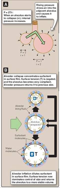
Figure 22.6
Surfactant stabilizes alveolar size.
P = intraalveolar pressure, r = alveolar radius; T = surface tension.
Clinical Application 22.1: Infant Respiratory Distress Syndrome
Infants born prematurely have underdeveloped lungs that are incapable of producing levels of surfactant necessary for stabilizing alveolar volume. Atelectasis is common, as are hyperexpanded regions of the lung. Collapsed alveoli cannot participate in gas exchange and the infant becomes cyanotic as a result. The infant's lungs also have low compliance, which increases the work of breathing. Infant respiratory distress syndrome (IRDS) is characterized by hypoxia, tachypnea, tachycardia, and exaggerated breathing movements. Ventilatory failure is a likely outcome in the absence of medical intervention. IRDS infants are supported with mechanical ventilation and by delivering surfactant to the lungs until the lungs are sufficiently developed to produce adequate surfactant.

Figure 22.7
Expanding and contracting an accordion's bellows creates airflow and musical notes.
c. Keeping lungs dry: The collapsing fluid bubble within an alveolus exerts a negative pressure on the alveolar lining. This pressure creates a driving force for fluid movement from the interstitium onto the alveolar surface. The presence of fluid within an alveolar sac interferes with gas exchange and negatively impacts lung performance. Surfactant reduces the pressure gradient and thereby helps keep lungs fluid free.
V. MECHANICS OF BREATHING
The blood–gas interface is separated from the external atmosphere by a distance of ~30 cm (i.e., the length of the trachea and other intervening airways). O2 cannot diffuse over such a distance fast enough to meet the demands of internal respiration, so air must be drawn into the lungs by an air pump.
A. Pump structure
The pump functions much like an accordion, a musical instrument comprising a bellows operated using two handholds (“manuals”) as shown in Figure 22.7. When the manuals are drawn apart, the bellows expand, and pressure inside them drops. This creates a pressure gradient that drives airflow over a set of reeds that give the instrument its familiar sound. Lung tissue (the pulmonary equivalent of bellows) is too fragile to be attached to the muscles and tendons that might function as manuals. Instead, they are hermetically sealed to the lining of the thoracic cavity. This allows the chest wall and diaphragm to expand the bellows while keeping the force per unit area applied to the lungs minimal (Figure 22.8). The seal relies on pleura and pleural fluid.
B. Pleurae
Pleurae are thin, serous membranes that cover the lungs. Similar membranes cover the heart (the pericardium) and viscera (the peritoneum). Pulmonary pleurae have two essential air-pump functions: creating the hermetic seal and secreting pleural fluid.
1. Hermetic seal: The lungs are enveloped in visceral pleura (Figure 22.9). Each lung is enclosed within its own pleural cavity, and there is no connection between the two. The chest wall, diaphragm, and mediastinum (heart, larger blood vessels, airways, and associated structures) are covered with parietal pleura. The visceral and parietal pleurae are physically attached to their respective underlying structures but not to each other, and the two membranes are separated by the intrapleural space. The pleurae effectively exclude air from the intrapleural space to hermetically seal the lungs to the diaphragm and rib cage.
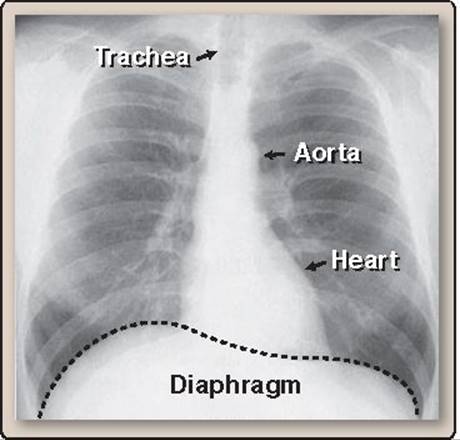
Figure 22.8
Normal posteroanterior chest x-ray.

Figure 22.9
Pleurae.
2. Pleural fluid: The parietal pleura is innervated and vascular. It is believed to be the source of a viscous pleural fluid that is secreted into the intrapleural space. Pleural fluid has two important functions: lubrication and cohesion.
a. Lubrication: Pleural fluid lubricates the pleural surfaces and allows the lungs to slide freely over the chest wall and diaphragm during normal breathing movements.
b. Aiding inspiration: Pleural fluid is secreted and reabsorbed constantly. The volume contained within the intrapleural space at any one time is ~10 mL total, but it spreads to create a thin film that covers all surfaces, making the two pleura almost inseparable under physiologic circumstances. The same cohesive force makes two glass microscope slides difficult to separate when a drop of water is caught between them. Cohesion allows forces generated by movement of the chest wall and diaphragm to be transferred directly to the lung surface.
C. Pump cycling
Respiration involves repeated cycles of inspiration and expiration. Inspiration draws air into the lungs and increases O2 availability at the blood–gas interface.
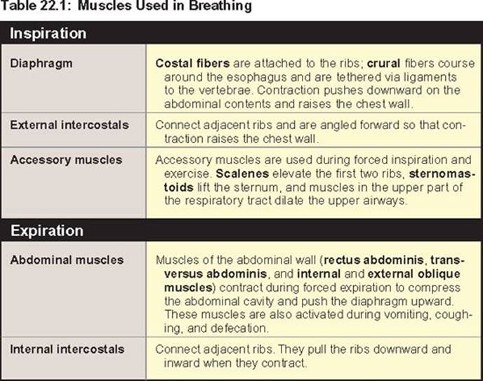
1. Inspiration: The air pump is operated by skeletal muscles (Table 22.1). Most important of these is the diaphragm (see Figure 22.9), a dome-shaped muscle that separates the thoracic and abdominal cavities that is innervated by the phrenic nerve. When the muscle contracts, intrathoracic volume increases.
a. Vertical dimensions: Diaphragm contraction pushes downward on the abdominal contents and increases the vertical dimensions of the thoracic cavity by between 1 and 10 cm, depending on activity level (Figure 22.10A).
b. Cross-sectional area: Contraction also increases cross-sectional area by pulling upward on the lower ribs (see Figure 22.10B). The ribs rise in a bucket-handle fashion, a motion aided by contraction of the external intercostal muscles.
2. Expiration: Expiration is generally passive and is driven both by the effects of surface tension on alveolar volume and energy stored in a lung's elastic elements during inspiration (elastic recoil). Elasticity reflects an abundance of elastin and collagen fibers in both airways and alveoli. Expiratory muscles are typically used during exercise or when airway resistance is increased by disease, for example (see Table 22.1).

Figure 22.10
Thoracic volume changes during inspiration.
VI. STATIC LUNG MECHANICS
Surface tension's influence on lung volume is tempered by surfactant, but it still remains a significant force that impacts lung behavior during normal breathing.
A. Forces acting on a static lung
A healthy lung at rest is subject to two equal and opposing forces, one directed inward and the other outward.
1. Inward: As discussed previously, a lung's elasticity and surface tension effects generate an inwardly directed force that favors smaller lung volumes (Figure 22.11A).
2. Outward: The muscles and various connective tissues associated with the rib cage also have elasticity. At rest, the elastic elements favor outward movement of the chest wall.
3. Net effect: The two opposing forces create negative pressure within the intrapleural space (intrapleural pressure, or Ppl). Ppl is measured relative to the atmosphere and averages several centimeters of water, depending on vertical position within the lung (discussed below). If either pleura is breached, air rushes into the pleural space, driven by the pressure difference between atmosphere and pleural space (pneumothorax) as shown in Figure 22.11B. Ppl falls to zero, elastic elements in the chest wall cause it to spring outward, and the lung collapses.
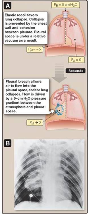
Figure 22.11
Pneumothorax. PA = alveolar pressure;
PB = barometric pressure;
Ppl = intrapleural pressure.
The human thorax contains two pleural cavities. In practice, this means that injury resulting in pneumothorax usually affects only one lung at a time. Thus, although pneumothorax is still a serious condition, it is not immediately fatal. By contrast, the American buffalo (bison) contains a single pleural cavity that is relatively easy to breach with a gunshot or arrowhead. This immense animal once roamed the plains of North America in large numbers, but its vulnerability to pneumothorax allowed hunters and farmers to systematically decimate herds during the late 1800s.

Figure 22.12
Pulmonary pressure–volume loop.
B. Pressure–volume curves
A collapsed lung can help us understand how much effort is required to inflate it during normal breathing.
1. Inflation: A collapsed lung can be inflated in one of two ways, both of which modify transpulmonary pressure (PL). PL is the difference between intraalveolar pressure (PA) and Ppl:
PL = PA − Ppl
A mechanical positive-pressure ventilator can be used to raise pressure inside the lung (PA > Ppl), inflating it much as one inflates a balloon. Alternatively, air can be withdrawn from the pleural space to create a negative pressure outside the lung (Ppl < PA). Both maneuvers raise PL. Lung collapse allows the airways to close off and seal with fluid. Restoring patency requires that the surface tension seal be broken, which requires considerable effort. PLmust be increased by several cm H2O before any significant increase in volume occurs (Figure 22.12, phase 1). Once PL exceeds ~7–10 cm H2O, airways pop open, and volume increases linearly with inflation pressure (phase 2). At ~20 cm H2O, the lung reaches its maximal volume, known as total lung capacity (TLC), as shown in Figure 22.12, phase 3.
2. Deflation: A lung that is allowed to deflate from TLC yields a different pressure–volume curve from that seen during inflation (a phenomenon known as hysteresis). This is because surfactant is recruited from pneumocytes to the alveolar surface film during lung inflation. The surfactant decreases elastic recoil and thereby resists lung deflation. Note that the hysteresis loop begins and ends at a positive volume (normally ~500 mL; see Figure 22.12). This is because the larger airways collapse at zero pressure and trap air within the more distal regions.
3. Normal breathing: Normal breathing involves changes in lung volume that are only a fraction of total, but the hysteresis is still evident (see Figure 22.12). Note also that inspiration normally begins at ~50% of TLC. When the lung is resting between breaths, the chest wall prevents it from collapsing, and, at 50% TLC, all alveoli are patent. The chest wall also positions the resting lung on the steepest portion of the pressure–volume curve, meaning that the increase in PL during inspiration is maximally effective in increasing alveolar volume.

Figure 22.13
Pressure–volume loop for a fluidfilled lung. TLC = total lung capacity.
4. Surface tension effects: Surface tension's influence on the pressure–volume loop can be estimated by filling lungs with fluid to eliminate air–water interfaces (Figure 22.13). Fluid-filled lungs are more compliant, and the hysteresis associated with surface tension disappears.
C. Gravitational effects
Lungs and blood have mass and, thus, are subject to the influence of gravity. Gravity causes significant regional differences in PL and alveolar volume.
1. Apex: When the thorax is positioned vertically, a lung within can be imagined to hang suspended by its apical pleura. Suspension creates a strongly negative Ppl (and strongly positive PL) locally and causes apical alveoli to inflate to ~60% of their maximal volume (Figure 22.14). Gravity similarly stretches the coils at the top of a Slinky (the famous toy) farther apart than those at the base (Figure 22.15). In practice, gravitational influences force the lung apex to function near the top of the pressure–volume curve, where the opportunity for further expansion during inspiration is very limited.
2. Base: The lung base supports the mass of pulmonary tissue above it. Alveoli in this region are compressed, much like coils at the base of the Slinky. The weight of tissue above also pushes outward against the chest. Ppl and PL both approach zero (see Figure 22.14). In practice, this means that alveoli at base of the lung respond to increases in PL with large changes in volume because they occur over the lower, steepest part of the pressure–volume curve.
D. Lung compliance
The amount that lung volume increases in response to changes in PL is a measure of its compliance. Lungs are highly compliant organs, increasing volume by ~200 mL for every cm H2O of transpulmonary pressure. Compliance is governed both by surface tension and the elastic properties of the lungs and chest wall. Lungs become less compliant with age due to connective tissue deposition.

Figure 22.14
Gravitational effects on alveolar volume. PA = intraalveolar pressure; Ppl = intrapleural pressure.
VII. LUNG DISEASES
Obstructive and restrictive pulmonary diseases are two broad disease groups that cause significant changes in the static lung properties. The two groups are typified, respectively, by emphysema and pulmonary fibrosis. We will revisit these diseases frequently to help illustrate the mechanical principles involved in normal breathing.

Figure 22.15
Gravitational effects on a Slinky.
A. Obstructive pulmonary disease
Emphysema, chronic bronchitis, and asthma all represent obstructive pulmonary diseases, which increase airway resistance to airflow. Because the former two examples often coexist and may be difficult to distinguish clinically, they are commonly grouped and discussed as chronic obstructive pulmonary disease (COPD). COPD is extremely common and has become the fourth leading cause of death in the United States. There are three general obstructive mechanisms: airway occlusion, wall thickening, and loss of mechanical tethering.
1. Airway occlusion: Airways may be occluded by foreign bodies or, more commonly, by secretions that are excessive or difficult to expel (Figure 22.16B). Occlusive diseases include chronic bronchitis, asthma, and bronchiectasis.
2. Wall thickening: When the airway wall hypertrophies or becomes edematous, it encroaches on the lumen and reduces its cross-sectional area (see Figure 22.16B).
3. Loss of mechanical tethering: All structures in the lung are linked mechanically. Together, they form a dependent network, much like the fabric of a nylon stocking (interdependence). Interdependence maintains airway patency when external forces may favor collapse. Emphysema develops when alveolar walls (the fabric of the lung) erode, allowing surrounding airways to collapse and obstruct airflow during normal breathing (see Figure 22.16C). Emphysema is commonly caused by heavy smoking.
Emphysema denotes anatomic tissue loss, although the term is sometimes used to describe smoking-related lung disease. It is a finding evident on computed tomography imaging of the lung. It is also a pathologic finding seen at autopsy or lung tissue biopsy. Postmortem examination of an emphysematous lung shows enlarged cystic air spaces replacing normal lung. Alveolar loss reduces elastic recoil, increases pulmonary compliance, and reduces the surface area available for O2 uptake.
B. Restrictive pulmonary disease
Pulmonary fibrosis is a restrictive pulmonary disease. Others include pleural diseases and problems affecting breathing muscles, all of which limit lung expansion. Pulmonary fibrosis (scarring) results from any one of a number of interstitial lung diseases. Scarring typically begins with an injury to the alveolar epithelium. Causes include any of several compounds inhaled in the workplace (e.g., asbestos, beryllium, coal dust, sawdust), circulating drugs (e.g., antibiotics and chemotherapeutic agents), systemic diseases (e.g., rheumatoid arthritis, lupus, scleroderma, and sarcoidosis), or may be idiopathic. The initial insult causes the alveolar wall to thicken and the alveolar space to fill with an exudate containing lymphocytes, platelets, and other immune effector cells (see Figure 22.16D). The space is then infiltrated by fibroblasts, which lay down bundles of collagen and other fibers between the alveolar sacs. Scar tissue is relatively noncompliant, so the lung becomes stiff and expands with difficulty during inspiration. Diseased lungs decrease O2 uptake, and hypoxemia may develop as scarring progresses.
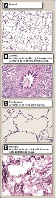
Figure 22.16
Normal and diseased lungs.
VIII. DYNAMIC LUNG MECHANICS
During inspiration, air moves from the external environment through a set of branching tubes of ever-decreasing diameter. Flow is driven by the pressure difference between the external atmosphere and alveolus (ΔP = PB – PA). Flow is inversely proportional to airway resistance (R):

where  is airflow (volume ÷ unit time).
A. Pressures driving airflow
Airflow occurs in response to pressure gradients set up between the alveoli and the external atmosphere. The body has no way of controlling PA directly. Instead, the diaphragm and other respiratory muscles manipulate intrapleural pressure. When Ppl falls, PL rises, and the alveoli expand. PA becomes negative because the product of pressure and volume of a fixed number of air molecules remains constant (as per the Boyle law).
PA1VA1 = ↓ PA2 ↑ VA2
Where PA1 and PA2 denote alveolar pressure before and after alveolar expansion (VA1 and VA2). Alveolar expansion thus creates a PB > PA pressure gradient that drives airflow into the lungs (Figure 22.17). Because flow occurs against a resistance, it takes time for air to move in or out of the lungs and for the pressure gradient to dissipate, particularly at the points farthest removed from the site of highest resistance.
B. Resistance to airflow
In a healthy individual, breathing is usually an effortless and unconscious act, so it seems surprising that airways can offer resistance to flow. The resistance has several origins. Resistance is proportional to airway length (l) and the viscosity (η) of the gas moving through it and inversely proportional to the fourth power of airway radius (r), as stated in the Poiseuille law:

1. Airway radius: Airway radius decreases with each successive generation within the bronchiolar tree. Decreasing radius increases resistance, but the negative impact on net airflow through the lung is more than offset by the gain in airway numbers with each successive generation. In other words, although individual bronchioles have a very high resistance, their combined resistance is almost negligible (calculated from the sum of reciprocals; see also 19·IV·B). The site of greatest resistance in the lung is in the pharynx and larger airways (generations 0 through ~7) as shown in Figure 22.18.

Figure 22.17
Pressure gradients driving airflow during inspiration. PB = barometric pressure; Ppl = intrapleural pressure. All values are given in cm H2O.
2. Air viscosity: Air viscosity is dependent on air density. Air density increases when compressed, as during a deep-sea dive, for example. Increasing density increases flow resistance and the work of breathing. Breathing an O2/helium mixture partly offsets this density increase. Helium has less density than atmospheric air and, therefore, reduces the work of breathing.
3. Turbulence: The Poiseuille law above assumes that airflow through the lungs is streamline, but this is generally not the case. The airways consist of a series of branching tubes. Each branch point creates a local eddy current that disrupts streamline flow and increases airway resistance. In practice, the eddy currents cause flow through the airways to be proportional to (ΔP + √ΔP) rather than

Figure 22.18
Resistance to airflow within the bronchial tree.
C. Factors affecting airway resistance
Airways are the primary source of resistance in the lung and, therefore, changes in airway radius can significantly impact lung function. Airway radius is governed by airway musculature and by lung volume.
1. Smooth muscle: Bronchioles are lined with smooth muscle cells. When the muscles contract, they decrease airway radius and increase resistance to airflow. Flow through the airways may decrease as a result. Smooth muscle relaxation and bronchiolar dilation reduces resistance and facilitates increased airflow. Airway muscles are regulated by the autonomic nervous system (ANS) and by local factors.
a. Autonomic control: Airways are controlled by both parasympathetic and sympathetic (SNS) branches of the ANS.
i. Parasympathetic: Parasympathetic nerve fibers from the vagus nerve release acetylcholine (ACh) from their terminals when active. ACh binds to M3 muscarinic ACh receptors and causes bronchoconstriction, which reduces airflow.
ii. Sympathetic: Sympathetic activation causes bronchioles to dilate, mainly by inhibiting ACh release rather than through direct effects on the musculature. SNS terminals release norepinephrine, which binds to a presynaptic β2-adrenergic receptor. This receptor is particularly sensitive to epinephrine release from the adrenal medulla during SNS activation. SNS-mediated bronchodilation is important for facilitating increased airflow to the blood–gas interface during exercise, for example.
b. Local factors: Local irritants and allergens constrict bronchioles and obstruct airways. Airway muscle contraction is a response to histamine and other inflammatory mediators.
2. Lung volume: Airway resistance is highly dependent on lung volume. Net airway resistance is low at high lung volumes and high at low volumes.
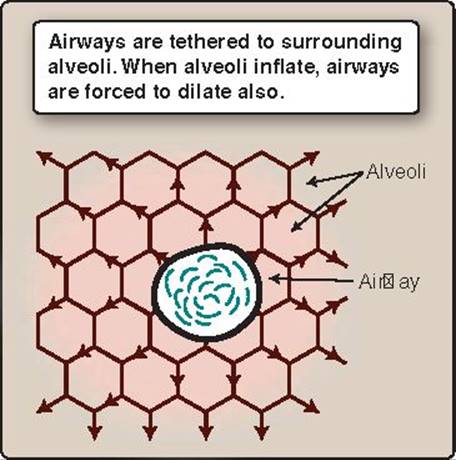
Figure 22.19
Radial traction on airways during lung inflation.
a. High volumes: The decrease in Ppl that establishes a gradient for airflow during inspiration is transmitted to the airways as well as to alveoli. This causes airway resistance to fall during lung expansion. Airways are also dilated by radial traction. Traction results from a mechanical tethering between the alveoli and all surrounding structures. In practice, when the alveoli are expanded, radial traction on intervening airways increases their radius and lowers their resistance (Figure 22.19).
b. Low volumes: At low lung volumes, radial traction is reduced, and airway resistance increases.
D. Airway collapse during expiration
Airways tend to collapse and limit flow during expiration, an effect known as dynamic compression of the airways. The reasons and consequences of collapse are easiest to appreciate during a forced expiration after a deep inspiration (Figure 22.20). Forced expiration begins with contraction of the abdominal muscles and internal intercostals, which forces the chest wall downward and inward, and causes Ppl to become positive. The positive pressure is transferred to and compresses the alveoli, decreasing their volume and causing PA to rise above PB. Compression thereby establishes the pressure gradient that drives expiratory outflow. The larger airways have a relatively high resistance to flow that limits lung-emptying rates, so there is a time period during which alveoli remain filled with pressurized air. High intraalveolar pressure maintains patency, even though Ppl may be positive and favoring alveolar collapse. Airway pressure falls with distance from the alveoli and proximity to the main site of resistance (bronchi and trachea). Thus, whereas intraalveolar pressure may be strongly positive (relative to PB), pressure within the larger airways may be much closer to zero (i.e., PB) and thus more susceptible to collapse by Ppl (see Figure 22.20[3]). The larger airways are equipped with cartilage that helps maintain patency during forced expiration, but it may be inadequate to prevent collapse. As air leaves the lungs and PA drops, the collapse zone moves distally and involves increasingly smaller airways. Compression and collapse of conducting airways is the self-regulating, limiting factor that determines how fast air escapes the lungs during expiration. If a subject attempts to speed outflow with a more forceful muscular contraction, the pressure gradient driving outflow is raised, but so are the forces favoring airway collapse with a net zero sum gain (Figure 22.21).
E. Work of breathing
Breathing requires that the respiratory muscles contract to expand the lungs against resistance. The work of breathing normally accounts for ~5% of total energy usage at rest, but it can rise to >20% of total during exercise. Such workloads are normally insignificant in a healthy individual, but some patients with pulmonary disease have difficulty expanding their lungs, and even resting breathing movements can fatigue their respiratory muscles and precipitate respiratory failure (see 40·VI).
1. Work components: Many factors contribute to the work of breathing. The two principal factors are elastic work and resistive work. Elastic work includes the work required to counter a lung's elastic recoil during inspiration, which is proportional to its compliance. Work is also required to displace the chest wall outward and the abdominal organs downward. Resistive work involves moving air through the airways against airway resistance.

Figure 22.20
Airway collapse during forced expiration. PA = intraalveolar pressure; PB = barometric pressure; Ppl = intrapleural pressure. All values are given in cm H2O.
Clinical Application 22.2: Pursed-Lipped Breathing
Chronic obstructive pulmonary disease (COPD) is characterized by airflow limitation (obstruction). Spirometry testing reveals a flow–volume loop contour that appears “scooped-out” (concave upward) in the expiratory limb of the loop. There may also be a long tail on the expiratory limb, which manifests because patients with COPD have a hard time exhaling due to loss of elastic recoil and airway collapse. Patients can partly compensate for the loss of mechanical support by pursing their lips (as if whistling) during expiration, a behavior known as pursed-lipped breathing, or puffing. This behavior is effective because it moves the site of main airway resistance closer to the mouth and extends the time during which airway pressure remains high and the airways patent. Patients with anatomic tissue loss (emphysema) in addition to airflow obstruction tend to hyperventilate and use accessory muscles to help with expiration, giving them a characteristic pink complexion (“pink puffers”). This contrasts with COPD patients whose disease is characterized by chronic bronchitis and excessive mucus production that interferes with oxygen uptake (these patients may be described as “blue bloaters”).

Effects of emphysema on airflow.
2. Measuring work: Work is calculated as the amount of force required to move an object a given distance. In pulmonary terms, the work of breathing is calculated from the product of the force needed to change the transpulmonary pressure gradient and the air volume moved per unit time. Work can be represented graphically as the area to the left of the inspiratory phase of the pressure–volume loop (Figure 22.22).
3. Pulmonary diseases: COPD and pulmonary fibrosis both increase the work of breathing (see Figure 22.22). Patients with COPD work harder to exhale against high airway resistance (increased resistive work). Pulmonary fibrosis stiffens the lung and requires that a patient generate higher transpulmonary pressures than normal to expand the lungs during inspiration (increased elastic work).
IX. LUNG VOLUMES AND CAPACITIES
Normal quiet breathing uses less than 10% of TLC. Exercise increases this amount significantly, but there is always a small residual volume that communicates with the ventilated space but does not itself participate in ventilation, even at maximal levels of exercise. Clinically, it is important to determine the contribution of this volume to the mix of gases in the lungs and to assess how lung volume(s) may be impacted by the progression of various pulmonary diseases. In addition to airflow measurement with spirometry, pulmonary function tests (PFTs) typically measure four primary lung volumes, which are then combined to derive several lung capacities (Figure 22.23). PFTs also assess the efficiency of the blood–gas interface (“diffusing capacity” is discussed in Chapter 23·V).
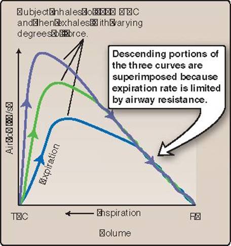
Figure 22.21
Airway resistance limits flow during forced expiration. RV = residual volume; TLC = total lung capacity.
A. Volumes
The volume of air inspired or expired with each breath, typically ~500 mL in an average adult, is called the tidal volume (TV). Inspiratory reserve volume (IRV) and expiratory reserve volume (ERV) are the volumes that can be inspired or expired, respectively, over and above TV. Residual volume (RV) is the volume of air remaining in the lung after a maximal expiration (~1.2 L in a normal individual). A spirometer is unable to provide information about RV, so pulmonary function testing often includes more specialized body plethysmography or techniques that monitor intrapulmonary concentrations of gases over time (i.e., helium-dilution and nitrogen-washout assays).
B. Capacities
The sum of all four lung volumes (TLC) amounts to ~6 L in a normal individual (see Figure 22.23). Functional residual capacity (FRC) is the volume remaining in the lungs after expelling a tidal breath. Inspiratory capacity(IC) is the sum of the TV and the IRV. Vital capacity (VC) is the sum of the TV, IRV, and ERV, and is the maximal TV achievable (i.e., the biggest breath one can take). Forced vital capacity (FVC) is the volume of air that can be forcibly expired after a maximal inspiration.
C. Forced expiratory volume
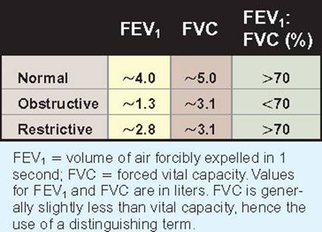
FEV1 is the volume of air that can be forcibly expired in 1 second following a maximal inspiration and is an important clinical measure of lung function (see Clinical Application 22.3).
X. VENTILATION AND DEAD SPACE
Gas exchange occurs at the alveolar surface. By the time that inspired air contacts the gas exchange interface, its O2 and CO2 concentration has been modified through mixing with gases lingering in the RV, which itself is influenced by how often the contents of the lung are refreshed (ventilation). Alveolar gas concentration is also influenced by the amount of inhaled air that does not participate in gas exchange because it fills dead space.
A. Dead space
The lung contains two types of dead space: anatomic and physiologic.
1. Anatomic: The pharynx, trachea, bronchi, and other conducting airways contain ~150 mL of air that is moved out during expiration without ever contacting the gas exchange interface. This represents anatomic dead space.

Figure 22.22
Effects of pulmonary disease on the work of breathing. COPD = chronic obstructive pulmonary disease.
Clinical Application 22.3: Pulmonary Function Tests
Pulmonary function tests are useful in detecting the presence of obstructive and restrictive pulmonary pathophysiology. Chronic obstructive pulmonary disease (COPD) is best identified by measuring airflow with spirometry and documenting obstruction (reduced forced expiratory volume in 1 second [FEV1] in a setting of an FEV1/FVC [forced vital capacity] ratio of <70%). Patients with COPD typically also operate at very high lung volumes because exhalation is impaired by airway obstruction. Patients with pulmonary fibrosis work at low volumes because the lung is noncompliant and difficult to expand, and, thus, total lung capacity is reduced. They typically take shallow breaths and breathe rapidly.
2. Physiologic: In a diseased lung, a proportion of the alveoli may be ventilated but unable to participate in gas exchange because the blood–gas interface is damaged or pulmonary blood flow to these regions has been interrupted. These regions represent dead space. The term physiologic dead space includes anatomic dead space and contributions from these nonfunctional alveoli. In a healthy person, physiologic and anatomic dead spaces are approximately equal. In a diseased lung, the physiologic dead space may be increased by 1,500 mL or more.
3. Calculating dead space: Dead-space volume (VD) can be calculated by measuring the amount of CO2 contained in expired air (PECO2). Dead space (by definition) does not participate in gas exchange and, thus, contains negligible CO2. The amount of CO2 in air originating from regions of the lung involved in gas exchange equals that of arterial blood (PaCO2), because blood gases equilibrate with alveolar gases during transit through the pulmonary circulation (i.e., PACO2 = PaCO2). Thus, dead-space volume can be determined from the extent to which the amount of CO2 in expired air has been decreased by CO2-free air originating from dead space:

where VT = total lung volume.
B. Ventilation: Ventilation can be expressed as minute ventilation or alveolar ventilation. Minute ventilation (VE) is the total volume of air inhaled and exhaled per minute:
Minute ventilation = TV × breaths/min
Alveolar ventilation (VA) is the volume of air per minute that enters the areas participating in gas exchange:
VA = (TV − VD) × breaths/min
Where VA represents alveolar ventilation and VD is dead space.

Figure 22.23
Spirometry. ERV = expiratory reserve volume; FRC = functional residual capacity; IC = inspiratory capacity;
IRV = inspiratory reserve volume;
RV = residual volume; TLC = total lung capacity; TV = tidal volume;
VC = vital capacity.
Chapter Summary
• Lungs facilitate exchange of O2 and CO2 between blood and air. The blood–gas interface is located within alveoli, thin-walled sacs that serve to amplify interface surface area and to bring the pulmonary circulation into close proximity to inhaled air.
• Alveoli are moistened with a thin fluid film that generates surface tension. Surface tension is a force that favors lung collapse and negatively impacts lung performance. The alveolar epithelium produces surfactant to counter this surface tension. Surfactant is a phospholipid complex that helps stabilize alveolar size and increases lung compliance.
• Breathing involves repeated cycles of inspiration and expiration. Air is drawn into the lungs by contracting the diaphragm and other respiratory muscles. Contraction increases the volume of the thoracic cavity and lungs.
• The diaphragm, chest wall, and lungs move as one unit. They are linked by a thin film of pleural fluid, which lubricates the visceral and parietal pleurae and provides the cohesive force required to expand the lungs.
• At rest, a lung is subject to two opposing forces. Surface tension and elastic elements in lung tissue favor collapse (elastic recoil). Elastic elements in the chest wall favor expansion and thereby prevent collapse. Introducing air between the two pleurae (pneumothorax) breaks the connection between lungs and chest wall and allows a lung to collapse.
• Gravity causes significant regional differences in alveolar size in an upright lung. The base of the lung is compressed by its own mass, whereas alveoli at the apex may be expanded to 60% of their maximal volume.
• Airflow between the alveoli and the external atmosphere is driven by pressure gradients. Flow occurs against a resistance that depends largely on an airway's internal radius.
• Airway resistance is modulated by the autonomic nervous system but also changes passively with lung volume. During lung expansion, the airways are forced to dilate by surrounding structures acting via mechanical tethers, and dilation causes airway resistance to fall. When lung volumes are low, the airways are compressed by the mass of surrounding tissue, and their resistance is high.
• Airways are also sensitive to transmural pressures developed during expiration, such that their resistance becomes a pressure-dependent limiting factor on outflow.
• Air movement between lungs and atmosphere is measured using spirometry, one of several pulmonary function tests (PFTs) used to assess lung health. PFTs derive four lung volumes (i.e., tidal volume, inspiratory reserve volume, expiratory reserve volume, and residual volume) and capacities (i.e., total lung capacity, functional residual capacity, inspiratory capacity, and vital capacity).
• Air that is enclosed within regions of the lung that do not participate in gas exchange is known as dead space.