
24
I. OVERVIEW
Normal breathing (eupnea) is usually an unconscious act driven by the autonomic nervous system. The cyclical pattern is established by a respiratory control center within the brain that coordinates contraction of the diaphragm and other muscles involved in inspiration and expiration (Figure 24.1). Because the needs of internal respiration change with activity level, the pattern generator must also change its output to match prevailing needs. Sensors located within the central nervous system (CNS) and throughout the periphery continuously monitor blood and tissue PCO2, PO2, and pH levels and feed this information back to the control center for processing (Figure 24.2). The center also receives information from mechanoreceptors located in the lungs and chest wall. The control center then adjusts ventilation as needed, using motor outputs to the diaphragm, the intercostals, and the other muscles involved in breathing. Although the precise location and functions of the various neurons involved in respiratory control remain ill-defined, it is clear that the principal goal is to maintain PaCO2 at a stable level while simultaneously ensuring adequacy of O2 flow to the tissues. The dominance of CO2 in respiratory control reflects the body's need to maintain extracellular fluid (ECF) pH within a narrow range.
II. NEURAL CONTROL CENTERS
Several regions of the brain influence breathing. The basic respiratory rhythm is established by a respiratory control center located within the brainstem medulla.
A. Medullary center
The medulla contains several discrete groups of neurons involved in respiratory control (Figure 24.3). Although functioning as a single unit, control-center neurons and their input/output pathways are mirrored on either side of the medulla. Either side is capable of generating independent respiratory rhythms if the brainstem is transected. Within the control center are two concentrations of neurons that fire in phase with the respiratory cycle and are assumed to have a key role in establishing the respiratory rhythm. These are called the dorsal respiratory group (DRG) and the ventral respiratory group (VRG).
1. Dorsal respiratory group: DRG neurons are active primarily during inspiration (Figure 24.4). Because inspiration is the only active phase of quiet breathing, it is likely that the DRG implements the respiratory rhythm, although the pacemaker (a central pattern generator, or CPG) probably resides elsewhere (see below).

Figure 24.1
The rhythmic inspiration–expiration cycle. ECG = electrocardiogram; EMG = electromyogram.

Figure 24.2
Respiratory control pathways.
a. Function: The DRG receives sensory inputs from the thorax and abdomen and processes incoming signals using interneuron networks. If the sensors report suboptimal CO2 or O2 levels, the DRG formulates and executes an appropriate response.
b. Output: DRG neurons communicate with premotor neurons controlling the diaphragm. They also project to a control-center area involved in forced inspiration and expiration (the VRG).
2. Ventral respiratory group: The VRG can be subdivided into rostral, intermediate, and caudal regions (see Figure 24.4).
a. Function: The VRG functions primarily to coordinate accessory muscles of inspiration and expiration. It is largely quiescent during quiet breathing but becomes highly active during exercise.
b. Output: The VRG's intermediate region contains motor efferents to accessory muscles in the pharynx and larynx that dilate the upper airways during inspiration. The caudal region contains premotor neurons that synapse within the spinal cord to control the internal intercostals and other accessory muscles of expiration. The rostral region (the Bötzinger complex) communicates via interneurons with the DRG and the caudal region of the VRG. It may be involved in coordinating VRG output.

Figure 24.3
Brainstem areas involved in respiratory control.

Figure 24.4
Respiratory control center organization. DRG = dorsal respiratory group;  = airway flow; V = voltage records from respiratory motor efferent nerve fibers; VRG = ventral respiratory group.
3. Central pattern generator: The VRG rostral region contains a small area known as the pre-Bötzinger complex composed of cells that exhibit pacemaker-like activity under experimental conditions. Ablation of the complex abolishes rhythmic breathing, suggesting that it constitutes a respiratory CPG.
Clinical Application 24.1: Sleep Apnea
Sleep is usually accompanied by a pattern of normal quiet breathing, but some individuals may cease breathing for prolonged periods (tens of seconds) several times per hour (sleep apnea). Not surprisingly, apnea usually manifests as excessive daytime sleepiness. There are multiple causes. Central sleep apnea results from a complete loss of respiratory drive and is relatively uncommon. Obstructive sleep apnea is more prevalent, especially in obese individuals. Loss of ventilation results from obstruction (by soft tissues, such as the tongue and uvula) and collapse of the upper airways during sleep. Airways normally lose an active dilator influence during sleep, which makes them more prone to collapse, even in a healthy person. Fat deposits around the airways greatly increase the likelihood of collapse, however, and may require use of a nighttime breathing aid (i.e., a continuous positive airway pressure mask) that provides relief by pneumatically splinting the airways to maintain patency.

Airway obstruction during sleep causes sleep apnea.
B. Pontine centers
The pons contains two areas that influence medullary output. The apneustic center resides in the lower pons. Transectioning the brain above this site causes prolonged inspiratory gasping (apneuses), suggesting that it normally limits lung expansion. Stimulating the pneumotaxic center (upper pons) shortens inspiration and increases breathing rate. A role for either center in normal breathing has not been established.
C. Cerebral cortex and other brain regions
Emotions such as fear, excitement, and rage can alter respiratory rate, reflecting the ability of the hypothalamus and limbic system to modulate the CPG. The CPG also readily succumbs to the cerebral cortex to allow for talking, playing a musical wind instrument, and other activities that require fine conscious control of breathing movements. A person can also consciously override the CPG and hyperventilate, hypoventilate, or cease breathing altogether. The effects of breath-holding on blood gas concentrations are tolerated for a relatively short time before chemical control feedback pathways override voluntary control.

Figure 24.5
Central chemoreceptors monitor arterial PCO2 through effects of CO2 on pH of cerebrospinal fluid (CSF) and extracellular fluid (ECF). CA = carbonic anhydrase.
III. CHEMICAL CONTROL OF VENTILATION
A primary mission of the respiratory system is to optimize ECF pH through manipulation of PCO2. It must also maintain steep O2 and CO2 partial pressure gradients to maximize transfer of these two gases between tissues and the external environment. Fulfilling these roles requires that information about the chemical composition of ECF be sensed and relayed back to the respiratory control centers so that ventilation might be modified accordingly. The body employs central and peripheral chemoreceptors for this purpose.
A. Central chemoreceptors
Ventilatory control under resting conditions is dominated by the central chemoreceptors, which are responsive primarily to changes in PaCO2. The chemoreceptors are CNS neurons located behind the blood–brain barrier (BBB) along the medullary surface. The BBB is impermeable to virtually all blood constituents except for lipid-soluble molecules, such as O2 and CO2. Once inside the barrier, CO2 dissolves to form carbonic acid, which acidifies brain ECF and cerebrospinal fluid (CSF) as shown in Figure 24.5. CSF contains minimal amounts of protein to buffer pH. The consequence is that even modest changes in PaCO2 cause significant CSF and ECF acidosis. The chemoreceptor neurons respond to acid with excitatory impulses that impel the respiratory center to increase breathing rate. The BBB acts as an important information filter because, by excluding bloodborne ions such as H+, it provides a way for the chemoreceptors to distinguish changes in PaCO2 from any background changes in ECF pH.

Figure 24.6
Peripheral chemoreceptors are contained within carotid bodies and aortic bodies.
Systemic changes in pH ultimately affect all tissues regardless of cause or BBB ion permeability. Central chemoreceptors play a critical role in the integrated response to changing blood pH, although reactions to metabolic acidosis or alkalosis may be slower and less intense than respiratory responses triggered by changes in PaCO2.
B. Peripheral chemoreceptors
Peripheral chemoreceptors were first introduced in reference to control of blood pressure (see 20·III·A·3), because they also relay information to the medullary cardiovascular control centers. The chemoreceptors with the greatest influence on respiration are located within carotid bodies located at the bifurcation of the two common carotid arteries (Figure 24.6). Chemoreceptors are also found within aortic bodies distributed along the underside of the aortic arch. Peripheral chemoreceptors monitor PaO2, PaCO2, and arterial pH.
1. Structure: Carotid and aortic bodies are notable for their small size (3–5 mm) and high rates of blood flow relative to their mass. The high flow rate minimizes the effect of chemoreceptor metabolism on blood gas content and, therefore, allows for a truer reading of arterial O2 and CO2 levels. Carotid bodies contain two types of cells arranged in clusters and in close apposition to fenestrated capillaries. Type I, or glomus, cells are the chemosensors (Figure 24.7). Type II, or sustentacular, cells play a supportive role similar to glia. Glomus cells signal the respiratory center via the carotid sinus and glossopharyngeal (cranial nerve [CN] IX) nerves (carotid bodies) or the vagus nerve (CN X; aortic bodies).
2. Sensory mechanism: Glomus cell membranes contain an O2-sensitive K+ channel whose open probability is PO2- dependent. When PaO2 is high, the channel is open and allows a K+ efflux that maintains glomus-cell membrane potential (Vm) at strongly negative levels. When PaO2 falls below 100 mm Hg, the channel closes, and Vm depolarizes. The change in Vm activates L-type Ca2+ channels and elicits a Ca2+ influx that stimulates neurotransmitter release onto the sensory afferents (see Figure 24.7). Glomus cells are also excited by increasing PaCO2 and H+ concentrations independently of changes in PaO2. They help fine-tune the information that the respiratory center receives from central chemoreceptors. Changes in PaCO2 and pH may also act by influencing O2-dependent K+ channel open probability, but the mechanism(s) has not been defined.
C. Ventilatory responses
The respiratory control center processes information about PaO2, PaCO2, and arterial pH. Changes in these variables rarely occur in isolation, which forces the respiratory center to make choices about an appropriate ventilatory response (Figure 24.8). Under most circumstances, the output from the respiratory center is designed to optimize PaCO2, but coincident changes in PaO2 and pH influence system sensitivity to changes in PaCO2.

Figure 24.7
Mechanism of glomus-cell response to falling PO2. CNS = central nervous system; Vm = membrane potential.
1. Changing carbon dioxide levels: The nature of control-center response to a rise in PaCO2 depends on whether the change is acute or chronic.
a. Acute: When tissue metabolism increases, PaCO2 rises, and the respiratory center compensates by increasing alveolar ventilation (see Figure 24.8). CO2 is an extremely potent ventilatory stimulus (Figure 24.9). The peripheral receptors are fast acting and trigger an immediate response. The central receptors take several minutes to activate fully, but their effects ultimately dominate the ventilatory response. In practice, ventilation increases linearly with any rise in PaCO2 above resting values, whereas ventilation decreases when PaCO2 falls below 40 mm Hg.
b. Chronic: Patients with chronic pulmonary disease may not be able to ventilate at levels required to hold PaCO2 at 40 mm Hg. Ventilation increases initially when PaCO2 begins to rise because the pH of the CSF falls, but the choroid plexus responds by secreting HCO3− into the CSF, which, over a period of 8–24 hours, largely offsets the effect of the higher PaCO2 on plasma pH. Thus, although PaCO2 may remain high, the medullary chemoreceptors no longer register the change, and the respiratory control system adapts to a new, higher PaCO2.
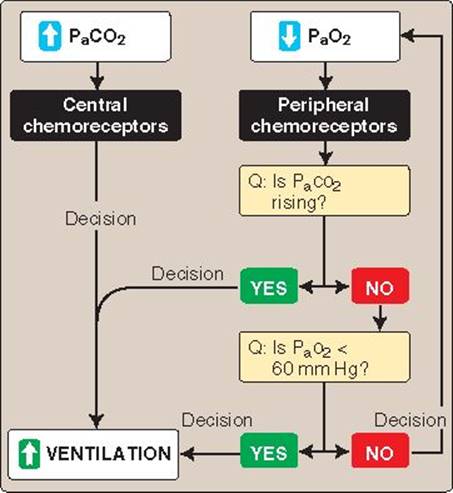
Figure 24.8
Ventilatory responses to changes in arterial PCO2 (PaCO2) and PO2 (PaO2).
Patients that have adapted to hypercapnia may rely on the effects of hypoxia on the peripheral chemoreceptors to sustain their ventilatory drive. When significant hypercapnia exists, administering O2 may help normalize PaO2 but it can also reduce the drive to breathe, potentially increasing PaCO2 further and inducing acute respiratory acidemia due to hypoventilation.
2. Changing oxygen levels: The peripheral chemoreceptors promote a rapid compensatory increase in ventilation if PaO2 falls to dangerously low levels (see Figure 24.10). More modest decreases in PaO2(between 60 and 100 mm Hg) have little effect on ventilation, even though spike frequency in the peripheral chemoreceptor nerve afferents increases in direct proportion to the drop in PaO2. The reason is that the peripheral and central chemoreceptor work against each other for respiratory-center control, with the central chemoreceptors retaining the upper hand until PaO2 falls to 60 mm Hg (see Figure 24.8). Control center output in response to changes in PaO2 is influenced both by blood pH and PCO2.
a. pH: When PaO2 falls, deoxyhemoglobin concentration rises. Deoxygenation makes hemoglobin (Hb) a more favorable substrate for H+ binding (the Bohr effect) and plasma H+ concentrations fall as a result. The rise in pH decreases chemoreceptor sensitivity to a fall in PaO2.
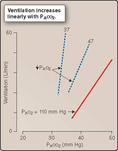
Figure 24.9
Ventilatory responses to changes in alveolar PCO2 (PACO2).
b. Carbon dioxide: When ventilation increases, PaO2 increases, and PaCO2 falls. Because the respiratory control centers are designed to optimize PaCO2, the response to mild hypoxia is overridden in favor of a stable CO2concentration.
The chemoreceptors begin to exert their influence at a PaO2 of 60 mm Hg, which, coincidentally, marks the point at which Hb begins to desaturate (i.e., the steep portion of the dissociation curve; see Figure 23.18). Thus, Hb's O2-binding properties allow the respiratory center to adjust ventilation over a wide range to maintain a stable PaCO2 with little adverse effects on O2 delivery.
3. Synergism: Conditions that decrease PaO2 typically cause a concomitant rise in PaCO2. Because PaCO2 equates with H+ concentration, pH falls also. Thus, it is not surprising to find that changes in PaO2, PaCO2, and arterial pH may act synergistically to elicit a ventilatory response that is greater than the sum of their individual actions.

Figure 24.10
Ventilatory responses to changes in alveolar PO2 (PAO2).
Hypoxia increases chemoreceptor sensitivity to hypercapnia, and rising PaCO2 and H+ concentrations sensitize the receptors to hypoxia.
Clinical Application 24.2: Cheyne-Stokes Breathing
Some disease conditions interfere with respiratory control-center function to cause abnormal resting breathing rhythms. Cheyne-Stokes breathing is a cyclic breathing pattern characterized by periods of apnea followed by a series of breaths of progressively increasing respiratory effort and airflow toward a maximum, and then again waning toward apnea. Although Cheyne-Stokes breathing is observed occasionally in normal individuals at high altitudes during sleep, it is common in patients with stroke and heart failure. Heart failure is associated with low perfusion rates, which impairs the brainstem's ability to monitor the effects of changing ventilation on blood gas composition, and may explain the abnormal rhythm. It is theorized that when the respiratory control centers increase ventilation to correct hypercapnia, there is a time delay before the centers are able to see the results of this change, during which they impel further increases in respiratory effort and cause a hypocapnic overshoot. The centers compensate by decreasing ventilation, with the same hypoperfusion delay causing respiratory effort to decline inappropriately and resulting in apnea. The cyclic breathing pattern is repeated with a variable periodicity of ∞30–100 sec.

Cheyne-Stokes Breathing.
IV. PULMONARY RECEPTOR ROLE
The lung and airways contain a variety of receptors that help protect the system from foreign bodies and provide the respiratory center with feedback about lung volume (Figure 24.11). Information flow from these receptors travels primarily in the vagus nerve.
A. Irritant receptors
The epithelium of the larger conducting airways contains sensory nerve endings that respond to irritants and noxious stimuli, such as ammonia, smoke, pollen, dust, and cold air. The receptors trigger bronchoconstriction, mucus secretion, and coughing, presumably to prevent foreign materials from reaching the respiratory zone. I rritant receptors have also been implicated in the bronchoconstriction that results from histamine release during an allergic asthma attack. These receptors are also known as rapidly adapting receptors, a reference to their behavior during lung expansion. When the lung is inflated, the irritant receptors are stretched by transmural pressure, causing a burst of action potentials in the afferent nerve fibers. The intensity of the bursting is proportional to the rate and degree of stretch, providing information about the rate and extent of lung expansion. The bursting behavior wanes if volume holds steady at the new level, an indication that the receptors adapt rapidly to a sustained stimulus.
B. Juxtapulmonary capillary receptors
The alveolar walls contain unmyelinated nerve fibers (C fibers) that have similar functions and response characteristics to the irritant receptors. Also known as juxtapulmonary capillary receptors, or J receptors, they are sensitive to lung inflation, injury, pulmonary vascular congestion, and certain chemicals. When stimulated, they cause bronchoconstriction, mucus secretion, and shallow breathing.
C. Stretch receptors
Embedded within the smooth muscle layers of conducting airways are myelinated sensory fibers. They respond to stretch, with output intensity reflecting the extent of inflation. In contrast to the other two classes of receptors described above, they adapt very slowly when the stimulus is sustained. If tidal volume is sufficiently high, the stretch receptors can terminate inspiration and prolong the exhalation that follows (the Hering-Breuer reflex). The function is unknown.

Figure 24.11
Chest wall and lung sensory receptors. J receptors = juxtapulmonary receptors.
The stretch receptors that mediate the HeringBreuer reflex relay sensory signals to the respiratory control centers via the vagus nerve. Lung-transplant patients breathe normally despite having lost this pathway, showing that feedback from the receptors is not required to sustain the breathing cycle. The Hering-Breuer reflex is prominent in newborns, however, suggesting that it may have a physiologic role in preventing lung overinflation during infancy.
D. Joint and muscle receptors
Stretch and tension receptors located in the chest wall sense wall movement and the amount of effort involved in breathing. Output from these receptors allows for increased force of inspiration and expiration when wall movement is impeded. Limb joints contain similar receptors that contribute to increased ventilation during exercise.
V. RESPIRATORY ADAPTATION TO ENVIRONMENT
O2 diffusion from the atmosphere to mitochondria is driven by a partial pressure gradient. Because atmospheric pressure decreases with height above sea level, moving to altitude forces the respiratory system to adapt to a reduced partial pressure gradient. Diving dramatically increases the pressure gradient driving uptake of O2 and other atmospheric gases, which can have severe physiologic consequences.

Figure 24.12
Effects of altitude on barometric pressure and PO2.
A. Altitude
The fractional composition of air does not change with altitude, but the partial pressures of its various constituents all fall with decreasing barometric pressure during ascent. The PO2 of air at the peak of Mt. Everest (8,848 m), for example, is 43 mm Hg (Figure 24.12). Alveolar PO2 is lower than in dry air because airways add water during inspiration, which reduces the fractional composition of the other components. PO2 is reduced by altitude to a greater extent than might be predicted based on barometric pressure alone, because the rate at which lungs and airways add water and CO2 to the lungs does not change with altitude. Falling PAO2 reduces the partial pressure gradient driving O2 uptake and causes hypoxia. Sensory and cognitive functions deteriorate rapidly with altitude as a result, reflecting the acute dependence of CNS neurons on O2 availability (Figure 24.13). The physiologic response to hypoxia can be divided into three phases: acute responses, adaptive responses, and long-term acclimation (Figure 24.14).
1. Acute (minutes): Hypoxia is sensed by the peripheral chemoreceptors. The respiratory center responds by increasing ventilatory drive to ensure that PAO2 remains high, but PaCO2 falls as a result, which activates the central chemoreceptors. Ventilatory drive is blunted as a consequence. The respiratory center also suppresses the cardioinhibitory center and allows heart rate to rise (see Figure 24.14A). Resting cardiac output increases, facilitating increased O2 uptake by increasing pulmonary perfusion. Hypoxia coincidentally causes pulmonary vasoconstriction, which increases pulmonary vascular resistance and forces the right heart to generate higher pressures to maintain output.
2. Adaptive (days to weeks): The central chemoreceptors adapt slowly over a period of 8–24 hours, allowing ventilation rates to climb in order to address the altitude-induced hypoxia (see Figure 24.14B). The resulting drop in PaCO2 causes respiratory alkalosis, but the kidneys compensate by decreasing acid excretion and blood pH renormalizes (see Figure 24.14C). Alkalosis also stimulates 2,3-diphosphoglycerate (2,3-DPG) production. 2,3-DPG decreases Hb's O2 affinity, causing the O2–Hb dissociation curve to shift rightward. This change enhances O2 unloading to the tissues.

Figure 24.13
Decrease in sensory and cognitive functions caused by the decrease in arterial O2 saturation that occurs with ascent to altitude.
3. Acclimation (months to years): Long-term acclimation to living at altitude involves changes in the properties of blood, the vasculature, and the cardiopulmonary system.
a. Blood: Hypoxia stimulates erythropoietin release from kidneys and promotes red blood cell production. Hb concentration increases proportionately, from ∞15 g/dL to ∞20 g/dL (see Figure 24.14D). Concurrent increases in circulating blood volume can yield an overall increase in blood's O2-carrying capacity by >50%.
b. Vasculature: Hypoxia stimulates angiogenesis. Capillary density increases throughout the body, allowing for improved tissue perfusion.
c. Cardiopulmonary system: The increase in pulmonary arterial pressures required to perfuse the lungs in the face of hypoxic vasoconstriction promotes vascular and ventricular remodeling. Smooth-muscle proliferation increases vascular wall thickness and the right ventricle hypertrophies to counter the increased afterload. Although the pressure increase stresses the pulmonary circulation, it is also beneficial in that it increases perfusion of the lung apex and allows apical alveoli to participate in O2 uptake.
4. Adverse effects: Many individuals develop acute mountain sickness when they ascend to high altitude, a temporary condition characterized by headache, irritability, insomnia, dyspnea, dizziness, nausea, and vomiting. Symptoms usually dissipate over a period of several days. Chronic mountain sickness develops after prolonged residence at high altitude and reflects the adverse cardiovascular consequences of the adjustments noted above. Polycythemia increases blood viscosity and resistance to blood flow, forcing both ventricles to operate at higher pressures (see 19·IV·C). Decreased PAO2 causes bronchoconstriction, which stresses the right side of the heart. If the hypoxia is sufficiently severe or prolonged, pulmonary veins also constrict, and the arteries become narrowed by vascular remodeling. Ultimately, this may cause pulmonary edema, right heart failure, and death.
B. Diving
Diving presents a number of challenges to the respiratory system, most of which are associated with external hydrostatic pressure at increased depth. Water is denser than air, so pressure rises quickly with depth beneath the surface. It takes a water column of only ∞10 m to exert a pressure equivalent to that of the atmosphere (760 mm Hg), so a diver at ∞30 m is subject to pressures approximating four atmospheres.
1. Effects of depth: Water squeezes and compresses a diver from all sides. It also compresses gases within the alveoli, which increases the partial pressures driving uptake of all gases and decreases alveolar volume, causing two significant challenges.

Figure 24.14
Changes in heart rate, ventilation, arterial pH, and hemoglobin (Hb) concentration after ascent and adaptation to altitude (3,000 m above sea level) shown as percentages relative to levels recorded prior to ascent.
a. Partial pressures: At sea level, O2 and CO2 are the only components of atmospheric air to dissolve in blood to any significant extent. Diving can increase the partial pressure on all constituents to such a degree that they all are forced to dissolve in potentially lethal excess.
b. Volume: Pressurizing a gas decreases its volume (Figure 24.15). At 30 m, 1 L of gas (sea level volume) occupies ∞250 mL. Conversely, 1 L of gas expands to fill 4 L when the diver surfaces from 30 m, which potentially can cause severe damage to any tissue that contains it.
2. Gas toxicity: Air consists principally of N2 (78%) and O2 (21%), both of which become toxic when inhaled under pressure. The CO2 composition of inspired air is insignificant and is not of concern unless the diver's breathing apparatus traps exhaled gas, allowing CO2 to rise.
a. Nitrogen narcosis: N2 has no significant effect on bodily function at sea level because it does not dissolve in tissues. However, at depths of ∞40 m and below, PN2 rises to the point where it dissolves in cell membranes in amounts sufficient to disrupt ion-channel function. Its effects are narcotic and similar to those of ethanol (nitrogen narcosis). The severity of its actions are related to depth and pressure, initially causing a feeling of well-being but ultimately causing loss of function at ∞80 m and below.
b. Oxygen poisoning: O2 is an inherently toxic molecule because of its tendency to form free radicals. At sea level, the amount of O2 being delivered to tissues is closely regulated by Hb, which acts both as a vehicle for transporting O2 through the circulation and also as an O2 buffer. The delivery system is essentially saturated under normal circumstances. Breathing O2 at high pressure causes it to dissolve in blood in amounts that exceed the buffering capacity of Hb. The tissues subsequently are exposed to a PO2 that exceeds the normal safe range (20–60 mm Hg), causing a variety of neurologic effects, including visual disturbances, seizures, and coma.
c. Deep-sea diving: Divers who work at depth breathe a helium/oxygen mix (heliox), with the percentage of O2 carefully tailored to yield a partial pressure that is supportive rather than harmful. Helium replaces N2 because it dissolves in body tissues less readily, is less narcotic, and has a density that is considerably reduced compared with N2 (14%). Inhaling the helium mix reduces airway resistance and decreases the work of breathing.
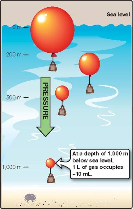
Figure 24.15
Changes in gas volume caused by water pressure at various depths below sea level (0 m).
Heliox can also be used clinically to support patients with anatomic or physiologic airway obstruction. The gas mix's decreased density allows it to slip past the obstruction site more easily than does atmospheric air and thereby helps improve patient oxygenation.
3. Decompression sickness: A diver breathing air at pressure for prolonged periods can accumulate significant amounts of N2 within their tissues. The average amount of N2 contained within the body at sea level is ∞1 L. A prolonged dive at 30 m raises this amount to as much as 4 L. N2 is taken up by diffusion across the blood–gas interface and then distributed by the circulation to all tissues, but it preferentially partitions in body fat. When a diver ascends back to the surface, N2 is no longer subject to the pressure that forced it to dissolve at depth, so it comes out of solution and forms pure N2 bubbles. The presence of bubbles in the blood stream causes decompression sickness, or “the bends.” Bubbles block blood vessels, and, as small bubbles coalesce to form large bubbles, progressively larger vessels are affected. Dependent tissues become ischemic, typically manifesting as pain in joints and in limb musculature. More severe symptoms may include neurologic deficits, dyspnea, and death. Slowing the rate of ascent gives the ∞3 L of excess gas dissolved in the aqueous phase more time to diffuse out of the tissues and into the circulation for transport to the lungs for exhalation (Figure 24.16). Fat is relatively avascular, however. This increases the distance over which N2 must diffuse before it can be carried away by the circulation, thereby slowing that rate at which it can be removed. Complete renormalization of tissue N2levels may take several hours after ascent.

Figure 24.16
Deep sea divers carefully time their ascent to avoid “the bends.”
Chapter Summary
• A cyclical pattern of inspiration and expiration is established and controlled by a central pattern generator (CPG). The CPG resides in a brainstem respiratory control center (medulla).
• The medulla contains two groups of cells involved in respiratory control. The dorsal respiratory group drives inspiration during quiet breathing. The ventral respiratory group coordinates accessory muscles and is believed to house the central pattern generator.
• Higher control centers are able to override unconscious breathing to allow for talking, coughing, and other voluntary acts that use the same musculature.
• Central and peripheral chemoreceptors relay information about the chemical composition of blood to the control centers. Central chemoreceptors monitor PaCO2. Peripheral chemosensors are sensitive to PaO2, PaCO2, and arterial pH.
• When PaCO2 rises, both the peripheral and central chemoreceptors are excited and cause the control center to respond with an immediate increase in ventilation. Decreases in PaO2 are a less effective stimulus for ventilation unless CO2 rises simultaneously.
• Other sensors feeding information to the medulla include irritant receptors, juxtapulmonary capillary receptors that are sensitive to lung injury, stretch receptors, and joint and muscle receptors.
• Atmospheric pressure decreases with altitude above sea level. PO2 also decreases, causing hypoxia. The respiratory and cardiovascular control centers help compensate in the short term by increasing ventilation and perfusion. Full acclimation to altitude requires months and involves an increase in lung perfusion, hematocrit, and capillary density in all tissues.
• Descent to depth in water increases the partial pressures of inspired gases. N2 and O2, which are poorly soluble at sea level, become toxic when forced to dissolve in tissues at depth. N2 preferentially partitions in fat and requires many hours to extract upon returning to the surface. Premature ascent results in the formation of pure N2 gas bubbles within the vasculature and causes severe pain known as “the bends. ”
Study Questions
Choose the ONE best answer.
V.1 A 55-year-old male with a history of interstitial pulmonary fibrosis undergoes pulmonary function testing. What parameter would most likely be decreased in this restrictive lung disease patient?
A. FVC (forced vital capacity)
B. Peak expiratory flow rate
C. FEV1 (forced expiratory volume in 1 second)
D. FEV1/FVC
E. Fraction of expired O2
Best answer = A. Restrictive lung disease is associated with lung stiffening, which limits lung expansion (22·VII·B). This manifests as a decrease in forced vital capacity (FVC) in pulmonary function tests. In practice, such patients can voluntarily inhale and exhale less air volume than a healthy person of comparable age, sex, and height. Peak expiratory flow rate and forced expiratory volume in 1 second (FEV1) may or may not be normal. The FEV1/FVC ratio is increased because FVC is typically reduced significantly. The fraction of expired O2 would not be changed by restrictive lung disease.
V.2 α-Adrenergic receptor agonists cause which of the following effects on pulmonary function?
A. Decreased forced vital capacity
B. Decreased total lung capacity
C. Increased diffusing capacity
D. Bronchiolar constriction
E. Bronchiolar dilation
Best answer = E. α-adrenergic receptor agonists relax airway smooth muscle, promoting bronchiolar dilation (22·VIII·C). Relaxation occurs due to inhibition of acetylcholine release from parasympathetic nerve terminals. The increased airway luminal diameter improves flow, as measured by the forced expiratory volume in 1 second (FEV1). Total lung capacity, forced vital capacity (maximal air volume that can be forcibly expired), and diffusing capacity (a measure of blood–gas barrier exchange capacity) are not affected by α-adrenergic drugs.
V.3 A 16-year-old boy presents with shortness of breath after his family adopts a new pet. His pulmonologist suspects an underlying allergy-induced asthma and orders a pulmonary function test. Which of following is most likely to have decreased in this boy?
A. Tidal volume
B. Expiratory reserve volume
C. Forced vital capacity
D. Inspiratory capacity
E. FEV1 (forced expiratory volume in 1 second)
Best answer = E. Allergy-induced asthma is associated with airway narrowing and obstruction (22·VII·A), which impairs the volume of air than can be forcibly expired per unit time. In most cases, forced vital capacity would be unaffected because this parameter is not time dependent. Tidal volume is similarly unaffected. Static lung volumes and capacities (expiratory reserve volume and inspiratory capacity) do not change appreciably with airflow obstruction, although residual volume may be increased by obstructive physiology when air-trapping occurs.
V.4 Pulmonary vascular resistance should be assessed when the effects of lung volume on pulmonary perfusion are minimal. When is this most likely to occur?
A. At high intrapleural pressures
B. At high alveolar pressures
C. At residual volume
D. At functional residual capacity
E. At total lung capacity
Best answer = D. Pulmonary blood vessels are thin walled, which makes then susceptible to extravascular compression (23·III). Pulmonary vascular resistance (PVR) is highest and perfusion is lowest when lung volumes are very high or very low. At total lung capacity and when alveolar pressures are high, PVR is high because the capillaries are stretched and compressed between adjacent alveoli. At residual volume and when intrapleural pressures high, arterial supply vessels are collapsed by external pressure. The nadir in the PVR-to-lung-volume curve occurs at functional residual capacity, because the combined effect of capillary and supply vessel compression is minimal.
V.5 A 58-year-old woman presents with a right-to-left shunt caused by a pulmonary arteriovenous malformation. Which of the following variables would you predict to be increased in this individual?
A. Arterial dissolved O2 content
B. Alveolar–arterial O2difference
C. Venous PO2
D. Arterial PO2
E. Oxyhemoglobin levels
Best answer = B. Right-to-left shunts allow blood to pass from the right to the left heart without being oxygenated (23·III·F). The shunted blood lowers the PO2 of arterial blood, thereby widening the alveolar–arterial O2difference. The amount of O2 that blood carries in dissolved form is minimal normally but would be decreased further by a shunt. A right-to-left shunt would decrease venous PO2and oxyhemoglobin levels.
V.6 A hypoxemic 50-year-old man with an increased alveolar–arterial O2 gradient is given 100% O2 via a face-mask, causing arterial PO2 to increase to >500 mm Hg. Results of a lung-diffusing capacity test were normal. What is the likely cause of the hypoxemia?
A. Diffusion limitation
B. Right-to-left shunt
C. Ventilation/perfusion mismatch
D. Hypobaric ambient conditions
E. Alveolar hypoventilation
Best answer = C. A hypoxemia with an enlarged alveolar–arterial O2 gradient (A–aDO2) could be due to either a ventilation/perfusion mismatch or a diffusion limitation (23·IV·D). However, the lung-diffusing capacity test eliminates a diffusion limitation. Right-to-left shunts also cause an increased A–aDO2, but 100% O2 does not increase arterial PO2 to the levels observed here. Hypobaric conditions and hypoventilation can result in hypoxemias, but they do not change A–aDO2.
V.7 A 75-year-old man with a history of interstitial pulmonary fibrosis presents complaining of increased dyspnea on exertion. A carbon monoxide (CO) uptake test is ordered. Which of the following best describes pulmonary CO uptake during this test?
A. Perfusion is limited.
B. Diffusion is limited.
C. Ventilation is limited.
D. Solubility is limited.
E. Binding is limited.
Best answer = B. Net carbon monoxide (CO) uptake by the lungs is limited by the rate at which it diffuses across the blood–gas barrier (23·V·A). It is relatively insensitive to changes in pulmonary perfusion (unlike uptake of a perfusion-limited gas such as N2O), which is why the test is used to assess lung-diffusing capacity. Uptake is not limited by ventilation under physiologic conditions. CO uptake binds to hemoglobin (Hb) with high affinity and, thus, uptake is not binding limited. The avidity with which Hb binds CO means that blood rarely carries appreciable amounts of gas in dissolved form.
V.8 A 25-year-old woman with normal lung function presents with anemia following childbirth (hemoglobin = 8.6 g/dL). Which of the following parameters is most likely to be reduced?
A. Arterial PO2
B. Arterial O2 saturation
C. Arterial O2content
D. Right ventricular output
E. Minute ventilation
Best answer = C. Anemia, as defined by blood hemoglobin (Hb) content (normal female Hb = 12–16 g/dL) reduces the total amount of O2 that can be carried by blood (23·VI·A). Arterial PO2 is a measure of dissolved O2concentration and is not significantly affected by Hb concentration. Arterial O2 saturation is a measure of Hb's O2 binding state, which is largely independent of blood Hb concentration under physiologic conditions. A decrease in arterial O2 content would stimulate compensatory increases in right ventricular output and minute ventilation.
V.9 A cerebrovascular accident that affects forced expirations during rest and exercise most likely damaged which neural area?
A. Apneustic center
B. Pneumotaxic center
C. Phrenic nerve center
D. Dorsal respiratory group
E. Ventral respiratory group
Best answer = E. Ventral respiratory group neurons are involved in forced expiration and coordination of labored inspiration and expiration (24·II·A). The pontine centers limit lung expansion (apneustic center) and cause rapid shallow breathing (pneumotaxic center), although the role of either center during normal breathing is uncertain. Dorsal respiratory group neurons regulate inspiration and implement the resting respiratory rhythm. The phrenic nerve contains motor neurons that control the diaphragm, which is a principal inspiratory muscle (22·V·C).
V.10 Carotid body glomus cells respond to low arterial PO2 with Ca2+ influx, causing release of neurotransmitters that stimulate sensory nerve afferents. An increase in which of the following most likely triggers Ca2+ influx in glomus cells?
A. Na+ conductance
B. Na+ equilibrium potential
C. K+ conductance
D. Membrane depolarization
E. Brain interstitial H+
Best answer = D. Arterial PO2 is sensed by an O2-dependent K+ conductance in glomus cells (24·III·B). A fall in arterial PO2 allows the K+ channel to close, decreasing K+ efflux and causing membrane depolarization. Depolarization activates voltage-gated Ca2+ channels and Ca2+ influx. Na+ conductances and changes in the Na+ equilibrium potential do not have a role in this response. Changes in brain interstitial H+ initiate ventilatory responses mediated by central chemoreceptors (24·III·A), not peripheral chemoreceptors.
V.11 A healthy 23-year-old woman reports coughing paroxysms when air temperature is below freezing. Which sensory receptors are most likely to have triggered this response?
A. Central chemoreceptors
B. Peripheral chemoreceptors
C. Irritant receptors
D. Pulmonary stretch receptors
E. Juxtapulmonary capillary receptors
Best answer = C. Irritant receptors protect the lung from noxious stimuli, such as dust, chemicals, and cold air (24·IV·A). These receptors may trigger coughing, bronchoconstriction, and mucus production when stimulated. Central and peripheral chemoreceptors respond to changes in arterial blood gas composition (24·III). Stretch receptors activate during lung inflation (24·IV·B), whereas juxtapulmonary capillary receptors respond to capillary engorgement and interstitial edema (24·IV·C).
V.12 A 29-year-old male living at sea level experiences headache and nausea after traveling to a ski resort (base = 2,500 m). Within a day, his symptoms improve, and he feels well enough to ski. Which of the following accounts for his physiologic accommodation?
A. Central chemoreceptor adaptation
B. Pulmonary stretch receptor stimulation
C. Red blood cell synthesis
D. Hemoglobin isoform alteration
E. Angiogenesis
Best answer = A. The initial response to hypobaric hypoxia at high altitudes is hyperventilation, which causes arterial PCO2 to fall, suppressing the normal drive to breathe (24·V·A). The hypoxemia that results causes the symptoms associated with acute mountain sickness. Central chemoreceptors adapt slowly to lowered arterial PCO2 over 8–24 hrs, allowing ventilation rate to rise and the symptoms improve. Red blood cell production and angiogenesis require weeks to months to compensate for the effects of hypoxia. There is no evidence that altitude causes a change in hemoglobin isoform or pulmonary stretch responses.