
31
I. OVERVIEW
In the upper portion of the gastrointestinal (GI) tube, food was liquefied and reduced in size but was not absorbed. The small intestine is where nutrients start to be removed in earnest. To facilitate this absorption, complex carbohydrates, lipids, and proteins are chemically digested into simpler forms for transport. Absorption requires transport across both the apical and basolateral membranes of enterocytes (absorptive cells of the intestinal epithelium). Not only macronutrients (carbohydrates, lipids, proteins, and water) are absorbed, but also ions, vitamins, and minerals. Once the necessary items are extracted, the remainder needs to be eliminated from the body. The distal colon, rectum, and anus participate in bowel movements, which involve both voluntary and involuntary components, to eliminate the remnants (Figure 31.1).
II. SMALL INTESTINE
The small intestine is the longest section of the GI tract, at about 6 m. It is divided into three functional segments: duodenum (first ~0.3 m), jejunum (next ~2.3 m), and ileum (final ~3.4 m). The majority of macronutrient, vitamin, and mineral absorption occurs in the small intestine. Absorption of the nutrients liberated by the digestive process is facilitated by the increase in epithelial surface area by villi (10-fold) and microvilli (20-fold) (Figure 31.2).
A. Motility and mixing
Intestinal motility not only propels chyme along the intestines via peristalsis but also allows for mixing of enzymes and other secretions from the pancreas and gallbladder (control and regulation of these secretions is discussed in Chapter 32). Segmentation is the back-and-forth mixing movement in the small intestine between adjacent segments. As in the stomach, smooth muscle contractions are initiated via slow waves. Slow waves in the small intestine are more frequent (~12 waves/min) than in the stomach, with the parasympathetic nervous system increasing this rate, and the sympathetic nervous system decreasing it. To aid in clearing residual contents during the fasting state, there are additional contractions regulated by motilin and known as migrating motor complexes (MMCs), which are initiated in the stomach and continue on through the small intestine at 60–120-minute intervals. MMCs sweep the small intestine clean. Feeding disrupts these complexes in favor of peristalsis and segmentation.

Figure 31.1
Intestines.

Figure 31.2
Villi and microvilli.
B. Intestinal secretions
Intestinal secretions include both aqueous solutions and mucus. Mucus is important for the lubrication of the chyme for intestinal protection and so that peristaltic contractions can better propel the chyme. In addition, a number of endocrine cells in the intestines secrete the hormones cholecystokinin (CCK), secretin, and glucose- dependent insulinotropic peptide.
C. Carbohydrate digestion and absorption
Carbohydrates provide a substantial energy substrate for metabolism (~4 kcals/g). Carbohydrates come in many forms (e.g., starch, dietary fiber, disaccharides, and monosaccharides), but they must be broken down into monosaccharides before they can be transported across the intestinal lumen.
1. Starch: Starch is classified as linear chained (amylose) or branch chained (amylopectin). The glucose bonds that form in linear configuration are α1,4 bonds, whereas those in the branched configuration are α1,6 bonds. Pancreatic amylase breaks α1,4 bonds. The products of the amylase reaction are maltose, maltotriose, glucose oligomers, and α-limit dextrin (Figure 31.3). These products are a substantial size reduction of starch and are further digested by di- and oligosaccharidases.
2. Dietary fiber: Dietary fiber can be divided into soluble (e.g., pectin) and insoluble (e.g., cellulose) varieties (Table 31.1). Dietary fiber contains bonds that human enzymes cannot break down in the small intestine. For example, cellulose contains linear β1,4 glucose bonds, whereas both salivary and pancreatic amylases only break α1,4 glucose bonds. Because dietary fiber cannot be adequately digested, these carbohydrates cannot be absorbed and serve to increase fecal bulk. Increased fecal bulk provides some beneficial effects, such as increased intestinal motility and increased frequency of defecation.
3. Disaccharides and oligosaccharides: Disaccharides are derived from the breakdown of starch and from direct dietary sources (e.g., sucrose and lactose). Amylase activity occurs in the intestinal lumen, whereas disaccharides and oligosaccharides are broken down into monosaccharides by membrane-bound disaccharidases. The disaccharidases can be specific for a substrate, such as lactase, or work on multiple substrates, such as sucrase and isomaltase to produce monosaccharide products (Table 31.2). Being membrane bound allows for a close association between enzyme products and absorption transporters. For example, lactose breakdown into glucose and galactose is facilitated by lactase, which is located close to cotransporters (SGLT1) for the products’ absorption (Figure 31.4).
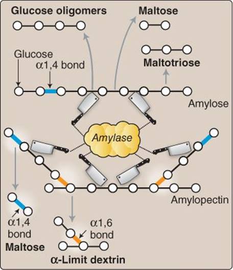
Figure 31.3
Carbohydrate digestion.


Clinical Application 31.1: Lactose Intolerance
Dietary lactose can be consumed in excess of lactase capacity in the small intestine. Excess nonabsorbed lactose is then broken down by intestinal microbes further down the gastrointestinal tract, which can then cause symptoms such as diarrhea, bloating, and cramping. Some persons are born with lower concentrations of lactase, but there is a progressive decrease in lactase expression across the lifespan, such that a person has higher concentrations early in life but lower concentrations later in life. Individuals with conditions such as inflammatory bowel disease are especially prone to lactose intolerance because of the associated small intestine inflammation. The ability to digest and absorb lactose can be measured by giving 100 g of oral lactose, followed by a blood draw every 30 min for 2 hr. Those with lactose intolerance exhibit an attenuated increase in blood glucose (<20 mg/dL) because glucose and galactose are not formed from lactase in sufficient quantities to be absorbed.

Figure 31.4
Relation between disaccharidase and apical transporters. SGLT1 = sodium-dependent glucose transporter 1.

Figure 31.5
Apical and basolateral monosaccharide transport. ATP = adenosine triphosphate; GLUT2 and 5 = members of the glucose transporter family; SGLT1 = sodium-dependent glucose transporter 1.
4. Monosaccharides: Monosaccharides, such as glucose, fructose, and galactose, are transported across the apical and basolateral membranes of small intestine enterocytes. Because monosaccharides are hydrophilic, transporters are needed to move these nutrients across these membranes.
a. Apical membrane transport: Glucose and galactose are transported across the apical membrane by SGLT1, a Na+-glucose cotransporter. The Na+-K+ ATPase provides a low Na+ environment within the enterocyte to allow for Na+ to be used as a driving force for the movement of glucose across the apical membrane. Fructose is transported by GLUT5 (glucose transporter) as shown in Figure 31.5.
b. Basolateral membrane transport: Transport of monosaccharides across the basolateral membrane from the inside of the enterocyte to the interstitium is facilitated by GLUT2 and GLUT5 transporters. GLUT2 transports both glucose and galactose, and GLUT5 transports fructose across the basolateral membrane. These nutrients can then diffuse into the portal circulation to be carried to the liver.
D. Protein digestion and absorption
Proteins also can be used for energy production (~4 kcal/g), but, in a fed state, proteins are primarily used as building blocks for reassembly into other proteins. Protein digestion begun in the stomach through the action of pepsinis then continued by several other proteases secreted in the small intestine.
1. Luminal proteases: A small amount of proteins and peptides are absorbed via phagocytosis across the apical membrane of enterocytes and specialized mucosal immune, or M, cells. However, the majority of proteins are broken down into amino acids and oligopeptides to facilitate absorption. End products of endopeptidases (trypsin, chymotrypsin, and elastase) are oligopeptides, peptides normally 6 or fewer amino acids in length. Exopeptidases ( carboxypeptidase A and B) cleave off single amino acids from oligopeptides (Figure 31.6). It is thought that these luminal actions convert about ~70% of proteins to oligopeptides and ~30% to amino acids.
2. Apical peptidases: Apical peptidases (also termed brush border peptidases) break down small peptides and oligopeptides into individual amino acids.
3. Apical dipeptide, tripeptide, and amino acid transport: Amino acids are transported across the apical membrane via different classes of amino acid cotransporters. Di- and trip-eptides are transported across the apical membrane by an H+-oligopeptide cotransporter (PepT1). Di- and tripeptides are then broken down by cytosolic peptidases into individual amino acids (Figure 31.7).
4. Basolateral amino acid transport: Individual amino acids are transported across the basolateral membrane without the need for cotransport. Many different amino acid transporters are located on the basolateral membrane and provide specificity (see Figure 31.6).
E. Lipid digestion and absorption
Fats are calorically denser (~9 kcal/g) than carbohydrates and proteins and are a substantial energy substrate for metabolism. Lipid absorption does not require the same transporter machinery because lipids are hydrophobic and can diffuse across the apical membrane. Lipids must be solubilized to ensure adequate mixing with enzymes. Lipid digestion begins in the mouth and stomach with lingual and gastric lipase, although the vast majority occurs in the small intestine. Assisting with lipid digestion, liver-derived bile salts surround and emulsify lipids so that lipase and colipase can interact with the lipid. Pancreatic lipase is the active enzyme that digests triglycerides into fatty acids and monoacylglycerols. Colipase functions to position and stabilize pancreatic lipase. Lipases do not digest phospholipids and cholesterol, requiring other pancreatic enzymes. Dietary cholesterol esters are digested into cholesterol and fatty acids by cholesterol esterase (carboxyl ester hydrolase) as shown in Figure 31.8. Phospholipase A2 breaks down phospholipids into fatty acids and lysolecithin.

Figure 31.6
Protein and peptide digestion.

Figure 31.7
Amino acid (AA) and di- and tripeptide transporters. ATP = adenosine triphosphate; NHE = Na+-H+ exchanger; PepT1 = H+-oligopeptide cotransporter.
Clinical Application 31.2: Hartnup Disease
Hartnup disease is an autosomal recessive disorder of neutral amino acid transport in gastrointestinal and renal systems. The specific transporter affected is an apical membrane Na+-amino acid cotransporter (SLC6A19 gene). These inherent disorders can lead to amino acid deficiencies, but it is possible that the other modes of protein absorption (i.e., PepT1 and phagocytosis) can partially accommodate this transport defect because some neutral amino acids can be absorbed by these routes. Deficiencies in neutral amino acids like tryptophan can lead to niacin availability issues because niacin is derived from tryptophan metabolism. This results in symptoms such as skin lesions and neurologic manifestations.
1. Free fatty acids: Fatty acid length (long, medium, or short) determines rate of absorption and assimilation. This length distinction is partly related to solubility: The longer the fatty acid, the less soluble it is in an aqueous environment.
a. Long-chain fatty acids: Long-chain fatty acids are concentrated into micelles in the small intestine lumen. Lipids often form micelles, in which hydrophilic portions face outward toward the water, and hydrophobic portions face the center. This is a stable conformation in aqueous environments and allows lipids to enter the unstirred layer surrounding the intestinal lumen in order to come in contact with the apical membranes of enterocytes. Close to the surface of the apical membrane, the micelle begins to disperse, possibly due to a pH change. Long-chain fatty acids are freed and can either diffuse directly across the apical membrane or be transported by fatty acid–binding proteins. These binding proteins speed absorption across the apical membrane. In the cytosol, long-chain fatty acids are attached to monoacylglycerols and diacylglycerols to form triglycerides within the enterocyte. Triglycerides are packaged in apoprotein vesicles called chylomicrons and, to a lesser degree, in very-low- density lipoproteins (VLDLs). Chylomicrons are then exocytosed through the basolateral membrane into the interstitial space. From the interstitial space, chylomicrons do not enter the circulation because of capillary fenestration size restrictions but, rather, move into the lymphatic system for transportation (Figure 31.9).
b. Medium-chain fatty acids: Medium-chain fatty acids (6–12 carbons long) are more soluble in water than are long-chain fatty acids. This allows them to cross the apical membrane by moving through the cytosol without the need to be repacked into a chylomicron. Medium-chain fatty acids cross the basolateral membrane into the interstitial space and then into the portal circulation. This is in contrast to long-chain fatty acids, which enter the lymphatic circulation (see Figure 31.9).
Medium-chain fatty acids can be used as dietary supplements to increase total absorbed kilocalories (energy). Due to both the solubility and transport method of these supplements, individuals with diseases such as bile duct obstruction are able to absorb these fats without the need for bile salts.

Figure 31.8
Lipid digestion.
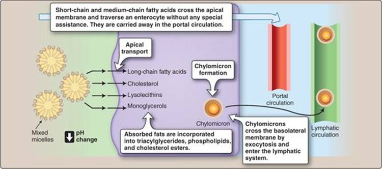
Figure 31.9
Lipid apical and basolateral transport.
c. Short-chain fatty acids: Short-chain fatty acids are less than 6 carbons in length. These fatty acids are absorbed and assimilated in a manner similar to that of medium-chain fatty acids.
2. Monoacylglycerols and glycerols: Monoacylglycerol is packaged in micelles (if there is a heterogeneous group of lipids, it is called a mixed micelle), released just prior to the enterocyte, and moves across the apical membrane through passive diffusion. In the enterocyte, monoacylglycerols are combined with long-chain fatty acids to form triglycerides and are secreted in chylomicrons (see Figure 31.9). Glycerol is absorbed directly across the enterocyte and is not repackaged. After glycerol exits the basolateral membrane of the enterocyte into the interstitial space, it can then diffuse directly into the portal circulation.
3. Cholesterols: Cholesterol esters are also packaged in micelles and released just prior to the enterocyte. Cholesterol esters appear to both diffuse through and be transported across the apical membrane. One of these transporters is NPC1L1 (Niemann-Pick C1 like 1), the pharmacologic blockade of which decreases cholesterol uptake and lowers circulating levels of cholesterol in some patients. In the enterocyte, cholesterol esters are esterified, packaged into chylomicrons, and secreted (see Figure 31.9).
4. Lysolecithins: Phospholipids are also packaged in micelles, released just prior to the enterocyte, and move across the apical membrane through passive diffusion. In the enterocyte, phospholipids are esterified into lysolecithin, packaged into chylomicrons, and secreted into the interstitial space to be picked up by the lymphatic system (see Figure 31.9).


F. Vitamin and mineral absorption
In addition to macronutrients, small amounts of vitamins and minerals must be in the diet to directly prevent disease (Tables 31.3 and 31.4).
1. Vitamins: Fat-soluble vitamins are incorporated in micelles and absorbed similar to long-chain fatty acids and packaged in chylomicrons. Watersoluble vitamins, with the exception of vitamin B12, are absorbed by Na+cotransport. Vitamin B12 is absorbed in a four-step process. First, vitamin B12 is liberated from dietary proteins. Second, vitamin B12 binds to haptocorrin released from G cells. Third, pancreatic secretions cause the release of haptocorrin, which is how intrinsic factor binds vitamin B12. I ntrinsic factor is released from parietal cells. Fourth, the intrinsic factor/vitamin B12 complex is absorbed by phagocytosis in the ileum.
2. Minerals: Monovalent ions and electrolytes will be discussed with the large intestine, later in this chapter. Divalent ions (Ca2+, Mg2+, Fe2+, Cu2+, and Zn2+) are absorbed in the small intestine. A good example of ion transport regulation can be seen with Ca2+, insofar as it can be absorbed through either a paracellular route (throughout the small intestine) or transcellular route (in the duodenum). The transcellular route involves an apical Ca2+channel, cytosolic binding by calbindin, and basolateral Ca2+ ATPase and Ca2+-Na+ exchanger (Figure 31.10). Vitamin D3 stimulates the expression of these four proteins, which allows for greater Ca2+ absorption through the transcellular route.

Figure 31.10
Calcium absorption. ATP = adenosine triphosphate.
G. Water absorption
The small intestine is the site of the majority of water absorption, nearly 80%. This fluid includes both what is eaten and drunk as well as secretions from the salivary glands, gastric, liver, pancreas, and intestinal lining. The majority of this absorption occurs via osmosis because of the apical transport of NaCl from the intestinal lumen.
III. LARGE INTESTINE
The large intestine comprises the cecum; ascending, transverse, descending, and sigmoid colon; rectum; and anus (Figure 31.11). The large intestine plays a lesser role in digestion compared to the small intestine but is intricately involved in ion and water absorption.
A. Motility
Motility is one of the prime functions of the large intestine. There are three main movement patterns in the large intestine: segmentation, peristalsis, and mass movement contractions. Besides the anatomic divisions, the large intestine can contract into smaller segments called haustra, which are seen as the beadlike appearance of the large intestine (Figure 31.12). Segmentation contractions increase the opportunity for contact between the luminal contents and intestinal epithelium, thereby allowing ion and water removal. Segmentation contractions do not propel chyme forward, but both peristalsis and mass movement contractions perform this function. Mass movement contractions occur a few times per day and involve a massive peristaltic wave that results in a significant chyme movement along the large intestine.
1. Ileocecal sphincter: The ileocecal sphincter prevents backflow from the large to small intestine (see Figure 31.11). Ileum distention and irritation (stimulation of chemical afferents) initiates ileum peristalsis and relaxes the sphincter, whereas cecum distention and irritation inhibits peristalsis and contracts the sphincter. Immediately following meal ingestion, the ileocecal sphincter relaxes and the ileum contracts. This response is known as the gastroileal reflex and is likely controlled by gastrin and CCK.
2. Other reflexes: The gastrocolic reflex is the urge to defecate shortly after food intake. It is thought both to have a neural component and be mediated by both mechanical- and chemical-sensitive neurons and functions to clear the colon and ready it for the remnants of the new meal. The orthocolic reflex is an urge to defecate after standing. This reflex is thought to be mediated by mechanosensitive neurons and the enteric nervous system via gravity-induced distention. For those on medical bed rest, this reflex should be periodically elicited to prevent constipation.
3. Anal sphincters: The anus comprises two sphincters: one internal, the other external. The internal sphincter is composed of smooth muscle. The external sphincter is composed of skeletal muscle that is under somatic control innervated by the pudendal nerve (Figure 31.13). Defecation is a multistep process involving both sphincters as well as both enteric and somatic regulation. A peristaltic wave from the large intestine forces feces from the rectum toward the anus. The internal sphincter then relaxes by inhibiting contraction of the smooth muscle within this area (termed the rectosphincter reflex). If the external sphincter is voluntarily relaxed, then defecation occurs. If the external sphincter remains contracted, then defecation is delayed, and feces is retained. Peristaltic waves may cause a sense of urgency, which may or may not lead to defecation depending on the external sphincter. Alternatively, a person may voluntarily increase thoracic and abdominal pressure by a straining maneuver (to push feces downward to start peristaltic waves, which involuntarily relax the internal sphincter) and then voluntarily relax the external sphincter in order to pass the feces.
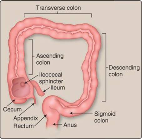
Figure 31.11
Large intestine.

Figure 31.12
Haustra.
B. Transport
The large intestine both absorbs and secretes ions. This ion transport also allows for water absorption and regulation during periods of water deprivation and dehydration. Finally, some fats are transported across the apical membrane.
1. Electrolytes: Na+ and water are absorbed via endothelial Na+ channels (ENaCs) in the distal colon (Figure 31.14). Cl− is passively absorbed paracellularly. In the proximal colon, Cl− crosses the apical membrane by Cl−-HCO3− exchangers. K+ is passively secreted in the distal colon (see Figure 31.14). Active secretion can also occur by the insertion of apical K+ channels in the large intestine with increased concentration of aldosterone or certain second messengers.
2. Short-chain fatty acids: Short-chain fatty acids are transported across the apical membrane to be used by colonic epithelial cells as an energy substrate (see Figure 31.14).

Figure 31.13
Innervation of the colon, rectum, and anus.
Clinical Application 31.3: Fecal Incontinence
Fecal incontinence is involuntary defecation. The severity can range from a partial ability to control defecation (except when there are increases in abdominal or thoracic pressure, such as during a cough or when straining to lift an object) to little or no voluntary control. The pathophysiology is often related to trauma, injury to the pelvic floor such as during childbirth or surgery, or a prolapsed rectum. Patients’ rectosphincter reflexes are typically normal, but the pathophysiology is associated with the external sphincter. Treatments for fecal incontinence are dependent on the cause and severity and include garments to collect feces, fecal bulking agents (because liquid stool is more difficult to contain), strengthening pelvic floor and sphincter muscles, and surgical procedures.

Figure 31.14
Ion and fatty acid transport. ATP = adenosine triphosphate; ENaC = endothelial sodium channel; SCFA = short-chain fatty acid; SMCT1 = short-chain fatty-acid transporter 1.
Clinical Application 31.4: Diarrhea and Metabolic Acidosis
Chronic (>4 weeks), persistent (2–4 weeks), or acute (<2 weeks) diarrhea, if severe enough, can result in excretion of large amounts of HCO3− and other ions. Diarrhea is a frequent semisolid or fluid stool that can have a number of causes (infection, toxins, etc.), but pathophysiology involves osmotic pressure being developed in the gastrointestinal lumen, which favors fluid being retained in the lumen or even dehydrating the surrounding large intestine interstitial spaces. This results in not only water loss but also a decrease in plasma HCO3−, thereby decreasing plasma pH. Because Na+ and Cl− are lost along with HCO3−, the anion gap does not appreciably change. Thus, this type of acid–base disturbance can be classified as metabolic acidosis with a normal anion gap.
3. Water: The large intestine also plays an important role in water absorption (Figure 31.15). Only 1% of fluid that is delivered (including that from both the diet and GI secretions) to the GI tract is excreted. The large intestine is responsible for ~20% of the fluid absorption. The capacity for water absorption can be increased twofold during hypohydrated states (e.g., when there is an aldosterone-mediated increase in Na+ transport that allows for more water to be osmotically absorbed).

Figure 31.15
Fluid intake, secretion, and absorption.
Chapter Summary
• Motility in the small intestine involves both mixing via segmentation and propulsion via peristalsis. Migrating motor complexes sweep the intestinal lumen free of residual particles between meals.
• Pancreatic amylase begins starch digestion by cleaving α1,4 glucose bonds, and apical membrane-bound disaccharidases convert the starch remnants to monosaccharides (glucose, galactose, and fructose) for absorption.
• Monosaccharide absorption involves cotransport of glucose and Na+ across the apical membrane, whereas fructose moves through without the aid of cotransport. Basolateral transport also does not involve cotransportation.
• Proteases secreted by the pancreas (trypsin, chymotrypsin, elastase, and carboxypeptidases) cleave amino acid bonds to form smaller peptides. These peptides are further digested by membrane-bound peptidasesforming amino acids, dipeptides, and tripeptides.
• Amino acids are transported across the apical membrane with Na+, and small peptides are transported with H+. Within the cytosol, small peptides are broken down into amino acids. Basolateral amino acid transport occurs by specific amino acid–class transporters.
• Bile acids emulsify lipids so that pancreatic lipase can cleave fatty acids from triglycerides. Dietary cholesterol esters are digested into cholesterol and fatty acids by carboxyl ester hydrolase. These products are then formed into micelles.
• Long-chain fatty acids and cholesterol both diffuse across the apical membrane. They are then reconstituted and repackaged into chylomicrons within the enterocyte. Chylomicrons are then secreted and enter the lymphatic circulation.
• The ileocecal sphincter regulates the amount of chyme entering the large intestine, and the internal and external anal sphincters regulate the feces exiting the gastrointestinal system. Motility in the large intestine consists of segmentation, peristalsis, and mass movement as well as a number of reflexes that control sphincter contraction and relaxation.
• The large intestine absorbs Na+, Cl−, and water and secretes K+ and HCO3−.