
32
I. OVERVIEW
The pancreas, gallbladder, and liver serve as accessory organs for the intestines (Figure 32.1), providing specialized secretions to digest carbohydrates, proteins, and lipids in the small intestine. Pancreatic secretions are highly regulated by neural and hormonal means both in anticipation of eating and in response to food in the gastrointestinal (GI) tract. Hepatobiliary secretions are steadily produced but then stored in the gallbladder for regulated secretion into the small intestine. Once digested and absorbed, most nutrients travel via the portal circulation to the liver to either be extracted and processed or to pass through to the systemic circulation (see Figure 21.11).
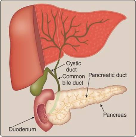
Figure 32.1
Gastrointestinal accessory organs.
II. EXOCRINE PANCREAS
The primary functions of the exocrine pancreas are to neutralize acid and deliver enzymes for macronutrient digestion within the duodenum. The acinar cells are the primary secretory cells. Small clusters of acinar cells are connected by intercalated ducts, which converge on the collecting duct (Figure 32.2). The cells lining the intercalated duct add ions and serous secretions to the enzyme and ion secretions of the acinar cells.
A. Regulation
Regulation of pancreatic secretions is dependent on the phase of digestion: cephalic, gastric, or intestinal.
1. Cephalic phase: During the cephalic phase, the vagus nerve stimulates pancreatic secretions by releasing acetylcholine (ACh) and vasoactive intestinal peptide (VIP) and is thought to account for about 25% of pancreatic secretions.
2. Gastric phase: The gastric phase accounts for about 10% of pancreatic secretions and is mediated by vagovagal reflexes stimulated by stomach distension.
3. Intestinal phase: The intestinal phase accounts for the majority of pancreatic secretions (~65%) and is controlled hormonally control via secretin and cholecystokinin (CCK) Secretin is released in response to H+, and CCK is released in response to amino acids, fatty acids, and monoacylglycerols. The primary inhibitors of pancreatic secretions are somatostatin and a decrease in chyme macronutrients.
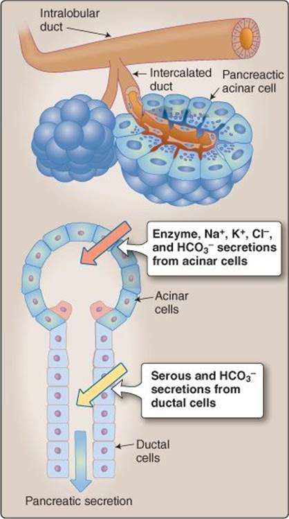
Figure 32.2
Pancreatic acinar and intercalated cells.
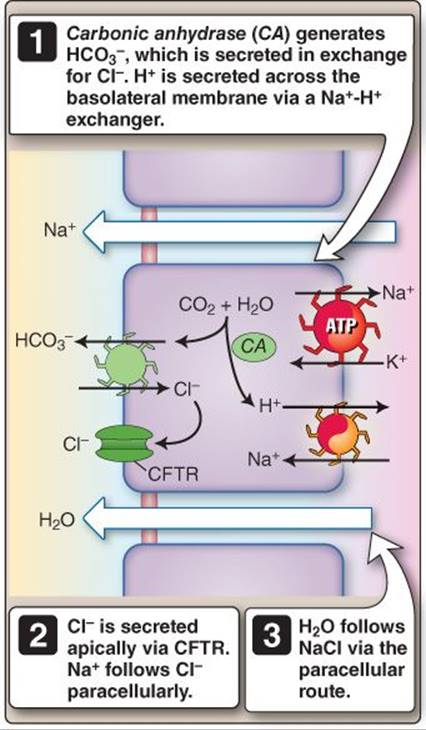
Figure 32.3
Intercalated ion secretion. ATP = adenosine triphosphate; CFTR = cystic fibrosis transmembrane conductance regulator.
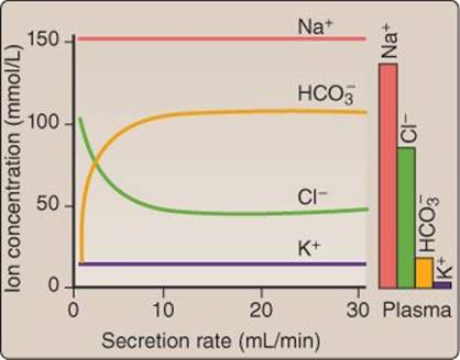
Figure 32.4
Effect of pancreatic secretion rate.
B. Enzyme secretion mechanisms
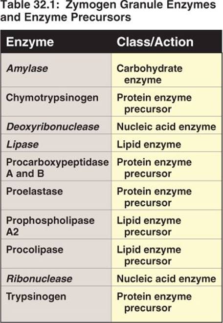
CCK is released from I cells in the small intestine. CCK and, to a lesser extent, VIP and gastric-releasing peptide, are the primary signals responsible for pancreatic enzyme secretion from acinar cells. Acinar cells contain zymogen granules that house some active but mostly inactive digestive enzymes (Table 32.1). When stimulated, acinar cells exocytose zymogen granules into the luminal space. Enzyme packaging occurs in the Golgi apparatus, and large vacuoles are condensed into zymogen granules prior to docking and fusing with the apical membrane. Exocytosis is regulated hormonally and neurally.
1. Classical hormonal signaling: CCK is released into the interstitial space and enters the bloodstream. It then travels in the circulation to pancreatic acinar cells where it binds to CCKA receptors.
2. Vagal afferent stimulation: CCK also binds to CCKA receptors on vagal afferents. This binding stimulates the afferents, eliciting the efferent stimulation of pancreatic acinar cells via VIP.
Clinical Application 32.1: Cystic Fibrosis
Cystic fibrosis leads to pancreatic insufficiency because of a gene mutation encoding for the cystic fibrosis transmembrane conductance regulator. By not having a functional version of this epithelial transporter, secretions are thickened, which can eventually partially block ducts and lead to pancreatic tissue damage. This inhibits the release of pancreatic enzymes, leading to malabsorption of proteins, fats, and fat-soluble vitamins.
C. Ion secretion mechanisms
Ion and serous fluid secretion occurs in both acinar and intercalated duct cells.
1. Acinar cells: Basolateral CCK and ACh binding stimulates Cl− transport across the apical membrane, which facilitates paracellular Na+ and water movement. Secretin release from S cells is stimulated in response to duodenal acidification.
2. Intercalated duct cells: Basolateral secretin and ACh binding in intercalated duct cells activates cystic fibrosis transmembrane conductance regulators (CFTRs), other Cl− channels, and Cl−-HCO3−cotransporters. These transporters recycle Cl− and secrete HCO3− (Figure 32.3).
3. Secretion rate: The secretion flow rate alters ionic concentration. As flow rates increase, HCO3− concentration increases and Cl− concentration decreases. Na+ and K+ are also secreted (at concentrations similar to plasma for Na+ and slightly above for K+) but are not affected by alterations in secretion flow rate (Figure 32.4).
III. HEPATOBILIARY SYSTEM
The liver produces and secretes bile (termed hepatic bile to distinguish it from bile from the gallbladder). Bile is secreted by hepatocytes into canaliculi, then traverses a series of bile ducts, which become less numerous but progressively larger in diameter until they form the common hepatic duct. The flow from hepatocytes is in the opposite direction (peripherally) of the blood from the hepatic artery and portal vein, which flow centrally (Figure 32.5). From this junction, bile can move through either the common bile duct into the duodenum or the cystic duct to the gallbladder. The sphincter of Oddi controls the path. When the sphincter is contracted, the common bile duct has high resistance to bile flow, and, thus, bile travels to the gallbladder. When the sphincter is relaxed, bile flows from the common hepatic duct and often from the gallbladder into the duodenum (Figure 32.6). Sphincter relaxation is regulated primarily by CCK.
A. Bile components
Bile components include bile acids, electrolytes, cholesterol, phospholipids, and bilirubin. Gallbladder bile is significantly more concentrated than hepatic bile, with the exception of osmotic ions, such as Na+and Cl− (Table 32.2).
1. Bile acids: Bile acids emulsify lipids to aid in their digestion by pancreatic lipase in association with colipase. Without bile acids, lipid digestion occurs slowly and is often incomplete because of a dramatic decrease in the surface area available for enzymes. Bile acids are formed from cholesterol, and there are two general forms of bile acids: primary and secondary.
a. Primary bile acids: Cholic and chenodeoxycholic acids are synthesized in hepatocytes via 7α- hydroxylase. Thus, the formation of these two bile acids is a principal route of cholesterol metabolism.
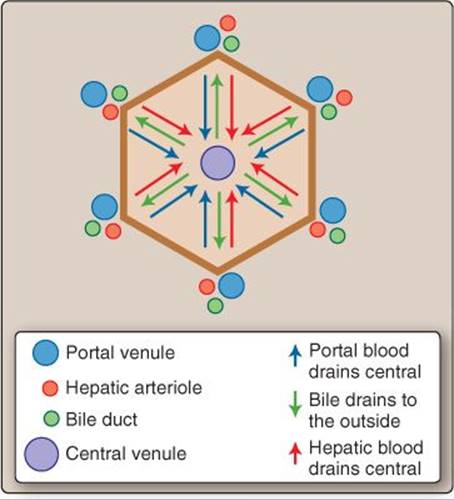
Figure 32.5
Hepatocytes and blood and bile flow.
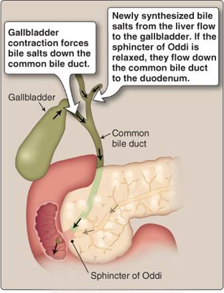
Figure 32.6
Bile storage and secretion.
b. Secondary bile acids: Deoxycholic and lithocholic acids are not synthesized in hepatocytes. Instead, bacteria in the large intestine and terminal ileum contain 7α-dehydroxylase, which converts cholic acid to deoxycholic acid and chenodeoxycholic acid to lithocholic acid. These bile acids are then passively reabsorbed and transported back to the liver via the enterohepatic circulation. These bile acids can be conjugated or unconjugated, where conjugation simply refers to a salt attachment. Bile acid type affects the specific intestinal or hepatocyte membrane transporter utilized.
2. Water and electrolytes: Ions including Na+, K+, Ca2+, Cl−, and HCO3− are secreted from hepatocytes isotonically. Some additional water and HCO3− are secreted by duct cells. Bile concentration is completed in the gallbladder, and this concentration can be quite dramatic (up to tenfold). This occurs via Na+ and Cl− reabsorption, which causes isosmotic water reabsorption that occurs paracellularly and cellularly via aquaporins (AQPs) 1 and 8. In the process of Cl− reabsorption, HCO3− is secreted (Figure 32.7).
3. Cholesterol and phospholipids: Besides the cholesterol converted to primary bile acids, small amounts of cholesterol are secreted in the bile. Phospholipids, primarily lecithin, are also secreted and help solubilize some of the bile constituents.
4. Pigments and organic molecules: The major pigment in bile is bilirubin. Bilirubin is formed from the catabolism of hemoglobin and is transported in the circulation in a complex with albumin. Hepatocytes secrete this bile pigment, which is either ultimately excreted directly in the intestines or temporarily reabsorbed and then excreted in the urine. Organic ions are also bile components, which serve as a method for the liver to excrete toxins, drugs, and related compounds.
B. Gallbladder
The liver constantly produces bile but not in sufficient quantity to properly emulsify lipids in the small intestine. The gallbladder serves as the bile storage and distribution center. Therefore, when needed, a large quantity can be released. The stored bile in the gallbladder is concentrated. (Think of it as concentrated dishwashing detergent: A little bit can go a long way.) The gallbladder can contract to propel out the bile with CCK stimulation. CCK is the same substance that causes the sphincter of Oddi to relax. This combined effect allows for sufficient quantities of bile acids to be secreted (Figure 32.8). Vagal stimulation also can cause weak gallbladder contraction. Both somatostatin and norepinephrine inhibit bile acid secretion.
IV. NONBILIARY LIVER FUNCTIONS
There are a number of integrative physiology processes of the liver related to metabolism, detoxification, and immune system function. One of the main functions is to provide energy substrates to other cells in the body, especially in times when food is scarce.
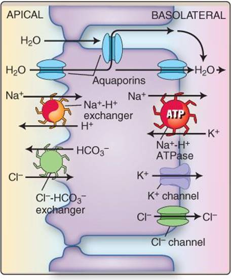
Figure 32.7
Gallbladder concentrations. ATP = adenosine triphosphate.
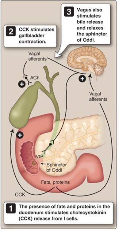
Figure 32.8
Neural and endocrine control of bile secretion. ACh = acetylcholine; VIP = vasoactive intestinal peptide.
A. Metabolism
The liver participates in carbohydrate, fat, and protein metabolism. The liver can also store and subsequently release large quantities of carbohydrates in the form of glycogen and certain vitamins and minerals.
1. Carbohydrate: The liver plays a major role in the storage and subsequent breakdown of glycogen. The average liver can store ~100 g of glycogen. Glycogen breakdown is called glycogenolysis, which liberates glucose for released into the systemic circulation. Besides releasing glucose, the liver can convert fructose and galactose into glucose as well as convert amino acids and triglycerides into glucose through a process known as gluconeogenesis.
2. Lipid: The liver contains the enzymes to undergo large amounts of lipid metabolism. Here the liver can mobilize fatty acids, through a process called lipolysis, to be released into the systemic circulation. The liver also produces lipoproteins, phospholipids, ketone bodies, and cholesterol and has the ability to convert amino acids and carbohydrates into new lipids.
3. Protein: The liver is involved in protein synthesis and amino acid uptake and metabolism. Proteins synthesised include plasma proteins, prohormones, clotting factors, apoproteins, and transport-binding proteins. The liver also has the capacity to deaminate amino acids.
4. Vitamins and minerals: Many vitamins and minerals are delivered to the liver by the portal circulation. The liver has the capacity to store lipid-soluble vitamins such as vitamins A, D, E, and K. This storage of fat-soluble vitamins allows for a short-term reserve for when dietary sources are not available. The liver also stores certain minerals, such as iron and copper.
B. Detoxification
The liver participates in a number of detoxification and removal reactions. Two of the most important of these processes are removal of ammonia and ethanol, and it also mediates various biotransformations.
1. Ammonia: The intestines (primarily the large intestine) are responsible for about ~50% of the ammonia produced. The liver receives the majority of this ammonia via the portal circulation. The liver removes most of the circulating ammonia through a series of reactions, which comprise the urea cycle. Urea is released into the systemic circulation, where the majority can be excreted by the kidney.
2. Ethanol: The liver contains alcohol dehydrogenase, which facilitates the conversion of ethanol into acetaldehyde and reduced nicotinamide adenine dinucleotide. These two products can then be converted into acetyl coenzyme A by peripheral tissues such as skeletal muscles.

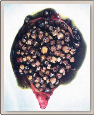
Clinical Application 32.2: Cholelithiasis
Cholelithiasis is the presence of gallstones. The stones can be of two primary types: calcium bilirubinate stones and cholesterol stones. Cholesterol stones are more common, and a number of processes contribute to the pathophysiology of stone formation involving genetic factors, bile stasis, and supersaturation of bile with cholesterol. Gallstones can lead to bile duct obstruction, thereby limiting the amount of bile secreted into the small intestine, which can lead to fat malabsorption.
3. Drug biotransformations: Biotransformation reactions involve two phases. These phases can be described as phase I, or oxidation, and phase II, or conjugation and elimination.
a. Phase I reactions: Phase I reactions utilize cytochrome P450 enzymes to oxidize organic molecules. These reactions metabolize the majority of drug classes, and there are only a few phase I cytochrome P450–independent reactions of amine-containing compounds. Phase I reactions can also be used to activate some drugs.
b. Phase II reactions: Phase II reactions conjugate the products to aid in solubility for release into the systemic circulation to be filtered and excreted in the kidney or to be secreted into the small intestine with bile for ultimate excretion.
The liver is involved in the first pass metabolism of oral pharmaceuticals via biotransformation reactions. Certain drugs are almost entirely metabolized in this first pass through the liver via the portal circulation. This is the reason why some drugs must be dosed and delivered in a topical, inhaled, or injectable form.
C. Immune functions
The portal circulation delivers nutrients from the intestines, but often bacteria are also among the sampling of portal blood. However, in the healthy person, there are no intestinal bacteria in the systemic circulation. Kupffer cellsare specialized phagocytic macrophages located in the liver, which engulf and digest these intestinal bacteria (Figure 32.9). The liver is also the major site of both lymph production and immunoglobulin A release.
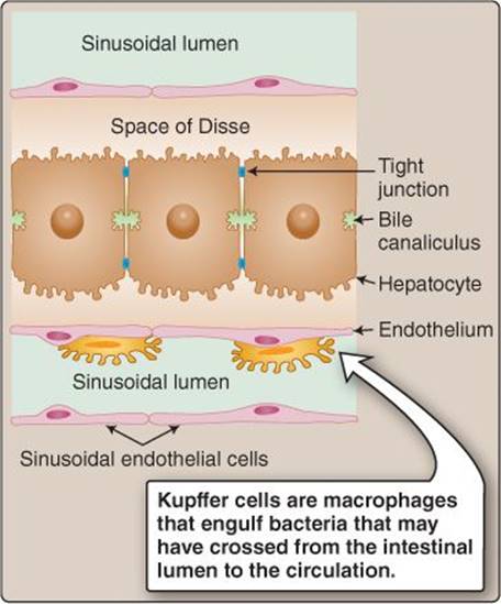
Figure 32.9
Kupffer cells.
Chapter Summary
• Exocrine pancreas regulation occurs via the stimulatory effects of secretin and cholecystokinin as well as the inhibitory effects of somatostatin. The exocrine pancreas secretes enzymes, ions, and serous solutions. Enzymes are secreted in their inactive forms to be activated in the small intestine, and HCO3− is secreted to aid in the neutralization of stomach acid.
• Bile components are bile acids (both primary and secondary), electrolytes, cholesterol, phospholipids, and bilirubin. The gallbladder is the primary bile storage site. Bile in the gallbladder is concentrated compared to the liver. Cholecystokinin causes gallbladder contractions to move bile toward the small intestine.
• The sphincter of Oddi is the narrowing that regulates bile release into the small intestine. Cholecystokinin causes the sphincter of Oddi to relax, thereby permitting bile to enter the small intestine.
• The liver participates in carbohydrate, lipid, and protein metabolism. The liver can either store or release these substrates depending on the fed versus fasted state. The liver also stores lipid-soluble vitamins and certain minerals.
• The liver participates in both the detoxification and removal of drugs, hormones, and ammonia. In addition, the liver produces large amounts of lymph and is involved in many immune-related functions.
Study Questions
Choose the ONE best answer.
VII.1 A 35-year-old woman with a recent breast cancer diagnosis reports gastrointestinal (GI) distress following chemotherapy treatments targeting fast-replicating cells. Which GI layer or signaling molecule is most likely affected by the treatment?
A. Longitudinal muscle
B. Submucosal plexus
C. Epithelium
D. Motilin
E. Vasoactive intestinal peptide
Best answer = C. Gastrointestinal (GI) epithelium has a very high turnover rate (29·II·A) and, thus, is most affected by chemotherapy. Longitudinal muscle and the submucosal plexus are important GI layers for gastric emptying and intestinal motility but have slower turnover rates. Motilin derived from M cells stimulates gastric and intestinal motility (29·III·C), and vasoactive intestinal peptide from parasympathetic nerves relaxes GI smooth muscle (29·IV·A), but such hormones and neurotransmitters do not originate from fast-replicating epithelial cells.
VII.2 Which of the following gastrointestinal signaling substances is released by sympathetic nerve terminals and decreases intestinal secretions?
A. Substance P
B. Vasoactive intestinal peptide
C. Gastrin-releasing peptide
D. Neuropeptide Y
E. Histamine
Best answer = D. Neuropeptide Y relaxes wall muscle and decreases intestinal secretions (29·III·B). Vasoactive intestinal peptide increases pancreatic and intestinal secretion but is released by parasympathetic and enteric neurons (29·III·A). Substance P increases secretions, partially in the salivary glands. Gastrin-releasing peptide increases gastrin secretion. Histamine increases gastric secretions and is released by enterochromaffin-like cells (29·IV·B).
VII.3 A 40-year-old man with uncontrolled Crohn disease undergoes an ileal resection to remove damaged tissue. Synthesis and release of which gastrointestinal hormone would be the most affected by this surgery?
A. Gastrin
B. Motilin
C. Glucose-dependent insulinotropic peptide
D. Prostaglandins
E. Cholecystokinin
Best answer = E. Cholecystokinin (CCK) is secreted by I cells throughout the small intestine, including the ileum (29·IV·A). CCK acts on the stomach, pancreas, and gall-bladder to promote secretion and gastric emptying. Motilin and glucose-dependent insulinotropic peptide are secreted by M and K cells, respectively, in the duodenum and jejunum, but not the ileum. Gastrin is secreted both in the stomach and the small intestine. Prostaglandins are not considered hormones but rather are classified as gastrointestinal paracrines.
VII.4 A 52-year-old woman taking scopolamine (a cholinergic antagonist) for motion sickness during airplane travel also develops symptoms consistent with xerostomia as a side effect. Which of the following changes is most consistent with xerostomia?
A. Increased parotid cell inositol trisphosphate
B. Stimulated parotid cell adenylyl cyclase C. Increased mucus production
D. Decreased salivary Cl− concentration
E. Decreased salivary K+ concentration
Best answer = D. Salivary secretion is controlled primarily by the parasympathetic nervous system (30·II·C). When active, acetylcholine release increases salivary secretion via the inositol trisphosphate (IP3) signaling pathway. Blocking cholinergic signaling reduces IP3 levels and decreases salivary flow. Salivary ionic composition is dependent on flow rates. When flow rate decreases, Cl− content decreases, whereas K+ concentration increases. Scopolamine does not stimulate adrenergic receptors, and, therefore, no change in adenylyl cyclase would be expected. Anticholinergics decrease mucus production by salivary glands.
VII.5 Which of the following best describes the gastrointestinal pacemaker cells known as interstitial cells of Cajal?
A. They generate 15–20 cycles/min.
B. They require voltage-gated Na+ channels.
C. They require voltage-gated Ca2+ channels.
D. Depolarization is initiated in the antrum.
E. Depolarization is initiated in the fundus.
Best answer = C. Interstitial cells of Cajal (ICC) are pacemakers located in the body of the stomach, not the antrum or fundus (30·IV·B). They generate waves of depolarization (slow waves) at a frequency of ~3–5 cycles/min. The slow waves generate action potentials and initiate waves of contraction that are mediated by voltage-gated Ca2+ channels rather than voltage-gated Na+ channels. The contractile waves are responsible for mixing and grinding stomach contents to help break down food prior to its delivery to the small intestine.
VII.6 If gastric D-cell function were impaired by immune or inflammatory mediators, acid secretion would increase through which of the following mechanisms?
A. Reduced potentiation
B. Increased acetylcholine release
C. Increased prostaglandin E2 synthesis
D. Decreased G cell secretion
E. Loss of parietal cell inhibition
Best answer = E. Gastric D cells secrete somatostatin, which normally inhibits H+ secretion from parietal cells (30·IV·C). Reducing somatostatin levels would set up the potential for increased H+ secretion. Prostaglandins also decrease H+ secretion normally, but through pathways that do not require D cells. G cells secrete gastrin, which stimulates H+ secretion from parietal cells. Acetylcholine (ACh) also increases H+ secretion by a number of direct and indirect routes. Potentiation refers to the observation that H+ secretion increases to a greater extent when two stimulatory factors bind simultaneously (e.g., gastrin plus ACh) than might be expected from the sum of their individual actions.
VII.7 A 35-year-old woman complains of heartburn and stomach pains, which frequently wake her at night. She is subsequently discovered to have a peptic ulcer. Which of the following best explains how the duodenum normally protects itself against ulcer formation?
A. It has a thick layer of viscous mucus.
B. It has a thick apical membrane.
C. S cells release secretin.
D. Enterochromaffin-like cells release histamine.
E. Peptidases are released in inactive form.
Best answer = C. Peptic ulcers occur in the stomach and duodenum (Clinical Application 30.3). They are often precipitated by Helicobacter pylori, but intestinal wall erosion is due to acid and enzymes. The duodenum's main defense against acid is secretin released from S cells when stimulated by acid. Secretin triggers HCO3− release from the pancreas (32·II·A). Unlike the stomach, the duodenum does not have a thick protective mucus layer, which makes it vulnerable to acid. It does not possess a thick apical membrane, which would impair nutrient absorption. Pancreatic peptidases are released in inactive form but activate immediately in the intestinal lumen. Histamine is a local gastric parietal cell control factor.
VII.8 Na+ is required for absorption of which of the following substances by the small intestinal epithelium?
A. Apical fructose uptake
B. Basolateral glucose transport
C. Apical dipeptide uptake
D. Basolateral amino acid transport
E. Apical glycerol uptake
Best answer = C. Apical absorption of dipeptides occurs via PEPT1, which is a cotransporter powered by an inward Na+ gradient (31·II·D). Apical fructose transport occurs via GLUT5, and basolateral glucose transport occurs via a GLUT2 transporter (31·II·C). GLUT family transporters facilitate diffusional uptake of substrates down their own concentration gradients and independently of Na+. Basolateral amino acid transport also occurs via either individual or group transporters independently of ion gradients. Apical glycerol uptake does not require the assistance of any ion or specialized transport protein. Glycerol uptake occurs by diffusion across the epithelial cell membrane.
VII.9 A 65-year-old woman on a strict 1,500-kcal meal plan presents with visceral pain and excessive fullness. A blood sample identifies elevated serum bilirubin, and an ultrasound of the right upper quadrant reveals gallstones obstructing the common bile duct. This obstruction would most affect the digestion and absorption of which of the following meal plans?
A. 55% carbohydrate, 15% protein, 30% fat
B. 20% carbohydrate, 30% protein, 50% fat
C. 70% carbohydrate, 10% protein, 20% fat
D. 40% carbohydrate, 40% protein, 20% fat
E. 50% carbohydrate, 20% protein, 30% fat
Best answer = B. The meal plan consisting of 20% carbohydrate, 30% protein, 50% fat contains the highest fat content and, therefore, would be the most difficult for this individual to digest and absorb. Fats require the emulsification properties of bile acids (31·II·E). Without this emulsification, lipid digestion is compromised, and steatorrhea (fat in the feces), pain, and bloating can result. The protein and carbohydrate portions of the meals will not be directly influenced by a reduction in bile acids.
VII.10 A 28-year-old woman recently gave birth to her second child by cesarean delivery. She is now experiencing both urinary and fecal incontinence during straining maneuvers. A pudendal nerve conduction test indicates that the pudendal nerve is the cause of the fecal incontinence. Which sphincter is most likely affected?
A. Pyloric
B. Ileocecal
C. Rectosigmoid
D. Internal anal
E. External anal
Best answer = E. The pudendal nerve innervates the external anal sphincter, which is a skeletal muscle under voluntary, somatic motor control (31·III·A). The internal anal sphincter is composed of smooth muscle and is innervated by the pelvic nerves and under involuntary control. The pyloric sphincter regulates gastric emptying into the duodenum. The rectosigmoid is a junction rather than a sphincter. The ileocecal valve controls movement of waste materials between the small and large intestines but is not directly involved in defecation.
VII.11 During a hepatic surgery, bile is sampled from the liver and then from the gallbladder. Compared with liver bile, how might the composition of the gallbladder contents differ?
A. Lower bile salt concentration
B. Lower fatty acid concentration
C. Lower cholesterol concentration
D. Higher bilirubin concentration
E. Higher Cl− concentration
Best answer = D. Bile is produced by the liver and stored by the gallbladder until needed to aid fat digestion (32·III·A). The gallbladder concentrates and modifies bile composition during storage, causing bilirubin levels to rise 10-fold. Bile salts, fatty acids, and cholesterol all increase concentration also. Cl− is reabsorbed along with some other ions during biliary concentration, its levels falling 10-fold.
VII.12 To evaluate possible cholecystitis, cholecystokinin (CCK) is given during a cholescintigraphy procedure in which bile constituents are radioactively labeled, and biliary secretions tracked. What is the primary function of CCK in this test?
A. To decrease primary bile salt formation
B. To decrease secondary bile salt formation
C. To stimulate local sympathetic efferents
D. To inhibit bicarbonate secretion
E. To contract the gallbladder
Best answer = E. Cholecystokinin (CCK) has several roles in gastrointestinal function, including to facilitate bile release into the intestinal lumen. Release is effected by relaxing the sphincter of Oddi and contracting the gallbladder (32·III·B). CCK also increases HCO3− secretion. Bile release is also stimulated by parasympathetic nervous system acetylcholine release. The sympathetic nervous system does not contribute to bile release, and norepinephrine is classified as an inhibitor of bile secretion. CCK does not regulate bile salt formation.